1. ®
Introduction
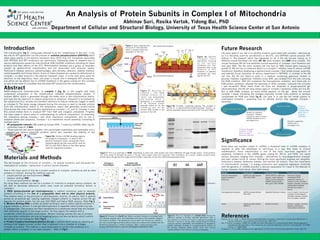
An Analysis of Protein Subunits in Complex I of Mitochondria
Abhinav Suri, Rasika Vartak, Yidong Bai, PhD
Department of Cellular and Structural Biology, University of Texas Health Science Center at San Antonio
The mitochondria (fig 1), colloquially referred to as the “powerhouse of the cell”, is the
key site for ATP generation via the process of oxidative phosphorylation (OXPHOS) which
takes place mainly in the electron transport chain (ETC) (Fig 1b). Complexes associated
with OXPHOS and ATP production are particularly interesting areas of research due to
various deficiencies caused by mitochondrial DNA (mtDNA) mutations encoding for these
proteins and their effects on the ETC. Mitochondrial diseases are a group of disorders
caused by dysfunctional mitochondria and are associated with seizures, ataxia,
Parkinson's disease, dystonia, opthalmoplegia, optic atrophy, cataracts, diabetic mellitus,
cardiomyopathy, and kidney failure. Some of these diseases are caused by deficiencies in
complex I, a major enzyme in the electron transport chain. It is the main entry point for
electrons to the respiratory chain and plays a crucial role in adequate ATP production,
one which can be altered due to mtDNA mutations in the genes coding for this complex
as well as the other 4 complexes that facilitate ATP production the mitochondria.
Abstract
NADH:ubiquinone oxidoreductase, or complex I (fig 2), is the largest and least
understood component of the mitochondrial oxidative phosphorylation system. It
oxidizes NADH, which is generated through the Krebs cycle in the mitochondrial matrix,
and uses the two electrons to reduce ubiquinone to ubiquinol. Ubiquinol is re-oxidized by
the cytochrome bc1 complex and transfers electrons to reduce molecular oxygen to water
at complex IV. The redox energy released during this process is used to transfer protons
from the mitochondrial matrix to the periplasmic space that generates proton-motive
force across the inner mitochondrial membrane at complex I, III, and IV. Complex V uses
this proton-motive force to produce ATP from ADP and inorganic phosphate. Progress
has been made in recent years in understanding its subunit composition, its assembly,
the interaction among complex I and other respiratory components, and its role in
oxidative stress and apoptosis. Complex I is a membrane bound assembly consisting of
multiple parts:
• 45 polypeptide subunits (38 coded by nuclear DNA, 7 coded by mtDNA). Refer Fig 2b.
• combined mass of 1 Mda
• These subunits are bound together into subcomplex assemblies and eventually into a
complex via various assembly proteins, which also maintain the stability of the
complex.
The lab focuses on the structure of complex I, its cellular functions, and discusses the
implication of complex I dysfunction in various human diseases.
Figure 2b: Complex I assembly pathway. The complex is
formed in a subunitàsubcomplexàcomplex fashion via
various assembly factors and such as B17.2L and CIA30.
1. PAGE (polyacrylamide gel electrophoresis): a method commonly used to separate
proteins according to the size of a polypeptide chain and no other physical property.
This method relies on the principle of electrophoretic mobility. An electric field is applied
across an acrylamide gel, causing negatively charged proteins to migrate across the gel
towards the positive anode. Our lab uses SDS-PAGE and Native PAGE analysis. Refer Fig 3.
2. Western Blotting (WB): a widely accepted technique used to detect specific proteins in
a given sample of extract. It uses gel electrophoresis to separate native proteins by size
of polypeptides. The proteins are then transferred to a membrane where they are stained
with antibodies specific to the target protein. For this reason the western blot is
sometimes called the protein immunoblot. Western blotting involves the use of primary
and secondary antibodies not only as targeting factors but also as factors which confirm
the protein we are looking for. Refer Fig 4.
Figure 4: This is an example of a western blot. During the
detection process a primary antibody (generated when a
host species of immune cell culture is exposed to protein of
interest) is applied to the gel. After rinsing the gel to remove
unbound primary antibodies, the membrane is exposed to
another antibody, directed at a species-specific portion of
the primary antibody (these usually come from animal
sources different from those of primary antibodies).
3. Protein Complex-Immunoprecipitation (Co-Ip): a method which works by selecting an
antibody that targets a known protein that is believed to be a member of a larger
complex of proteins. This method is used traditionally to confirm the existence of a
protein within a complex, in our case complex I. Refer to Fig 5.
Future Research
One of the major parts of this lab is protein analysis of complex I proteins as well as other
proteins of interest. Among the methods used are
• polyacrylamide gel electrophoresis (PAGE)
• western blotting (WB)
• co-immunoprecipitation (Co-Ip).
By using these methods (as well as a number of controls) to analyze various proteins, we
are able to decisively determine which ones could be potential formation factors in
complex I.
Materials and Methods
The main goal of our lab is to identify proteins associated with complex I deficiencies
as it is the entry point for all electrons in the ETC, and OXPHOS cannot produce ATP
without it. The research which has happened so far in the lab focuses mainly on 3
different mouse fibroblast cell lines: A9, 4A clone isolated, and 4AR clone isolated. The
mouse fibroblast A9 cell line exhibited normal assembly of complex I and therefore had
wild type DNA. The 4A clone isolated cell line had an ND6 mutant gene (caused by
growth of A9 cell line on rotenone which is a complex I inhibitor used to induce mtDNA
mutations), therefore developing a dysfunctional complex I that lacked proper assembly
and stability. As an indication of serious impairment in OXPHOS, in contrast to the A9
cell line, the 4A cell failed to grow in a medium containing galactose instead of
glucose. However, a galactose- resistant clone, 4AR, was isolated from the cells carrying
the ND6 mutation. 4AR still contained the homoplasmic mutation, and there was no
ND6 protein synthesis, whereas the assembly of other complex I subunits into complex
I was recovered. When these three cell lines are observed in polyacrylamide gel
electrophoresis, the A9 cell lines shows signs of complex I existence unlike cell line 4A.
But in 4AR PAGE analysis, an extra streak appears on the gel above the normal
complex I streak indicating the changed assembly formed more proteins to perhaps
compensate for ND6 loss (refer fig 4). Our goal is to use the cell model system to
understand Complex I assembly and to identify factors that regulate Complex I
assembly.
Disorder Genetic Origin Clinical phenotype Functional defects
Classical mitochondrial
diseases (complex I
deficiency)
mtDNA-encoded subunits:
ND1-ND6, ND4L
LHON, MELAS, adult-onset
dystonia, Leigh syndrome,
CPEO, exercise intolerance,
MERRF, bipolar disorder,
MCI, ptosis, MW, NIDDM
Increased or altered
mitochondrial ROS
production, reduction in
complex I activity, disrupted
complex I assembly, decreased
mitochondrial membrane
potential and complex I
activity
Neurodegenerative disorder mtDNA-encoded subunits:
ND1,ND5
Idiopathic Parkinson’s disease Loss of complex I activity and
a tendency toward apoptotic
cell death, reduction of protein
level of complex I subunits
Cancer Nuclear-encoded subunits:
GRIM-19
Hürthle cell tumors Defective complex I assembly,
apoptosis, and defective
mitochondrial metabolism
Figure 1: Basic anatomy and
structure of mitochondria
which is located within the
cytoplasm of the cell.
Found to
possess a
mutated ND6
gene; loss of
complex I
assembly
Mouse fibroblast A9 cell
line
Growth on rotenone
(complex I inhibitor;
commonly used to induce
mtDNA mutations) for a
prolonged time.
4A clone isolated
4AR clone isolated
Contains the same
mutation in ND6
gene; Complex I
assembly restored.
Growth in galactose
medium for a
prolonged time.
Figure 3: This is an example of PAGE. Depending on their size, each protein will move differently through the gel matrix, small proteins
will more easily fit through the pores in the gel, while larger cones will encounter more resistance. As a result, smaller proteins travel
farther down the gel than the larger proteins.(http://www.sfu.ca/bisc/bisc-429/electrophoresis.html)
Figure 2b: Human mtDNA showing regions
encoding for complexes I- V used in the
electron transport chain. Among the most
essential genes are the ones which code for
ND1-6, and NDL4. Most of the other genes
code for chaperone proteins.
Significance
Since there are multiple copies of mtDNA, a threshold level of mtDNA mutation is
required to alter the phenotype of cell/tissue in a way that leads to clinical
manifestation. Since mitochondria is one of the most important regulators of
apoptosis, mutations in the mtDNA genes coding for ND1-6, and ND4L can cause
various disorders ranging from complex I deficiencies to neurodegenerative diseases
and even certain forms of cancer. Among the more significant diseases are idiopathic
Parkinson's disease, Alzheimer disease, and Hürthle cell tumors. Thus the importance
of mitochondrial complex I in energy production and apoptosis regulation combined
with the genetics of mtDNA provides a rational explanation for many of the features of
human diseases listed above. (See table below)
Figure 5: Process of a Co-IP test. When a protein mixture is incubated with
an antibody coupled resin, the antibodies will bind to their specific antigens
and various proteins. The separation occurs when the mixture as a whole is
put into a centrifuge and the proteins which are unbound are washed and the
remaining substance is further analyzed. (http://www.piercenet.com/
browse.cfm?fldID=9C471132-0F72-4F39-8DF0-455FB515718F)
Complex I
Figure 1b: Electron
t r a n s p o r t c h a i n
specifically focusing
o n t h e v a r i o u s
complexes and their
respective roles in the
synthesis of ATP.
Complex I is typically
considered the entry
way for the ETC.
Figure 2: Structure of
complex I. The 1Mda
complex consists of 2
“arms” one hydrophobic
and another which is
hydrophilic. It is comprised
of 45 individual protein
subunits coded for by the
mtDNA as well as the
nuclear DNA located in the
nucleus of the cell.
ReferencesSharma, R; Lu, J; Bai, Y. (2009) Mitochondrial Respiratory Complex I: Structure, Function and Implication in
Human Diseases. Current Medicinal Chemistry,16, 1266-1277
Bai, Y; Attardi, Giuseppe. (1998) The mtDNA-encoded ND6 subunit of mitochondrial NADH dehydrogenase is
essential for the assembly of the membrane arm and the respiratory function of the enzyme. The EMBO
Journal, 17, 4848-4858
Anderson, S. et al. (1981) Sequence and organization of the human mitochondrial genome. Nature, 290, 45