Lupus case report
•
2 likes•2,823 views

1) A 16-year old female presented with a 3 month history of oral ulcers. Examination also revealed a butterfly rash on her face and a history of joint pain. 2) Based on these findings along with laboratory tests, she was diagnosed with systemic lupus erythematosus (SLE). Oral ulcers can be an early sign of SLE. 3) Treatment with steroids and immunosuppressants led to improvement of her oral and skin lesions. This case highlights the importance of thorough medical history and examination of extra-oral sites in accurately diagnosing systemic conditions that present with oral manifestations.

Recommended
More Related Content
Similar to Lupus case report
Similar to Lupus case report (20)
More from Abu-Hussein Muhamad
More from Abu-Hussein Muhamad (20)
Recently uploaded
Recently uploaded (20)
Lupus case report
- 1. 117 INDIAN JOURNAL OF DENTAL EDUCATION The Indian Journal of Dental Education (ISSN 0974-6099, Editor-in-Chief Registered with Registrar of Balwant Rai Newspapers for India: Assoc. Prof, Commander, ILWEG Moon Mars Mission, 100 B DELENG/2008/32281) Kepler Space University, USA published quarterly, is dedicated to the dissemination of new Executive Editor knowledge and information Rajnish K. Jain on all fields of dentistry. The IJDE publishes original peer- reviewed articles that examine Deputy Editors all phases of dental treatment, Jasdeep Kaur, India reports on unusual and interesting case presentations Maria Catalina, Astronaut Teacher Alliance, NASA JPL Solar System and invited review papers. Ambassador, USA The aim of this journal is to Editor Emeritus Managing Editor convey scientific progress in S.C. Anand A. Lal dentistry for the benefit of the dental health of the community. The journal serves valuable tool for helping clinicians, general International Editorial Advisory Board practitioners, teachers and Abbas Taher, London, UK Hamid Jafarzadeh, Iran administrators involved in the Abu-Hussein Muhamad,Athens, Greece Aous Dannan, Germany prevention and treatment of Adebola Oluyemisi Ehizele, Nigeria Lojain Jibawi, Syria dental disease. Alexandra Roman, Romania Marit Vandenbruane , Clement Chinedu Azodo, Nigeria Netherlands Dean Boss, USA Helen James, USA G. Wellam, Belgium L. James, UK Jaipaul Singh, UK Marcelo Carlos Bortoluzzi, Brazil Abdel Rahman Mohammad Said, Al-Tawaha,Jordan Natheer H Al-Rawi, UAE Inayatullah Padhiar, Karachi Patrick T, Belguim Florent Richy, Belgium Rafael Manfro, Brazil Ulrich Suchner, Germany Sandrine Brunel-Trotebas, Figen Cizmeci Senel, Turkey Romania Bora Bagis, Turkey National Editorial Advisory Board Printed at B.K. Behra, Rohtak Deepti Dwivedi, Lucknow R.V. Printing Press Manaswin Tripathi, New Delhi C-97, Okhla Industrial Area Naveen Gupta, New Delhi Phase-1 R. K. Sharma, Rohtak New Delhi - 110 020 S. C. Narula, Rohtak Surendra Nath, Delhi All right reserved. The views and opinions expressed Corresponding address are of the authors and not of the Indian Journal of Red Flower Publication Pvt. Ltd. Dental Education. The Indian Journal of Dental 41/48, DSIDC, Pocket-II, Mayur Vihar Phase-I Education does not guarantee directly or indirectly P.O. Box 9108, Delhi - 110 091(India) the quality or efficacy of any product or service Phone: 91-11-65270068/48042168, Fax: 91-11-48042168 featured in the advertisement in the journal, which E-mail: redflowerppl@vsnl.net, Web:www.rfppl.com Volume 3 Number 3, July-Sept 2010
- 2. 119 Indian Journal of Dental Education Volume 3 Number 3 July-Sept 2010 Contents Role of oral medicine specialist in disclosing systemic lupus erythematosus: Adiagnostic dilemma, Running title: systemic luapus erythematosus 121 Zahra Delavarian, Maryam Amir Chaghmaghi, Pegah Mosannen Mozafari, Mohamad Reza Hatef Fard, Rheumatologist Oral Health Care of elderly in India: Present Scenario and Future Concerns 127 Pankaj Datta, Sonia Sood A review on repair of fracture porcelain 133 Roseline Meshramkar The effect of developer age and file thickness on diagnostic accuracy of Kodak insight (F-speed) and Ektaspeed plus (E-speed) films in position assessment of file tip to radiographic apex 139 A. Dabaghi, M. Lomee, S. Saati Instructions to authors 146 Volume 3 Number 3, July-Sept 2010
- 3. 121 Indian Journal of Dental Education Volume 3 Number 3, July-Sept 2010 Role of oral medicine specialist in disclosing systemic lupus erythematosus: A diagnostic dilemma Zahra Delavarian* Maryam Amir Chaghmaghi** Pegah Mosannen Mozafari*** Mohamad Reza Hatef Fard**** ABSTRACT Systemic lupus erythematosus(SLE) is a connective tissue disease in which organs such as liver, kidney and heart in addition to skin and mucosa are involved. Oral findings are one of the diagnostic criteria which can be presented with ulcer or red and white lesions. In this article we report a case of SLE that is diagnosed by oral medicine specialist on the basis of oral ulcers. A 16-year-old female was referred to oral medicine department with 3 months lasting oral ulcers. There was a history of transient artheralgia in review of systems. In extra oral examination a butterfly diffuse erythema was observed on nasal bridge and malar prominences. Oral ulcers had different forms and involved different parts of oral mucosa. Due to chronic oral ulcers , malar rash and history of artheralgia ,a presumably diagnosis of SLE was affirmed. She was referred and hospitalized to rheumatology department. Oral and skin lesions were improved significantly in follow up examination. Oral findings may be the first diagnostic presentation of SLE. It is important for dentists to pay attention to medical history and different systemic symptoms to achieve accurate clinical diagnosis. Key words: Systemic lupus erythematosus, Oral ulceration, Case report, Oral Medicine, Iran INTRODUCTION several factors such as autoantibodies, immune complex, tissue damage, genetic factors (e.g. specific HLA types and gene loci), Systemic lupus Erythematosus (SLE) is one environmental factors (e.g. exposure to sun of the most important immunity related light and infections) , endocrine agents and diseases with unknown etiology, although drugs can predispose an individual to this disease (1,2) .There are four main clinic- Author’s Affilation: *Associated Professor of Oral pathological forms: systemic, discoid (chronic Medicine , Dental Research Center of Mashhad Dental Faculty, cutaneous),acute cutaneous and subacute Mashhad University of Medical Sciences, Mashhad, Iran, **Assistant Professor of Oral Medicine , Dental Research cutaneous(3).Organ damage occurs as a result Center of Mashhad Dental Faculty, Mashhad University of of direct attachment of autoantibodies to host Medical Sciences, Mashhad, Iran, ***Assistant Professor of antigens or precipitation of immune complex Oral Medicine , Dental Research Center of Mashhad Dental Faculty, Mashhad University of Medical Sciences, Mashhad, in small vessels and tissues (vasculitis). (1,2,4) Iran, ****Associated Professor of Rheumatology School of Medicine, Mashhad University of Medical, Sciences, Imam There is a female predominance (10:1) and Reza Hospital, Mashhad, Iran. blacks are involved more frequently. Reprint’s request: Pegah Mosannen Mozafari, Assistant SLE typically arises in adults aged 15 to 45. Professor of Oral Medicine, Dental Research Center of Mashhad Dental Faculty, Mashhad University of Medical (1,2,5)Only 15% of individuals with SLE are Sciences, Mashhad, Iran. Email: Mosannenp@mums.ac.ir, younger than 18 year at the time of Tell:0098 511 8829501-15, Fax :0098 511 8829500, Mobile number:0098 915 306 0496. diagnosis(6). (Received on 24.08.2010, accepted on 26.010.2010) Volume 3 Number 3, July-Sept 2010 © Red Flower Publication Pvt. Ltd
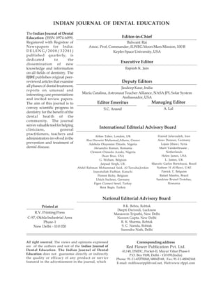
- 4. 122 Zahra Delavarian, Maryam Amir Chaghmaghi, Pegah Mosannen Mozafari, Mohamad Reza Hatef Fard Clinical manifestations of this disease vary The affected sites are the buccal mucosa, upon specific organ involvement. Fever, gingiva, vermilion border of the lips, palate fatigue and weight loss are of clinical and tongue (1, 8) .Usually diagnosis of SLE is components. (7) Arthritis is the most common performed by physicians but at least one case clinical manifestation of this disease (in 90% report exists about diagnosis of SLE by dentist of patients) (1,7) and usually appears (11). symmetrically. Interphalanges, knees, wrist This article, presents a case of SLE, in which and metacarpal joints are affected more the diagnosis was made based on oral frequently.(1) Malar rash, dry pruritic skin, manifestations. Despite a history of skin lesions gasterointestinal disorders and muscle spasms and articular pain, the patient had received are of other clinical signs.(1) Diagnosis of this improper treatments and Oral Medicine disease can be made via clinical and Specialist could reveal the disease. paraclinical findings.. If four of 11 criteria become evident simultaneously or consecutively in the course of this disease CASE REPORT diagnosis can be made with a 75% sensitivity and 95% specificity. (1) A 16-year-old female was referred to Oral Oral manifestations can be the first Medicine Department of Mashhad Dental presentation of disease, and may lead to Faculty in OCT 2008. She complained of oral diagnosis. Incidence of oral manifestations, ulcers with three months duration. In review was first reported by Monach (1931)as 50%, of systems, there was a history of transient (8) and Vasculitis is considered as the main artheralgia in knees, elbows and wrists in 6 etiology of oral lesions (1,7,8,9). months before initiation of oral manifestations. These manifestations include: nonspecific Ibopruphen, calcium and vitamin D was chronic ulcers, erosion, inflammation, prescribed for her by an internist, and partial erythema and keratotic white lesions (papule, relief was obtained after this therapy. There plaque …) or even granulomatous lesions and was also a history of hair loss. In extra oral malignant transformation of oral ulcers. examination, generalized erythema was seen (1,3,7,8)Candidosis, periodontal disease and on nasal bridge and malar region (Butterfly temporomandibular disorders and rash) with exfoliation of skin in some areas desquamative or marginal gingivitis are other (Fig 1) accompanied by a thick crust on the oral findings of SLE. (1, 8, 9, 10,11) In advanced lower vermilion border. SLE , xerostomia may appear (1).Sometimes delayed primary and permanent tooth The patient noted exacerbation and eruption and twisted root formation can be exfoliation of Malar rash after sun exposure. encountered as a result of corticosteroids She was advised to use sunscreen by a treatment (10). dermatologist and a few resolution was acquired. Fig 1: Butterfly rash and exfoliation of cheeks and nasal bridge skin and lip crust. Indian Journal of Dental Education
- 5. Role of oral medicine specialist in disclosing systemic lupus erythematosus: Adiagnostic dilemma 123 In intraoral examination multiple ulcers and palatal aspects of maxillary gingiva with different patterns were seen in several (premolar and molar region) (Fig 2) areas of oral mucosa such as palate, buccal Multiple small , clustered ulcers were region, gingiva and tounge. observed in right lateral side of hard palate Diffuse map like ulcers were present adjacent to first premolar and molar, involving bilaterally on buccal aspects of mandibular an area of 1.5×1.5 cm diameter. there was no attached gingiva (canine, premolar region) keratotic lesion with reticular pattern Fig 2: A large deep ulcer of 1×3 cm diameter was present on marginal gingiva of first right permanent molar extending to hard palate. (lichenoid reaction). SLE was considered as a dl and lymphopenia. CRP, RF and renal possible clinical diagnosis by an oral medicine function tests were normal. specialist due to chronic oral ulcers, butterfly Our patient’s condition satisfied Six criteria rash and history of articular involvement. So, for a diagnosis of SLE: 1)Malar rash 2)Oral because all of these signs represent a systemic ulcers 3)Photosensitivity 4)Lymphopenia disease, there was no need for biopsy of oral 5)Positive anti ds DNA 6)Positive ANA, so lesions (especially when there was no evidence the diagnosis of SLE was confirmed. of lichenoid pathology). The Patient was referred to Rheumatology clinic for further The treatment was initiated by diagnostic tests and appropriate therapy. Prednisolone, Hydroxychloroquine, Calcium D, Cephteriaxon (due to uretral infection). She was admitted to Imam Reza hospital After 2 weeks of flare up control, she was with provisional diagnosis of SLE. Laboratory discharged with instructions to continue her tests such as CBC, Rheumatoid factor, ANA prior medications. No topical treatment was (Antinuclear antibody), Anti ds (double needed for oral ulcers, due to rheumatologic strand) DNA, CRP (C-reactive protein) and clue. ESR (Erythrocyte sedimentation Rate) and kidney function tests were ordered for the After 48 days, the patient was examined in patient. The results included: positive ANA, Oral Medicine Department. Malar rash was Elevated ESR, Anti ds DNA>300, Hgb=9gr/ relatively faded out and there was no exfoliation.( Fig 3)The lip ulcers were Fig 3: Significant improvement in Butterfly rash and lip crust Volume 3 Number 3, July-Sept 2010
- 6. 124 Zahra Delavarian, Maryam Amir Chaghmaghi, Pegah Mosannen Mozafari, Mohamad Reza Hatef Fard completely healed and a mild facial edema was after appropriate treatment (9).In one research evident (possibly due to corticosteroid on Venezuelan patients oral lesions were therapy). found in first two years after diagnosis (9) Oral Nine months later (Aug 2009) the patients ulcer was the main oral manifestation in our was admitted once more for ten days, with a case. Rhodous has reported other oral findings complaint of extreme fatigue, arthralgia and such as xerostomia (%100 of cases), mucositis myalgia . Oral ulcers were not evident in this and glossitis (81/3%), glossodynia (87.5%) and visit. There was no lupus nephritis, avascular angular cheilitis (87.5%), in evaluated necrosis and cardiopathy. Appropriate patients(7) . the severity of these symptoms is treatment was administered for her by compatible with disease flare up(7,13) rheumatologist. although no significant changes in titers of c3,ANA or Anti ds DNA has been attributed In October 2009 she was called and no by some authors(13) Lymphadenopathy and complication was emerged. focal parotid necrosis (14) are another occasional findings in head and neck area. DISCUSSION Fernandes et al (10) attempted to address oral health an TMJ dysfunction in JSLE patients. They understood that JSLE patients Although this case represents an unusual had poor oral hygiene , higher incidence of diagnostic dilemma, but it seems that in gingivitis and TMJ dysfunction especially in Juvenile SLE(JSLE) ,this kind of error is not so those on long corticosteroid and rare(6) .In JSLE the presenting signs are immunosuppression treatments. protean and many of them are common complaints among adolescents.(e.g. fatigue , In our patient, because histopathologic artheralgia),so inexperienced physician may examination of oral ulcers had no benefit and fail to considered SLE in differential diagnosis systemic involvement would lead to diagnosis, of transient artheralgia and a facial rash in an biopsy was not performed. There are other adolescent female. differentiated diagnoses for extra oral manifestations of this patient. Similar malar Children and adolescents have a more lesions can be seen in achne rosacea, seboreic severe disease presentation (6) and develop dermatitis and achne vulgaris and some kinds severe organ damages more quickly. So early of viral infections (15,6) Although other diagnosis and intervention is a crucial point systemic signs are not compatible with these to improve overall outcome of treatment. Our diagnoses. Other systemic diseases such as case had at least nine months diagnostic delay Behçet’s syndrome and dermatomyositis were despite articular and cutaneous symptoms. also mentioned for this case. Tucker reported a summary of common Absence of recurring oral and genital ulcers presenting signs of JSLE with mucocutaneus and presence of malar rash excluded Behçet’s ulceration as a relatively rare presenting signs syndrome. Absence of muscular and in this age group.(6) He speculated that every pathognomonic skin involvement ruled out the adolescent who appear to have unexplained diagnosis of dermatomyositis. “un wellness” with vague symptoms of SLE should be further evaluated for diagnosis of DLE was also included in differential this entity. It is more fundamental in a prone diagnosis. But in DLE, the lesions are limited ethnic group (e.g. Asian adolescents) to skin and mucosa (with no systemic involvement) and oral involvement appears Prevalence of oral manifestations of SLE has as lichenoid reactions in combination with been reported as %7 to 87.5% (7,12) in different skin discoid rash (a finding not observed in studies. The difference can be due to lack of our case.) (16) diagnosis of SLE at the time of oral presentation or resolution of these findings Immunologic findings also are of diagnostic criteria for SLE. Elevated Anti Nuclear Antibody (ANA) titer (1/40 or high) is the most Indian Journal of Dental Education
- 7. Role of oral medicine specialist in disclosing systemic lupus erythematosus: Adiagnostic dilemma 125 sensitive diagnostic criterion for SLE in kept in mind by general dentists to reveal an serologic tests, and was positive in this case. undetermined systemic condition. Elevated ANA titers can be found in %99 of SLE patients; however, in early stage of REFERENCES disease, it can be negative. ANA is not an specific test for SLE since one study revealed elevated ANA titers in %32 of normal adults 1. Albilia JB, Lam DK, Clokie CML, Saìndor GKB. (5,16) Systemic lupus erythematosus: A review for dentists. J of the Can Den Assoc, 2007; 73(9): ANA is positive in other diseases such as 823-28. Sjögren’s syndrome (%68), sclerodermy (40- %75) and rheumatoid arthritis (25-50%) but 2. Sharon GC. The pathogenesis of systemic lupus erythematosus. Orthopedic Nursing, 2006; 25(2): lower titers and different immuno fluorescent 140-5. pattern are observed in these cases. 3. Compilato D, Cirillo N, Termine N, Kerr AR, Anti ds DNA survey has high specificity Paderni C, Ciavarella D,Campisi G.Long- and low sensitivity for SLE and in JSLE is a standing oral ulcers: Proposal for a new ‘S-C-D prominent laboratory profile. In this patient it classification system’. J Oral Pathol Med, 2009; was increased. Although complement levels 38(3): 241-253. (C3, C4, C5) are normal in variable kinds of 4. Ramos-casals,M.Nardi,n.Lagrutta,m.Brito- vasculitis, they are decreased in SLE, as a result zeron,P.Bove,A.Delgado,G.(et al) vasculitis in of consumption. In inflammatory process in systemic lupus Erythematosus: prevalence And second administration (flare up) of this patient, clinical characteristics in 670 patients. Medicine, C3 and C4 levels were low. SLE owns episodes 2006; 85(2): 95-104 of flare up and remission (2, 7) and decreased 5. Oral lupus Erythematosus[on line].Available levels of complement is the sign of disease flare from URL:http://www.eaom.net/app/prvt/ up. (17) VediNotizia.d/Notizia-96 .accessed sept 20, 2006. The aim of treatment for SLE in acute phase is management of acute attacks. And because 6. Tucker LB. Making the diagnosis of systemic of multiple organ involvement, treatment plan lupus erythematosus in children and adolescents. Lupus, 2007; 16: 546-9. is based on clinical presentation. (1) 7. Rhodus, L.Johnson D K. the prevalence of Oral Management regimen in these patients manifestations of systemic lupus erythematosus. include NSAIDS, corticosteroids, Anti malaria Quintessence International, 1990; 21(6): 461-5. drugs and Immunosuppressants.(1) Prognosis 8. Fernandes, R,L. Review of systemic lupus depends on severity and extent of organ Erythematosus. Oral Surg Oral Med Oral Pathol involvement and complications of treatment. Oral Radiol Endod, 2001; 91(5): 512-6. poor prognostic factors are young age at onset, male gender, poor socioeconomic status and 9. López-Labady J, Villarroel-Dorrego M, González N,Pérez R, Mata De Henning M .Oral positive titers of antiphospholipid antibodies. manifestations of systemic and cutaneous lupus (1) Since oral lesions respond well to systemic erythematosus in a Venezuelan population . J therapy, no additional treatment is necessary. Oral Pathol Med, 2007; 36(9): 524-527. 10. Fernandes, RL,Savioli C,Siqueira JTT,Silva CAA. CONCLUSION Oral health and masticatory system in juvenile systemic lupus Erythematosus. Lupus, 2007; 16: 713-9. SLE is a systemic disease with multiple organ 11. Jayakumar ND, Jaiganesh R, Padmalatha O, involvement and variable diagnostic features. Sheeja V. Systemic lupus erythematosus.Ind J So one may be referred to a dentist with Dent Res, 2006; 17: 91-3. chronic oral ulcers, with an undiagnosed SLE. 12. Meyer V, kleinheinz J, Handschel J, kruse-losler Importance of achieving a complete “review B, weingart D, joos V. Oral findings in three of systems” and accompanying signs must be different groups of immuno compromised patients. J Oral pathol med, 2000; 29(4): 153-8. Volume 3 Number 3, July-Sept 2010
- 8. 126 Zahra Delavarian, Maryam Amir Chaghmaghi, Pegah Mosannen Mozafari, Mohamad Reza Hatef Fard 13. Urman JD, Lowenstein MB, Abeles M, Weinstein 15. Zuber MA.Butterfly rash: No lupus. Zeitschrift A. Oral mucosal ulceration in systemic lupus fur Rheumatologie, 2009; 68(5): 409-10. erythematosus. Artheritis Rheumatism, 2005; 21: 16. Gill JM, Quisel AM, Rocca PV, Walter DT. 58-61. Diagnosis of systemic lupus erythematosus. Am 14. Carron J, Karakla DW, Watkins DV. Focal Fam Physician, 2003; 68(11): 2179-86. parotid necrosis in systemic lupus 17. Roane DW, Griger DR. An approach to erythematosus: Case report and review of the diagnosis and initial management of systemic literature. Oral Surgery Oral Medicine Oral vasculitis Am Fam Physician, 1999; 60(5): 1421- Pathology Oral Radiology Endodontology, 1999; 30. 88: 455-460 . Indian Journal of Dental Education
- 9. 127 Indian Journal of Dental Education Volume 3 Number 3, July - Septemebr 2010 Oral Health Care of Elderly in India: Present Scenario and Future Concerns Pankaj Datta* Sonia Sood** ABSTRACT India has a rapidly growing elderly (60 +) population of 77 million which is likely to rise up to 300 million by 2050. For the most of this rapidly growing geriatric population there are no specialized oral health services. The elderly suffer from multiple oral health problems. The Indian population in the 21st century requires an in-depth understanding of the co-relation between oral health and general well being.1 Viewing these issues through the lens of oral health care provider allows an analysis of current oral health care status of the elderly in India; understand the cause of their poor oral health, their attitudes and treatment needs. The unique combination of growing age, physical disability, personal habits, socio- economic status and our oral healthcare system presents challenges for appropriate oral health care.2 The present article highlights on the need to understand the shortfalls in its current oral heath status in elderly and formulate strategies to improve its oral healthcare structure as well as education policy in geriatric dentistry to help resolve problems of oral health care for the elderly in India. Key words: elderly, oral healthcare, dental treatment needs, geriatric dentistry INTRODUCTION million and 177 million elderly in the year 2001 and 2025 respectively, which will rise to 300 million in 2050 3. Special features of the elderly Demographic ageing is a global in India are: 52% of elderly in the country are phenomenon. India’s booming population of women. 71% of the elderly reside in rural above one billion people and improved life areas. Nearly 75% of the elderly are expectancy (63 for males and 65 for females) economically dependent. 30% of the elderly 1,3 has led to rapidly increasing number of are below the poverty line. 73% of the elderly elderly people (>60 years age group). This are illiterate. It is estimated that 90% of the includes both healthy adults and adults who old people belong to unorganized sector (i.e. are cognitively and physically challenged without gratuity, pension etc.) 4. With such and/or medically compromised. large population estimates of the elderly, out The 20.2 million population of elderly was of which most of them are underserved, 5.60% (4) (5.6%) in 1951 which climbed to considerable efforts are required to support the 7.63% in 2001 and is likely to climb to 14% in geriatric oral health. 2025 2. In absolute numbers there are 77 EXTENT OF POOR ORAL HEALTH Author’s Affilation: *Vice-Principal & Head, Deptt. Of Prosthodontics, Inderprastha Dental College & Hospital, Sahibabad, Ghaziabad UP, **Post-graduate Student, Department of Public Health Dentistry, ITS-CDSR, As per the National Oral Health Survey Muradnagar, Ghaziabad , UP (2004), poor oral health among elderly has Reqrint’s request: Dr. Pankaj Datta, C-86, Anand Vihar, resulted in a high level of tooth loss (29.3%), Delhi – 110 092, Mob; 9811774350/9811274799, E-mail: dental caries status (84.7%), periodontal pankajdatta97@rediffmail.com. (Received on 15.08.2010, accepted on 25.11.2010) Volume 3 Number 3, July-Sept 2010 © Red Flower Publication Pvt. Ltd
- 10. 128 Pankaj Datta, Sonia Sood disease (79.4%), mucosal lesions (10%) and drugs prescribed for these chronic diseases can oral cancer (0.5%) 5. cause adverse effects to the oral mucosa, lichenoid reactions, hypersensitivity and xerostomia 18, 19. A LINK TO SYSTEMIC HEALTH Elderly are especially at risk for caries and periodontal diseases if they suffer from The ill effects of poor oral conditions are xerostomia. It may be caused by illness, particularly significant among older people in radiation therapy and chemotherapy apart the form of caries, periodontitis and from medication. edentulousness. Direct ill effects cause a state Dental professionals must understand that of partial or complete edentulousness. the elderly must be considered under the Extensive tooth loss/ Ill-fitting prostheses category of “special needs and care” for reduces chewing performance and affects treatment due to their social, psychological, food choice; edentulous people tend to avoid physical and medical conditions 20. Thus, at dietary fiber and prefer refined foods leading times it may necessitate alterations in the to poor nutrition 6, weight loss 7 and problems treatment objectives, deviating from the in communication besides low esteem 8. standard norms with the prime objective to Poor oral health is a common risk factor for “compress morbidity and chewing disability” many systemic diseases; severe periodontal and keeping oro-dental apparatus in a state disease is associated with diabetes mellitus 9, of reasonable function. ischemic heart disease 10, 11, chronic respiratory disease 12 and osteoporosis 13. The challenge of maintaining oral health for the nursing CURRENT SHORTFALLS IN ORAL elderly holds additional danger of aspiration HEALTH CARE OF THE ELDERLY pneumonia 14, 15. As more epidemiological evidence links To have and maintain oral health, there are dental infections and systemic complications, three basics tenets that must be in place. For it should be clear that dental and health older adults, one or more of these tenets may benefits should not be compartmentalized be absent. As a result, the prevalence and rather it should be replaced with a new severity of oral diseases and conditions in older paradigm—that of including dental care in adults are a significant public health concern. comprehensive medical care improve our 1. Knowledge of the importance of oral geriatric patients’ quality of life and outlook.16 health and its value to overall health. There are compounding factors such as CHALLENGES OF ORAL HEALTH CARE deficiencies in knowledge, attitudes, practices IN ELDERLY and socioeconomic status which predispose the elderly to oral health problems. Fear of surgical nature of work may make them As they age, older people are more likely to apprehensive of dental care, and may deter live alone, may be socially isolated and some them from seeking it. Many may not realize are unable to manage walking without the benefits of good oral health as the effects assistance, have failing eyesight and other at times may not be evident instantly. physical infirmities. The high prevalence of oral cancer in India The maintenance of oral health becomes is related to behavioral risk factors such as more difficult if the elderly person is also poor oral hygiene, improper diet, alcohol and suffering from other systemic illness e.g. tobacco abuse. arthritis, diabetes, cardiovascular disease, osteoporosis, neurological diseases associated 2. Physical ability to maintain oral health with age such as stroke, Alzheimer’s disease through oral hygiene practices. and Parkinson’s disease. 17. Many systemic Indian Journal of Dental Education
- 11. Oral Health Care of Elderly in India: Present Scenario and Future Concerns 129 Most elderly due to poor manual dexterity education and promotion of oral healthcare have difficulty in performing routine oral of elderly in underserved communities needs hygiene procedures, which increases the to be implemented by outreach activities of prevalence of dental decay, periodontal public health professionals. disease and edentulism in this population 21. There is need of setting up of mobile oral 3. Ability to access professional oral health health care services involving multidisciplinary services. teams to provide domiciliary services to the In India, primary health centres do not have elderly in the rural areas. Regular preventive the provision for dental care. This has left oral dental care with portable dental equipment health far behind other health services. It can be used to serve the functionally appears that oral health is not a priority in dependent elderly at home/nursing homes to our health care system. Except those in reduce the development of harmful oral health organized sectors like in government jobs, conditions. railways, defense services and public sector, Use mass media (particularly TV) to raise majority of the elderly have no oral health the public awareness and understand the security. importance and benefits of good oral hygiene. Most services for geriatric patients are on a Educate the public about the harmful effects “fee-for-service” basis in the private clinics of tobacco and alcohol abuse on the oral health which is expensive and not within the reach as it predisposes them to a high risk of of most of the elderly with reduced retirement periodontal disease and precancerous oral income. With the paucity of government lesions. Oral cancer is more common after age dental colleges/ dental departments of sixty and early detection is among a major government hospitals in the country; most of approach to prevention of the disease. the elderly patients do not get comprehensive treatment either due to lack of facilities or long waiting period 22 . There are no health TRAINING IN GERIATRIC DENTISTRY insurance plans which cover dental treatment except in an emergency (trauma). With an increasing awareness in the society Improper distribution of dental manpower about oral health and treatment needs, there in India has created a void in the desired has been a greater demand for geriatric healthcare status in the elderly. Older adults specialists in dentistry. To serve them better, are often at risk of limited access to oral health it is important to understand the physical, care because of transportation, economics, mental and socioeconomic background of the medical illness, social and personal reasons. elderly, their illnesses, medication and age- related disabilities. Thus, special training in geriatric dentistry is required 22. However, RESPONDING TO GERIATRIC ORAL there is no institute to provide it in India. Till HEALTH NEEDS the time we have geriatric dentists there will INCREASING THE AWARENESS AND remain an urgent need of specialists in KNOWLEDGE AT COMMUNITY LEVEL endodontics, periodontics, prosthodontics and public health to club together as a part of rehabilitative team to minimize the oral About 70% of the rural population does not disability and restore the oral health of elderly. have access to dental facilities 23. Currently, only 2% 24 of the specialists are being trained In the current scenario, the dental education in public health dentistry, whereas in a needs to be reframed with the rising need of country like India, there is a greater need for preparing students to care for the increasing these specialists to emphasize on the numbers of medically complex, dentate importance of oral health among elderly. elderly. It is time for a new model of dental Primary prevention, imparting dental health education to be implemented at undergraduate level so that it is more Volume 3 Number 3, July-Sept 2010
- 12. 130 Pankaj Datta, Sonia Sood 27 integrative with a variety of elderly patients, , there is acute shortage of dental manpower health care providers and individuals who are in the rural areas due to significant geographic involved in health care management of older imbalance in the distribution of dental population. colleges. This has resulted in two unfavorable Apart from people involved in dentistry outcomes. other health professionals must be provided (1) Though it improved the overall dentist oral health training and information on the to population ratio, there has been a great specific needs of older adults. variation in the dentist to population ratio in rural and urban areas. The dentist: population Lastly, there is an urgent need to educate ratio is 1:13,000 in the urban areas 23 and caregivers in families, assisted living, 1:250,000 in rural areas 28. supportive housing and nursing homes on how they can effectively assist older adults for (2) It left a big void in the geriatric oral health oral hygiene practices. care services in rural areas. Since, most of the dental colleges provide free dental treatment to people in nearby periurban and rural areas. NEED TO IMPROVE ORAL HEALTH To cover up the shortage of dentists to serve SERVICES the underserved populations in rural area there is an urgent need of expanding the use To fill up the desired level of oral health of dental auxiliaries in the provision of dental amongst elderly in India, National Oral Health services. Dental auxiliaries can provide Policy needs to be implemented. The negative services to rural patients without much impact of poor oral health on the quality of financial impact on the health agencies. When life of elderly is an important public health hygienists are utilized to the full scope of issue which must be addressed by policy- preventive practice, they can free time for makers. The need of dentists and dental restorative procedures by dentists. Denturists auxiliaries in National Health Program was can be utilized for directly providing removable suggested for providing oral health care at prostheses to the elderly. primary health care (PHC) and community Lastly, we need support other than dentists’ health care (CHC) as per the Bajaj Committee to help us to lobby government for geriatric Report 25 . This was further recommended by dental care. It is time for us to look after the National Oral Health Care Program, but generation which brought us to this level and unfortunately still the implementation part is let them feel proud of themselves for raising missing at PHC and CHC level 26. Till the time us. any positive step is taken by the government it is incumbent on us, as oral health professionals, to deal with this need and CONCLUSION provide access to care for elderly patients. The major cause of poor oral health due to There is a growing demand for oral health the absence of primary health care approach care among elderly in India. India needs a in dentistry is the prime area of focus where comprehensive gerontological oral health care oral health professionals (dentists and dental program with the following objectives. First, auxiliaries) should be increased. In 1990 there there is deficient data about the current oral were 3,000 registered hygienists and 5,000 health status and disease trends. Second, we laboratory technicians in India. There are no need to learn more about the efficacy of the registered dental nurses, chair side assistants current treatment modalities. Third, the future and denturists 24. dental needs and demands of the elderly needs To improve the shortage of dental to be explored. Fourth, the organization of the professionals, permission to open new dental dental health care delivery system to catch and colleges was granted. Despite increased address the changing and probably new oral number of dental colleges (291) in the country health problems of the elderly needs to be expanded. Fifth, to meet these challenges, Indian Journal of Dental Education
- 13. Oral Health Care of Elderly in India: Present Scenario and Future Concerns 131 geriatric dentistry needs to be developed to 13. Clare Van Sant. Preparing your office and team create a trained and dedicated workforce for the care of geriatric patients. Available at which can effectively plan and administer http://www.dentistrytoday.com/ME2/ geriatric oral healthcare delivery, education dirmod.asp (accessed on 5th March 2010). and research in India. Finally, the relationship 14. Abea S, Ishihaara K, Adachib M, Okuda K. Oral between oral health and general health must hygiene evaluation for effective oral care in preventing pneumonia in dentate elderly. be understood, if oral health care is to have a Archives of Gerontology and Geriatrics, 2006; reasonable chance of success. l43(1): 53-64 15. Awano S, Ansai T, Takata Y, Soh I et al Oral REFERENCES Health and Mortality Risk from Pneumonia in the Elderly. J Dent Res, 2008; 87(4): 334-339. 16. Rubinstein Helena Gail. Access to oral health 1. Available at http://www.prb.org/pdf08/ care for elders: mere words or action? Journal of 08WPDS_Eng.pdf (accessed on July 27th 2010) Dental Education, 2005; 69(9): 1051-1057. 2. Swami H M, Bhatia V. Primary geriatric health 17. Scully C, Ettinger RL. The influence of systemic care in India needs initiative in the new diseases on oral health care in older adults. J millennium. Indian J Prev. Soc. Med, 2003; 34(3): Am Dent Assoc, 2007; 138:7S-14S. 4. 18. Abdollahi M, Radfar M. A review of drug- 3. Peterson P E, Yamamoto T. Improving the oral induced oral reactions. J Contemp Dent Pract, health of older people: the approach of the WHO 2003; 4(1): 10-31. global oral health programme. Community Dent Oral Epidemiol, 2005; 33: 81-92. 19. DeRossi SS, Hersh EV. A review of adverse oral reactions to systemic medications. Gen Dent, 4. Shah N, Tank P. Rehabilitation and Residential 2006; 54(2): 131-8. Care Needs of the Elderly. Indian journal of psychiatry-CPG-2007. 20. Sandra Nagel Beebe. The special needs of elderly patients. Available at http:// 5. National Oral Health Survey and Flouride www.irishdentist.ie/articles/articles_ Mapping, 2002-2003. New Delhi: Dental (accessed on July 24th 2010). Council of India, Ministry of Health and Family Welfare, Govt. of India, 2004. 21. Talwar M, Chawla HS. Geriatric dentistry: Is rethinking still required to begin undergraduate 6. Walls AWG, Steele JG, Sheiham A, Marcenes W, education? Indian J Dent Res, 2008; 19: 175-7. Moynihan PJ. Oral health and nutrition in older people. J Public Health Dent, 2000; 60: 304–7. 22. Shah N. Geriatric dentistry: The need for a new speciality in India. The National Medical Journal 7. Ritchie CS, Joshipura K, Silliman RA, Miller B, of India, 2005; 18(1). Douglas CW. Oral health problems and significant weight loss among community- 23. DCI Perspective. Dentistry India. Sep 2007 — dwelling older adults. J Gerontol A Biol Sci Med Vol. 1, Iss. 1 available at http:// Sci 2000;55: M366–71. www.dentistryindia.net/article.php?id=1010, 8. Smith JM, Sheiham A. How dental conditions 24. Tandon S. Challenges to the Oral Health handicap the elderly. Community Dent Oral Workforce in India. J Dent Educ, 2004; 68 (7). Epidemiol, 1979; 7: 305–10. 25. Bajaj Committee report available at http:// 9. Shlossman M, Knowler WC, Pettitt DJ, Genco nihfw.org/NDC/DocumentationServices/ RJ. Type 2 diabetes and periodontal disease. J Reports/Bajaj%20Committee%20report.pdf Am Dent Assoc, 1990; 121: 532–6. (accessed on 2nd August 2010). 10. Trichopoulos D, Ascherio A, Willett WC. Poor 26. National Oral Health Care Programme oral health and coronary heart disease. J Dent (NOHCP) Implementation Strategies. Indian Res, 1996; 75: 1631–6. Journal of Community Medicine, 2004; XXIX(1). 11. Joshipura KJ, Hung H-C, Rimm EB, Willett WC, 27. Available at http://mohfw.nic.in/ Ascherio A. Periodontal disease, tooth loss and Adental.html. (accessed on 21st July 2010). incidence of ischemic stroke. Stroke, 2003; 34: 28. Industry Insight Indian dental industry; 47–52. available at http://www.cygnusindia.com/ 12. Scannapieco F. Role of oral bacteria in pdfs (accessed on 3rd Jan 2010). respiratory infection. J Periodontol, 1999; 70: 793–802. Volume 3 Number 3, July-Sept 2010
- 14. 132 Indian Journal of Dental Education Library Recommendation Form If you would like to recommend this journal to your library, simply complete the form below and return it to us. Please type or print the information clearly. We will forward a sample copy to your library, along with this recommendation card. Please send a sample copy to: Name of Librarian Library Address of Library Recommended by: Your Name/ Title Department Address Dear Librarian, I would like to recommend that the library subscribe to the Indian Journal of Dental Education. I believe the major future uses of the journal for our library would be: 1. As useful information for members of my specialty. 2. As an excellent research aid. 3. As an invaluable student resource. 4. I have a personal subscription and understand and appreciate the value an institutional subscription would mean to our staff. 5. Other Should the journal you’re reading right now be a part of your University or institution’s library? To have a free sample sent to your librarian, simply fill out and mail this today! Stock Manager Red Flower Publication Pvt. Ltd. 41/48, DSIDC, Pocket-II, Mayur Vihar, Phase-I P.O. Box No. 9108, Delhi - 110 091 (India) Tel: 91-11-65270068, 22754205, Fax: 91-11-22754205 E-mail: redflowerppl@gmail.com, redflowerppl@vsnl.net Website: www.rfppl.com Indian Journal of Dental Education
- 15. 133 Indian Journal of Dental Education Volume 3 Number 3, July-Sept 2010 A review on repair of fractureon repair A review porcelain of fracture porcelain Roseline Meshramkar ABSTRACT Because of their high esthetic qualities and mechanical stability in the oral environment, ceramic restorations are commonly used in daily dental practice. Due to the inherently brittle nature of porcelain restorative materials, failure of metal ceramic restorations under intraoral conditions is not uncommon. The majority (65%) of failures are observed in the anterior region (60% in labial, 27% in buccal, 50% in incisal and 80% in occlusal regions). Clinical studies show failure rates upto 90% for ceramic veneers. Because it is arduous to remove the ceramic restorations from the mouth, they are repaired intraorally, using a bonding system and composite resins. It is necessary to know the possible causes of fracture of porcelain, the various bonding systems and the composites resins used for repairing. The current review takes into account the majority of papers published in the last few decades concerning the issue of bonding composite resins to porcelains. Key Words: Porcelain fracture, bonding agent, ceramic repair, composite. INTRODUCTION porcelain fused to metal restorations are commonly used in daily dental practice.[7] It is to be expected that with increased application Since 1728, when Fauchard [1] first of this technique the number of failures also suggested the use of porcelain, the art and will increase.[8] Clinically failures often begin science of ceramics in restorative dentistry has as porcelain fractures that may be caused by evolved into a revolutionary method for inappropriate coping design, poor abutment aesthetically treating dental needs of a wide preparation, technical errors, contamination, variety. Porcelain inlays and crowns as well physical trauma or premature occlusion. [9] as the use of porcelain facial veneers are These fractures are mainly in the maxilla [75%] reported several decades ago. [2-4]Although and predominantly at the labial surface.[10] It aesthetically satisfactory, the brittle nature of is necessary to assess the possible cause of the early porcelain restorations limited their fracture so that the most suitable treatment wider application.[5] can be recommended. Depending on the In the 1960’s gold was used as a reinforcing extent of the area to be restored, cost and time under structure. [6], followed by MacLean’s available treatment may range from making application of high aluminous ceramic a new prosthesis, faceting or overcastting to substructure for fixed partial dentures. resin composite repairs.[11] Replacement of a Because of their high aesthetic qualities and failed restoration is not necessarily the most mechanical stability in the oral environment practical solution for obvious economic reasons and because of the complex nature of the restoration. [12] Because it is arduous to Author’s Affilation: Professor, Dept of Prosthodontics, SDM Collge of Dental Sciences, Dharwad E-mail: remove these restorations from the mouth roselinemeshramkar@yahoo.co.in, Received on 01.12.2010, ceramic restorations are repaired intra- accepted on 06.01.2010 orally.[10] Reprint’s request: Dr. Roseline Meshramkar, Professor, With development of the composite Dept of Prosthodontics, SDM Collge of Dental Sciences, Dharwad E-mail: roselinemeshramkar@yahoo.co.in. restorative materials and the introduction of (Received on 01.12.2010, accepted on 06.01.2010) organosilanes by Bowen [13] in 1962; solutions VolumeFlower Publication Pvt. Ltd © Red 3 Number 3, July-Sept 2010
- 16. 134 Roseline Meshramkar to the repair problem were possible. Two types from his experiments that cold-cured resins of bond, metal-resin and porcelain-resin are produced a stronger bond than mechanically involved in the repair process of ceramo-metal retained porcelain teeth, but that thermal restorations. Surface configuration, reactivity cycling was detrimental to the bond. A study of the bonding surface and the use of adhesive of porcelain teeth in cold-cured dentures by resins are important for metal-resin and Duhaney HN [27] in 1970 indicated that porcelain-resin bond. [14] To achieve a retention by bonding with silane solution was satisfactory bond between porcelain and as satisfactory as mechanical retention. composite resin several mechanical and Jochen and Caputo [28] reported that the chemical retention systems were developed. abrasion of the surface of porcelain with a Mechanical roughening of porcelain surfaces diamond rotary instrument increased the with a coarse diamond, Air-abrasion retention of the repair material. In 1978, (sandblasting) and acid etching with Eames et al [29] evaluated the composite resins hydrofluoric acid [15], acidulated phosphate utilizing silane coupling agents for repair of fluoride [16] , Ammonium biflouride [17] or porcelain. Porcelain denture teeth were used phosphoric acid[18] are some of the commonly in this study and acceptable bond strength for used methods to achieve retentive porcelain temporary repairs was reported. In 1978, surface texture. The organosilane repair Newburg and Pameijer [8] also studied the materials enhance the adhesions of the repair bond strength of composite resin to porcelain resin to the porcelain surface.[19,20] Within the denture teeth utilizing a silane coupling agent, last few years, several types of porcelain repair and reported that the samples produced a systems have been developed for use by the reliable bond. Highton et al [30] 1979 also dental profession. The purpose of this article studied the effects of silane coupling agents is to review the treatment pertaining to the on the composite resin/porcelain bond. The various porcelain repair systems. study indicated that the repair system using a bonding agent with acrylic resin was THE EVOLUTION OF PORCELAIN significantly stronger than the repair system REPAIR SYSTEMS using a composite resin. Nowlin et al [31] reported that fusion plus concise (3M Co. Dent products Div., st. Paul Historically, intraoral repair of fractured Minn) was superior to Dent-mat and 18% of porcelain restorations has required the original porcelain strength was regained. roughening of the porcelain surface with a rotary abrasive, application of silane fallowed In 1983, Ferrando et al [32] concluded that by composite to replace the contour of the Enamalite (Lee pharmaceuticals, South El restoration. [21,22] Early in the 1960s Monte, Calif.) was superior to Fusion plus manufacturers’ reinforced plastics with Adaptic (Johnson and Johnson Dental particles of glass treated with silane bonding products co., East Windsor, N.J.), Adaptic, agents, Bowen (1962)[13] used these materials Dent-mat porcelain repair kit and cyano- in the development of composite resins that veneer (Ellman International Manufacturing were reported to the dental profession in Inc., Hewlet., N.Y) in tensile strength and had 1963. [23] the least leakage at the resin-porcelain interface. Paffenbarger et al 1967[24] bonded porcelain teeth to acrylic resin using silane solution as The adhesion of resin to dental porcelain the coupling agent. In 1968, Semmelman and was enhanced by etching the porcelain surface Kulp [25] reported results of bonding porcelain with hydrofluoric acid (Horn 1983[33]; Calamia denture teeth to acrylic resin with a silane 1983 [34] ) and using silane coupling agents coupling agent. The study indicated that (Calamia and Simonsen, 1984). [35] failure occurred not at the tooth resin interface, Combination of hydrofluoric acid etching and but within the body of the porcelain indicating the application of silane coupling agent was a true bonding. In 1969 Myerson [26] concluded shown to be an effective method for improving Indian Journal of Dental Education
- 17. A review on repair of fracture porcelain 135 the adhesion of resin. (Stangel et al 1987; retention of resin composite.[42] The mechanical Shetch et al 1988[5]; Aida et al 1990) bonding always poses an inherent As an alternative to hydrofluoric acid, disadvantage of microleakage.[43] acidulated phosphate fluoride (Lacy et al[25] Chemical bonding to ceramic surface is 1988) or phosphoric acid (Newburg and achieved by silanization with a bifunctional Pameijer [8] 1987; Okamoto et al [36] 1989; coupling agent.[44] Silane coupling agents can Matsumara et al[37] 1989) were investigated. improve the bonding of composite resin to However, neither etching with hydrofluoric porcelain by approximately 25%. [5] Silane acid nor adding silane resulted in an adequate coupling agents possess the general chemical resin bond to some new high-strength structure X-(CH2)3 Si-(OR)3 and have ability ceramics. [38] High-alumina [39] or Zirconia- to bond chemically to both organic and reinforced ceramics[40] cannot be roughened inorganic surfaces.[45] The coupling agent at by hydrofluoric acid etching since such one end chemically bonds to the hydrolyzed ceramics do not contain a silicon dioxide (silica) silicon dioxide of the ceramic surface and a phase. methacrylate group at the other end For this reason, special conditioning systems polymerizes with the adhesive resin.[44] The are indicated for these newer types of ceramics. type of resin composite also effects of bond Modern surface conditioning methods utilize strength to porcelain. It is assumed that larger air-particle abrasion for achieving sufficient particle size resin composites or hybrid.[16] bond strength between the resins and high strength ceramics that are reinforced either THE MATERIALS AND THE TESTING with alumina or Zirconia.[40] In this technique METHODS USED FOR THE BOND TEST the surfaces are air abraded with aluminium oxide particles modified with silicic acid with different particle sizes ranging from 30 to Material selection and clinical 250µm.[40] The blasting pressure results in the recommendation of resin bonding to ceramics embedding of silica particles on the ceramic are based on mechanical laboratory tests that surface, rendering the silica-modified surface show great variability in materials and chemically more reactive to the resin through methods.[7,46] Many methods of measuring the silane coupling agents.[41] in-vitro bond strength affected by porcelain repair systems have been described. These include torsion, flexural,[19] tensile and shear THE BOND BETWEEN PORCELAIN bond strength tests.[47] The most commonly AND THE RESIN COMPOSITE employed is the shear bond strength test. The crosshead speed used for testing the samples Bonding of resin to a ceramic surface is range from 0.5 mm/min to 5 mm/min. But as based on the combined effect of yet there is no universally accepted bond micromechanical interlocking and chemical strength tests for resin composite bonded to bonding. The bond strength of composite to ceramic. porcelain is affected by the surface preparation The ceramic-composite bond is susceptible and the type of bonding agent.[42] to chemical, [48] thermal,[49] and mechanical[50] Mechanical roughening of porcelain influences under intraoral conditions. A surfaces with coarse diamond has notable feature of some studies [51] is the demonstrated improved repair strength. [28,32] observation that, the failure mode is often Sandblasting with aluminium oxide (Al2O5) cohesive within the ceramic bases rather than is another method of surface roughening[15] at the adhesive interface. On the basis of which and porcelain can also be etched with it has been suggested that the bond strength hydrofluoric acid, ammonium biflouride, exceeds the cohesive strength of the ceramic. phosphoric acid or acidulated phosphate But this ignores the nature of the stresses fluoride gel to facilitate micromechanical generated and their distribution within the Volume 3 Number 3, July-Sept 2010
- 18. 136 Roseline Meshramkar adhesive zone which can have a profound durable resin bond to zirconium oxide influence on the mode of failure. Finite element ceramic [56] The equipments for airborne stress analysis (FEA) has been used to study particle abrasion are recently simplified and the sensitivity of bond strengths to specimen brought to the chairside.[41] design and changes in testing conditions. [52] These studies show that there is need for a more critical approach on the design of DISCUSSION appropriate tests for evaluating the bond strength of resin composite to ceramic if the Intraoral repair of fractured porcelain design for a standardized test procedure is to restorations with resin composite presents a be achieved. substantial challenge for clinicians. Newer generation multipurpose adhesive systems RECENT DEVELOPMENTS involve several treatment steps and agents for porcelain repair with resin composite. [57] Several studies focus on mechanical retention, Bonding to traditional silica based ceramics chemical agents and the combination of these is a predictable procedure yielding durable two methods.[10,51,42] Because of the insufficient results when certain guidelines are bonding characteristics of the chemical agents, followed. [45] The physical properties and physical alteration of the porcelain surface composition of high strength ceramic materials must be used together with these agents to like aluminium oxide-based [40,53] and promote adhesion. Wolf et al[45] concluded that Zirconium oxide-based ceramics [41] differ sandblasting with Al 2O 3 or roughening by substantially from silica based ceramics and burs achieve satisfactory bond strength but require alternative bonding techniques to when more durable and higher bond strength achieve a strong, long term and durable resin is desired, hydrofluoric acid etching is the most bond.[40] significant step in the surface treatment Modern surface conditioning methods because of deep acid penetration. require airborne particle abrasion of the The silane coupling agents achieve a surface before bonding in order to achieve chemical link between the resin composite and high bond strengths. One such system is silica porcelain; moreover they promote wetting of coating. In this technique the surfaces are air the porcelain surface so that it enhances the abraded with aluminium oxide particles flow of the low-viscosity resin composites. modified with silisic acid. [54] The blasting They improve the bond of resin composite to pressure results in the embedding of silica porcelain by approximately 25%. [22] particles on the ceramic surface, tending the Aluminium oxide and Zirconium oxide-based silica modified surface chemically more ceramics require the use of special resin cement reactive to the resin through silane coupling along with airborne article abrasion. agents. Silane molecules after being Compared with silica-based ceramics, the hydrolyzed to silanol can form polysiloxane number of in vitro studies on the resin bond network or hydroxyl groups cover the silica to high-strength ceramics is small. Further surface. Monomeric ends of the silane controlled clinical trials are required to test molecules react with the methacrylate groups specific treatment modalities and their long- of the adhesive resins by free radical term durability. polymerization process, when a ceramic exhibits chemical states of silicone and oxygen. The siloxane bond will be achieved as these REFERENCES represent the bonding sites for the coupling agent to the ceramic surface.[55] 1. Jones DW. Development of dental ceramics: a The phosphate modified resin cement after historical perspective. Dent Clin North Am, airborne particle abrasion provide a long-term 1985; 29: 621-44. 2. Ernsmere JB. Porcelain dental work. Br J Dent Indian Journal of Dental Education
- 19. A review on repair of fracture porcelain 137 Sci, 1900; 43: 547. on the bond of resin to dental porcelain. Dental 3. Wells JO. The evolution of the artificial crown. Mater, 1992; 8: 238-40. Dent Sci J, 1901; 44: 540. 19. Bailey JH. Porcelain to composite bond strengths 4. Pincus CL. Building mouth personality. J Cal using four organosilane materials. J Prosthet Dent Assoc, 1938; 14: 125. dent, 1989; 61: 174-7. 5. Sheth J, Jensen M, Tolliver D. Effect of surface 20. O’Brien WJ. Dental porcelains. In Craig RG ed. treatment on etched porcelain bond strength to Dental Materials Review. Ann Arbor University enamel. Dent Mater, 1988; 4: 328-37. of Michigan Press, 1977; 123-35. 6. Dunswarth FD. Porcelain fused to gold. J 21. Friedman MJ. A 15-year review of porcelain Prosthet Dent, 1958; 8: 635-9. veneer failure- a clinician’s observations. Compend Contin Educ Dent, 1998; 19: 625-36. 7. McLean JW. A higher strength porcelain for crown and bridge work. Br Dent J, 1965; 119: 22. Hsu CS, Strangel I, Nathanson D. Shear bond 268-72. strength of resin to etched porcelain. J Dent 8. Newburg R, Pameijer CH. Composite resins Res, 1985; 64: 296. bonded to porcelain with silane solution. J Am 23. Bowen RL. Properties of a silica-reinforced Dent Assoc, 1978; 96: 288-91. polymer for dental restorations. J Am Dent 9. Kelly JR, Nishumura I, Campbell SD. Ceramics Assoc, 1963; 66: 57-64. in dentistry: Historical roots and current 24. Paffenbarger GC, Sweeney WT, Bowen RL. perspectives. J Prosthet Dent, 1996; 75: 18. Bonding porcelain teeth to acrylic resin denture 10. Appeldoorn RE, Wilwerding TM, Barkmeier bases. J Am Dent Assoc, 1967; 74: 1018-23. WW. Bond strength of composite resin to porcelain with newer generation porcelain 25. Semmelman JO, Kulp PR. Silane bonding repair systems. J Prosthet Dent, 1993; 70: 6-11. porcelain teeth to acrylic. J Am Dent Assoc, 1968; 11. Kussaono CM, Bonfante G, Batista JG, Pinto 96: 69. JHN. Evaluation of shear bond strength of 26. Myerson RL. Effects of silane bonding of acrylic composite to porcelain according to surface resins to porcelain on porcelain structure. J Am treatment. Braz Dent J, 2003; 14: 132-5. Dent Assoc, 1969; 78: 113-9. 12. Thurmond JW, Barkmeier WW, Wilwerding TM. 27. Duhaney HN. A clinical and laboratory study Effect of porcelain surface treatments on bond of silane bonding of porcelain teeth to the auto- strengths of composite resin bonded to curing methyl methacrylate base, thesis. Boston porcelain. J Prosthet Dent, 1994; 72: 355-9. University School of Graduate Dentistry, 1970. 13. Bowen RL. Dental filling material comprising 28. Jochen DG, Caputo AA. Composite resin repair vinyl silane-treated fused silica and a binder of porcelain denture teeth. J Prosthet Dent, 1977; consisting of a reaction product of bisphenol 38: 673-9. and glycidyl acrylate. US Patent no. 3,066, 12 Nov. 1962. 29. Eames WB, Rogers LB, Feller PR, Price WR. Bonding agents for repairing porcelain and 14. Kato H, Matsumura H, Tanaka T, Atsuta M. Bond strength and durability of porcelain gold: an evaluation. Oper Dent, 1977; 2: 118-24. bonding systems. J Prosthet Dent, 1996; 75: 163- 30. Highton RM, Caputo AA, Maryas J. 8. Effectiveness of porcelain repair systems. J 15. Bertolotti RL, Lacy AM, Watanable LG. Adhesive Prosthet Dent, 1979; 42: 292-4. monomers for porcelain repair. Int J 31. Nowlin TP, Barghi N, Norling BK. Evaluaiton Prosthodont, 1989; 2: 483-9. of the bonding of three porcelain repair systems. 16. Llobell A, Nicholls JI, Kois JC, Daly CH. Fatigue J Prosthet Dent, 1981; 46: 516-8. life of porcelain repair systems. Int J Prosthodont 32. Ferrando J MP, Gracer GN, Tallents RH and 1992; 5: 205-13. Jarvis RH. Tensile strength and microleakage 17 . Lacy AM, Laluz J, Watanabe LG, Dellinges M. of porcelain reapir materials. J Prosthet Dent, Effect of porcelain surface treatment on the bond 1983; 50: 44-50. to composite. J Prosthet Dent, 1988; 60: 288-91. 33. Horn HR. Porcelain laminate veneers bonded 18. Hayakawa T, Horie H, Aida M et al. the influence to etched enamel. Dent Clin North Am, 1983; 27: of surface conditioners and silane agents 671-84. Volume 3 Number 3, July-Sept 2010
- 20. 138 Roseline Meshramkar 34. Calamia JR. Etched porcelain facial veneers: a 46. Fradeani M. Six year follow-up with Empress new treatment modality based on scientific and veneers. Int J Periodontics Restorative Dent, clinical evidence. NYJ Dent, 1983; 53: 255-9. 1998; 18: 216-25. 35. Calamia JR, Simonsen RJ. Effect of coupling 47. Chadwick RG, Mason AG, Sharp W. Attempted agents on bond strength of etched porcelain. J evaluation of three porcelain repair systems. Dent Res, 1984; 63: 197. What are we really testing? J Oral Rehabil, 1998; 25: 610-5. 36. Okamoto A, Kobayashi Y, Nakai T et al. The study of silane coupling agent for reaparing 48. McKinney JE, Wu W. Chemical softening and fractured porcelain. Jpn J Conserv Dent, 1989; wear of dental composites. J Dent Res, 1985; 64: 32: 978-85. 1326-31. 37. Matsumura H, Kawahara M, Tanaka T, Atsuta 49. Palmer DS, Barco MT, Billy EJ. Temperature M. A new porcelain repair system with a silane extremes produced orally by hot and cold coupler, ferric chloride and adhesive opaque liquids. J Prosthet Dent, 1992; 67: 325-7. system. J Dent Res, 1989; 68: 813-8. 50. Harrison A, Moores GE. Influence of abrasive 38. Rosenstein SF, Gupta PK, Van der Sluys RA, particle size and compressive stress on the wear Zimmermann MH. Strength of a dental glass- rate of dental restorative materials. Dent Mater, ceramic after surface coating. Dent Mater, 1993; 1985; 1: 14-8. 9: 274-9. 51. Diaz-Arnold AM, Schneider RL, Aquilino SA. 39. Kern M and Thomson VP. Bonding to glass Bond strengths of intraoral repair materials. J infiltrated alumina ceramic: adhesive methods Prosthet Dent, 1989; 61: 305-9. and their durability. J Prosthet Dent, 1995; 73: 52. Von Noort R, Noroozi S, Howard IC, Cardew G. 240-9. A critique of bond strength measurements, J 40. Kern M, Wegner SM. Bonding to Zirconia Dent, 1989; 17: 61-7. ceramic: adhesion methods and their durability. 53. Zeng K, Oden A, Rowcliffe D. Evaluation of Dent Mater, 1998; 14: 64-71. mechanical properties of dental ceramic core 41 Amaral R, Õzcan M, Bottino MA, Valandro LF. materials in combination with porcelains. Int J Microtensile bond strength of a resin cement to Prosthodont, 1998; 11: 183-9. glass infiltrated zirconia-reinforced ceramic: The 54. Wegner SM, Kern M. Long-term resin bond effect of surface conditioning. Dent mater, 2006; strength to zirconia ceramic. J Adhes Dent, 2000; 22: 283-90. 2: 139-47. 42. Suliman AA, Swift EJ, Perdigao J. Effects of 55. Piconi C, Maccauro G. Zirconia as a ceramic surface treatment and bonding agents on bond biomaterial. Biomaterials, 1999; 20: 1-25. strength of composite to porcelain. J Prosthet 56. Strub JR, Stiffler S, Scharer P. Causes of failure dent, 1993; 70: 118-20. following oral rehabilitation: biological versus 43. Lu R, Harcourt JK, Tyas MJ, Alexander B. An technical factors. Quintessence Int 1988; 19: 215- investigation of the composite resin/porcelain 22. interface. Aust Dent J, 1992; 37: 12-9. 57. Guler AU, Yilmaz F, Ural C, Guler E. evaluation 44. chwatz RS, Summit JB, Robbins JW. of 24-Hr shear bond strength of resin composite Fundamental of operative dentistry. to porcelain according to surface treatment. Quintessence Publication Co. Inc, 1996. Int J Prosthodont, 2005; 18: 156-60. 45. Wolf DM, Powers JM, O’Keefe KL. Bond strength of composite to porcelain treated with new porcelain repair agents. Dent Mater 1992; 8: 158-61. Indian Journal of Dental Education
- 21. 139 Indian Journal of Dental Education Volume 3 Number 3, July-Sept 2010 The effect of developer age and file thickness on diagnostic accuracy of Kodak insight (F-speed) and Ektaspeed plus (E-speed) films in position assessment of file tip to radiographic apex A. Dabaghi* M. Lomee** S. Saati*** ABSTRACT Objectives : To determine the effect of developer age and file thickness on diagnostic accuracy of E and F- speed films in position assessment of file tip to radiographic apex., Study design: Endodontic files size 10 and 15 were placed in mandibular first molar and second premolar up to the root apex or 1.5 mm shorter. Ten series of radiographs were made with two types of film: Kodak insight (F-speed film) and Ektaspeed plus (E-speed film) in different positions of file in apex or 1.5 mm short. The films of each series were processed manually on each day, using Champion chemicals. The films were assessed by four endodontists. They rated the position of file tip to radiographic apex using a 3-point confidence scale in the questionnaires (tip to tip, 1.5 mm under, can not diagnose). The diagnostic performance of observers was compared with true diagnosis., Results : Mean Az value of E and F-speed films, that shows the diagnostic accuracy is 0.986 and 0.983 for E and F-speed films respectively, that was not significantly different (P = 0.777). Also diagnostic accuracy of films processed during 10 days was not statistically different (P = 0.726). Assessment of files tip size 10 and 15 in lower molar and premolar canals was not significantly different (P = 0.712)., Conclusion : The performance of the F-speed film was not statistically different from E-speed for assessment of file tip to radiographic apex. Because of less required exposure, we suggest to use Kodak insight (F-speed film) in clinical examination. Key words: Diagnostic accuracy, Intra oral radiographic film, Working length INTRODUCTION is the best technique for assessment and measuring the working length [2]. Ektaspeed plus films usually are used to determine the Radiography is an essential tool in routine working length because of their excess dental practice for caries diagnosis and root availability [3,4]. canal treatment [1]. Assessment of the working length is an initial important stage in root A major objective of diagnostic radiology is canal therapy. Parallel periapical radiography to provide images of optimal quality at a radiation dose as low as reasonably achievable [1]. One of the most effective ways to reduce Author’s Affiliation: *Assistant Professor of Oral and patient radiation exposure is using more Maxillofacial Radiology, Faculty of Dentistry, Ahvaz Jundi Shapur University of Medical Science, **Post Graduated sensitive films (Kodak insight F-speed film). Student of Endodontics, Faculty of Dentistry, Mashhad These films have larger silver halid grains and University of Medical Science, ***Post Graduated Student of thicker emulsion, thus need lower x-ray Oral and Maxillofacial Radiology, Faculty of Dentistry, Ahvaz Jundi Shapur University of Medical Science. radiation (20% less x-ray exposure compared Reprints’s request: A. Dabaghi, M. Lomee Assistant with E-speed films) to produce equal Professor of Oral and Maxillofacial Radiology, Faculty of diagnostic accuracy [5,6]. According to former Dentistry, Ahvaz Jundi Shapur University of Medical Science. studies, the conditions of processing influence (Received on 17.03.2010, accepted on 25.08.2010) VolumeFlower Publication Pvt. Ltd © Red 3 Number 3, July-Sept 2010
- 22. 140 A. Dabaghi, M. Lomee, S. Saati the sensiometric characteristics of F-speed films consisted of eight different modes and [7,8]. therefore 32 radiographs were obtained. To Today in dental office, developers are used assessment the effect of developer age on in little volume (about 250 cc) and about one diagnostic accuracy of radiographs, this week. If the diagnostic accuracy of E and F- procedures was done 10 days and finally 320 speed films in low volume developer was radiographs were made. similar, it would reduce patients and The films were processed manually with personnel dose and increase x-ray tube life. In Champion chemicals (X-ray Iran Co, Tehran, addition, the effect of developer age and file Iran) in the same condition (1min developing, thickness on diagnostic accuracy of E and F- 15s washing, 2min fixing and 5min final speed films to determine the position of file washing) at 25 C ± 1. For similarity to dental tip to radiographic apex, have been studied. office, little volume developer (250cc) was used. Every day, 32 radiographs randomly MATERIALS AND METHODS were divided into eight groups of four. Each group was processed in individual set (eight similar set for developer, fixer and water). To In order to simulate clinical examination, a similarity the number of processed dried human mandibular segment containing radiographs with dental office and helping premolar and molar teeth was used. The root developer aging, eight radiographs of a step- canal of the left second premolar and the wedge were processed until day 9 th . Four mesiobuccal and distobuccal canals of the first endodontists were asked to rate the position molar were accessed. The radiographic length of file tip in relation to the apex of the tooth of the root canals was determined under the on a three-piont scales: U:1.5mm under, T: tip supervision of endodontist with a #20 K-file to tip and CD: can not diagnose. (Dentsply-Maillefer, Ballaigues, Switzerland) Data analysis was performed with ROC, T- and Kodak Ektaspeed dental film(Eastman test and ANOVA tests. Kodak Co. NY, USA). Endodontic #10 and Az 15 K-files (Dentsply-Maillefer, Ballaigues, Switzerland) were placed at the apex or 1.5 RESULTS mm shorter. A light-cured composite resin stop was used, so that, the files could be reused in the same position. A series of radiographs with Mean value (showing diagnostic different combinations of correct and short file accuracy) of Kodak insight (F-speed) and length was made with Planmeca Prostyle x- Ektaspeed plus (E-speed) films were ray unit (Planmeca Oy, Helsinki, Finland) determined with ROC analysis and were operating at 63 kvp, 8 mA and 36 cm SID. compared with each other (Table 1- Figure1). The parallel technique with a endodontic film value of E-speed film was 0.986 and for F- holder (Endoray , Rinn Densply) was used to speed film was 0.983. According to the minimize the magnification and distortion. analysis, there was no significant difference The exposure time was 0.20s for E-speed between two types of films in determining the dental film and 0.16s for F-speed dental film position of file tip in relation to the apex (P = according to the manufacturer’s 0.777). recommendation (about 20% less exposure for Diagnostic accuracy of determining the F-speed film compared with E-speed film). position of size #10 and 15 K-files, was not 1.7cm selfcure acrylic resin was used as statistically different in mandibular second scattering agent to stimulate soft tissue. premolar and first molar canals (P=0.712). Four identical series of radiographs were Also, the comparison of mean Az value from obtained with each type of films and file sizes. day 1 st to day 10th showed that, diagnostic because of different file length (tip to tip or accuracy of processed films in 10 days of 1.5mm shorter) in three canals, each series Indian Journal of Dental Education
- 23. The effect of developer age and file thickness on diagnostic accuracy of Kodak insight (F-speed) and 141 Ektaspeed plus (E-speed) films in position assessment of file tip to radiographic apex Figure 1: Az mean values of four observers with E and F-speed films Table1: Az values of four observers with E and F-speed films, and comparison them with T-test analysis Az of E Az of F film film No.1 observer 0.994 0.992 No.2 observer 0.969 0.956 No.3 observer 0.994 0.992 No.4 observer 0.990 0.994 A Mean 0.986 0.983 z value developer aging, was not significantly different DISCUSSION (P = 0.726) (Table 2). Az Az Az Az Az Az Az Az Az Az Thir Sixt Tent First Secon Fourt Fifth Sevent Eight Nint d h h day d day h day day h day h day h day day day day No.1 0.97 0.99 1.00 observe 0.990 0.979 0.990 1.000 1.000 1.000 1.000 9 0 0 r No.2 0.96 0.99 0.96 observe 0.948 0.990 0.938 0.938 0.990 0.958 0.938 9 0 9 r No.3 0.97 1.00 1.00 observe 0.990 0.969 0.990 1.000 1.000 1.000 1.000 9 0 0 r No.4 0.97 1.00 1.00 observe 0.958 1.000 1.000 1.000 1.000 0.990 0.990 9 0 0 r Az 0.99 0.99 0976 0.971 0.984 0.979 0.984 0.997 0.987 0.982 Mean 5 2 value Volume 3 Number 3, July-Sept 2010