VIP Call Girls Lucknow Nandini 7001305949 Independent Escort Service Lucknow
4 u1.0-b978-1-4160-4224-2..50035-1..docpdf
1. Chapter 32
Post-term Pregnancy
Jamie L. Resnik, MD, and Robert Resnik, MD
In 1902, Ballantyne1 described the problem of the post-term pregnancy screening examination performed between 17 and 22 weeks’ gestation
for the first time in modern obstetric terms. Although the language was reported in one recent study to be more accurate in predicting the
used to describe the entity in early 20th-century Scotland was different delivery date than a first-trimester screen,5 most reports have tended
from that of today, Ballantyne’s words clearly reflected the thinking of to agree with the findings of Bennett and associates.6 These authors
his time: “The postmature infant . . . has stayed too long in intrauterine randomly assigned women to either a first-trimester (n = 104) or a
surroundings; he has remained so long in utero that his difficulty is second-trimester (n = 92) ultrasound examination; 5 of the women in
to be born with safety to himself and his mother. The problem of the first group underwent labor induction for a post-term gestation,
the . . . postmature infant is intranatal.” compared with 12 of those in the second group. In any case, it is clear
During the ensuing years, the issue of post-term pregnancy, its that use of the LMP alone tends to substantially overestimate the
risks, and its management generated great interest and controversy. An number of post-term gestations and that the widespread use of first-
abundance of older as well as more recent data have firmly established trimester ultrasound examinations, now used for noninvasive genetic
that the fetal risk associated with a prolonged pregnancy is real, albeit screening, will have a great impact on the diagnosis and subsequent
small. Consequently, the pregnancy that continues beyond 42 weeks management of this entity.
requires careful surveillance.
Pathogenesis
Definition and Incidence Knowledge of the mechanism of parturition is increasing rapidly, and
By definition, a term gestation is one that is completed in 37 to 42 the current understanding of the pertinent molecular, biochemical,
weeks. Pregnancy is considered prolonged, or post-term, when it and physiologic findings are reviewed in Chapter 5. It is clear that the
exceeds 294 days from the last menstrual period (LMP), or 42 weeks. normal timing of parturition requires the integration and synchrony
The frequency of this occurrence has been reported to range from 4% of numerous factors, including the fetal hypothalamic-pituitary-
to 14%, with only 2% to 7% of pregnancies completing 43 weeks. The adrenal axis, the placenta and its membranes, and the myometrium
chances that parturition will occur precisely at 280 days after the first and cervix. Although it is not known specifically why some pregnancies
day of the LMP (40 weeks) is only 5%. are abnormally prolonged, clues exist from interesting observations of
One of the major problems in delineating the extent of risk beyond aberrant timing of labor in humans and other species. For example,
term is the limited reliability of the LMP as a basis for accurately pre- it has long been known that fetal pituitary defects in Holstein cattle
dicting gestational age. Traditionally, and until the 1990s, most epide- may lead to failure of normal delivery timing.7 In humans, congenital
miologic studies pertaining to fetal and neonatal risks of delayed primary fetal adrenal hypoplasia and placental sulfatase deficiency
parturition were based on the LMP. Since that time, the use of ultra- leading to low estrogen production may result in delayed onset of labor
sound, particularly in the first trimester, has led to much greater preci- and failure of normal cervical ripening.8,9
sion in pregnancy dating, and data confirm that the LMP is a much Whether the primary defect in delayed parturition involves aberra-
less reliable predictor of true gestational age. For example, as early as tions in fetal endocrine signaling or abnormalities in the setting of the
1988, Boyd and colleagues2 showed that the incidence of post-term “placental clock” (as was suggested by McLean and colleagues10), or
gestation fell from 7.5% when based on menstrual dating to 2.6% whether the myometrial contractile and cervical softening mechanisms
when early ultrasound examination was used. In a subsequent study are at fault, it is clear from the abundant data currently available that
by Gardosi and colleagues,3 the post-term delivery rate among women the timing of parturition is determined by complex interactions at the
dated by LMP was 9.5% but decreased to 1.5% if ultrasound dating maternal-fetal interface.
was used. In their study, 71.5% of “post-term” inductions as dated by
LMP were not post-term according to ultrasound studies. This finding
is consistent with the observations of Taipale and Hiilesmaa,4 who
performed ultrasound examinations at 8 to 16 weeks’ gestation in
Risk Factors
17,221 women. When ultrasound biometric criteria rather than the Primiparity has long been known to be more frequently associated
LMP were used to determine gestational age, the number of post-term with post-term gestation than multiparity. However, there also appears
pregnancies fell from 10.3% to 2.7%. Although a second-trimester to be an increased frequency of recurrence among women who have
2. 614 CHAPTER 32 Post-term Pregnancy
had a previous post-term pregnancy. One large cohort study from
6
Denmark has demonstrated that women who delivered post-term in
Stillbirth
their first pregnancy had an almost threefold increase in the incidence
Neonatal death
of subsequent post-term pregnancy, compared with those whose first Postneonatal death
delivery was at term.11 These findings were recently confirmed by 5
Kistka and coworkers12 in a study of 368,633 births in Missouri, in
Mortality per 1000 ongoing pregnancies
which mothers with an initial post-term birth were at increased risk
for a subsequent post-term pregnancy (relative risk [RR], 1.88; 95%
confidence interval [CI], 1.79 to 1.97). These findings also suggest the 4
possibility of a genetic predisposition, inasmuch as the risk of recur-
rent post-term pregnancy in the Danish study was not observed if the
first and second children had different fathers.
3
Perinatal Risks 2
Morbidity and Mortality
Almost all reports up to the present time, even those with inherent
limitations imposed by inaccuracies in gestational age determination, 1
suggest an increase in perinatal morbidity and mortality when preg-
nancy goes beyond 42 weeks’ gestation. One of the earliest and most
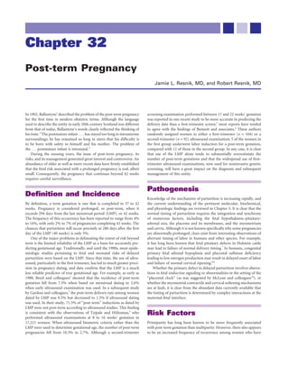
frequently cited studies was provided by the National Birthday Trust 0
of Britain in 1958, which undertook a detailed examination of more 28 29 30 31 32 33 34 35 36 37 38 39 40 41 42 43
than 17,000 births in the United Kingdom from March 3 to March 9 Gestational age (weeks)
of that year.13 Their data demonstrated that the perinatal mortality rate
began to increase after 42 weeks’ gestation, doubling by about 43 weeks, FIGURE 32-1 Perinatal mortality as a function of gestational age.
and was four to six times higher at 44 weeks than at term. A more The rates of stillbirth, neonatal, and postneonatal death increase with
advancing gestational age beyond 41 weeks. The perinatal mortality
recent study showed that the risks begin to accelerate between 41 and
is expressed per 1000 ongoing pregnancies. (From Hilder L, Costeloe
42 weeks and rise more sharply after that point (Fig. 32-1).14 Numerous K, Thilaganathan B: Prolonged pregnancy: Evaluating gestation-
other reports have confirmed this increase in risk.15-17 Alexander and specific risks of fetal and infant mortality. BJOG 1998;105:169-173.)
associates18 retrospectively evaluated outcomes of more than 27,000
pregnancies with 41 or 42 weeks’ gestation, compared with approxi-
mately 29,000 completed at 40 weeks’ gestation. Length of labor, inci-
dence of prolonged second-stage labor, forceps use, and cesarean pared with term infants,17,23 and birth injuries can occur as a result of
delivery were all increased with the longer gestation period. It is not difficult forceps deliveries and shoulder dystocia. Morbidity also
clear, however, whether the observed increase in complications was due includes cephalohematomas, fractures, and brachial plexus palsy.24
to prolonged gestation, routine use of induction at 42 weeks, or Study of fetal growth characteristics in 7000 post-term infants con-
both. firmed a gradual shift toward higher birth weights and greater head
In a more recent Norwegian study, in which 17,493 pregnancies circumference between 273 and 300 days of gestational age.25 These
with confirmed dates by second-trimester ultrasound were analyzed, findings were further reinforced by a study of 519 pregnancies extend-
1336 were found to be post-term. The post-term group had twice the ing beyond 41 weeks, in which 23% of the newborns weighed more
perinatal mortality rate (CI, 0.9 to 4.6); the RR of having an Apgar than 4000 g and 4% weighed more than 4500 g.26
score lower than 7 at 5 minutes was 2.0 (CI, 1.2 to 3.3), and the RR of Although the majority of post-term infants are appropriately grown
requiring neonatal intensive care was 1.6 (CI, 1.3 to 2.0).19 Another or macrosomic, the risk of a small-for-gestational-age (SGA) infant is
prospective cohort study of 27,514 pregnancies from the same country also increased in post-term pregnancy. In a population-based study of
demonstrated that maternal and fetal risks were lowest at 39 weeks’ 510,029 singleton pregnancies from the Swedish Birth Registry, the rate
gestation, with increasing rates of maternal and neonatal complica- of SGA infants increased from 2.2% in term infants to 3.8% in post-
tions, as well as operative deliveries, as pregnancy proceeded past term infants.27
term.20 Similar findings were reported in a Danish population.21
Meconium Staining and
Abnormal Fetal Growth Pulmonary Aspiration
Since the report of Clifford22 and his description of the postmature- Almost all studies of post-term gestation report a markedly higher
dysmature neonate with wasting of subcutaneous tissue, meconium incidence of meconium-stained amniotic fluid, compared with term
staining, and peeling of skin, many have focused their attention on the pregnancies, and the greater risk of meconium aspiration syndrome in
problems of the undernourished post-term fetus. In fact, only 10% to these infants is well recognized.17 Among those infants defined by
20% of true post-term fetuses exhibit any of the findings described by ultrasound-estimated fetal growth curves to be appropriately sized for
Clifford. Macrosomia is actually a far more common complication, gestational age (AGA), those delivered post-term had a threefold higher
because, under most circumstances, the fetus continues to grow in incidence of meconium aspiration and twice the risk of an Apgar score
utero. Twice as many post-term fetuses weigh more than 4000 g, com- of less than 4 at 5 minutes, compared with term AGA infants.27 The
3. CHAPTER 32 Post-term Pregnancy 615
presence of oligohydramnios further complicates the risks of meco- However, a cohort study done in Sweden showed no correlation
nium staining because of the lack of fluid to dilute the meconium, between an AFI of less than 5 cm and adverse outcome.38 Similarly,
which results in thicker, more tenacious material in the oropharynx Divon and associates,39 in a longitudinal assessment of AFI in 139
and lower in the respiratory tract. women with post-term pregnancy, found an increased frequency of
abnormal fetal heart rate tracings and meconium staining but no other
significant adverse fetal outcome. Alfirevic and colleagues40 compared
both methods with respect to pregnancy intervention in post-term
Fetal Evaluation and pregnancies and found more frequent abnormal AFIs than abnormal
Management vertical pocket depths, leading to more inductions and fetal monitor-
ing but no difference in perinatal outcome. Morris and colleagues41
When one considers the rapidly accelerating risk of fetal morbidity and conducted a prospective, double-blinded, cohort study to determine
mortality between 42 and 43 weeks’ gestation and again between 43 whether an AFI of less than 5 cm or a single vertical pocket of less than
and 44 weeks’ gestation (see Fig. 32-1), it becomes apparent that no 2 cm was superior in predicting adverse perinatal events. They found
historically derived or laboratory-measured fetal age provides the pre- the AFI to be significantly more associated with birth asphyxia and
cision required in the management of the post-term pregnancy. Tra- meconium aspiration, but with poor sensitivity. More recently, Zhang
ditional landmarks, such as LMP, uterine size, and first auscultation of and associates,42 using data from the Routine Antenatal Diagnostic
fetal heart tones, can miscalculate gestational age by 2 weeks or more. Imaging with Ultrasound (RADIUS) study, compared a large popula-
Even sensitive sonographic determinations, such as crown-to-rump tion of women screened by ultrasound to control subjects and observed
length in the first trimester, demonstrate a range of several days. In that women with isolated oligohydramnios had no greater adverse
fact, in any given gestation, the actual fetal age is known only if the perinatal events or impaired growth. Another study comparing the two
time of ovulation and conception have been studied, as in ovulation techniques showed that the single vertical pocket method used for
induction and in vitro fertilization. Therefore, a gravida thought to be antepartum surveillance led to less frequent diagnosis and intervention
at 41 to 42 weeks or further in gestation, in current practice, either for oligohydramnios, but without any difference in adverse perinatal
is induced and delivered or undergoes meticulous antenatal outcomes.43
monitoring. Given these disparate findings, it is not difficult to understand why
there is no consensus as to the reliability or superiority of either tech-
nique for identification of the fetus at risk in prolonged pregnancy.
Antenatal Fetal Monitoring Therefore, it is reasonable to conclude that an AFI of less than 5 cm,
Despite the lack of randomized clinical trials, it is generally accepted particularly if it has been falling sharply over a short time interval, or
that careful antepartum and intrapartum fetal monitoring can virtu- the absence of a single identifiable vertical pocket of greater than 2 cm,
ally eliminate fetal post-term mortality and reduce fetal morbidity.28-32 indicates that delivery is warranted. Conversely, it is also reasonable to
However, a careful evidence-based literature analysis concluded that consider that the finding of a normal amniotic fluid volume implies
data were insufficient to determine whether routine antenatal surveil- little fetal risk.
lance before 41 weeks’ gestation improves outcome or which type of There does not appear to be any value in monitoring Doppler flow
monitoring and frequency are most appropriate.33 Consequently, most velocity in fetal vessels, inasmuch as there is no correlation between
obstetricians initiate antenatal testing at 41 weeks’ gestation and repeat the findings and outcome.44 Zimmerman and associates45 demon-
the testing twice weekly. This testing consists of either a biophysical strated that the sensitivity of umbilical artery velocimetry for predict-
profile (BPP) or a nonstress test and assessment of amniotic fluid ing poor outcome was 7%.
volume.
In a study of 307 women whose pregnancies had proceeded beyond
294 days, a normal twice-weekly BPP that included normal amniotic
Fetal Monitoring versus
fluid volume resulted in no perinatal mortalities, and morbidity was Induction of Labor
equivalent to that observed in a comparison group undergoing elective Even though antenatal monitoring can virtually eliminate perinatal
labor induction with a favorable cervix.32 Based on a cumulative expe- mortality in the post-term gestation, some morbidity—including
rience with 19,221 high-risk pregnancies, the same investigative group meconium staining, increased cesarean delivery for a diagnosis of fetal
recommended delivery if amniotic fluid volume decreases.34 distress, and macrosomia with its associated complications—still
The technique used to assess amniotic fluid volume and its role in exists. Although the frequency of morbid events is very low, the
evaluation of the prolonged gestation remains controversial because of continuing concern has been addressed by an alternative approach—
conflicting studies regarding which of the two tests of volume (amni- that of cervical ripening followed by induction at 41 or 42 weeks’
otic fluid index [AFI] or single vertical pocket) is the better predictor gestation.
of outcome and the possibility that the AFI may lead to too many Comparison of these two management approaches in several ran-
unnecessary interventions. Oligohydramnios is thought to be a marker domized controlled trials has yielded generally similar results. Hannah
for fetal complications, including umbilical cord compresssion, hypox- and coauthors46 studied 3407 women with uncomplicated pregnancies
emia, and meconium aspiration, as well as fetal heart rate abnormali- at 41 or more weeks’ duration, who were randomly assigned to
ties and risk of neonatal admission to an intensive care unit.35-37 either elective induction after cervical ripening with prostaglandin E2
Bochner and coworkers36 observed an almost 24-fold increase in cesar- (PGE2) gel or serial antenatal monitoring (fetal kicks, nonstress test,
ean delivery for the indication of fetal distress when the maximum amniotic fluid). In the monitored group, labor was induced only if
vertical amniotic fluid pocket depth was less than 3 cm. The incidence there was evidence of compromised fetal status. The authors observed
of meconium-stained amniotic fluid in the post-term gestation was a lower rate of cesarean delivery for a diagnosis of fetal distress in the
37% among those women with adequate amniotic fluid volume but induction group but no significant difference between the two groups
increased to 71% if the amniotic fluid volume was decreased.31 in fetal mortality or morbidity. The same investigators subsequently
4. 616 CHAPTER 32 Post-term Pregnancy
reported that routine induction was more cost-effective than serial including a significant risk of postpartum hemorrhage and an increased
antenatal monitoring.47 The Maternal-Fetal Medicine Network pro- risk of cesarean delivery.
spectively evaluated 440 patients, comparing induction with serial The Bishop score,53 or some suitable modification of it, can be used
monitoring.48 They observed no fetal deaths in either group, and rates as a guide to select the most appropriate induction technique. This is
of neonatal morbidity and cesarean delivery were similar. A more especially true in primigravid women. If the Bishop score is lower than
recent study from Norway, in which 254 women at 41 weeks’ gestation 5, amniotomy and oxytocin infusion are associated with an unaccept-
were randomly assigned to an induction or expectant manage- ably high incidence of unsuccessful inductions as well as fetal and
ment group, found no differences in neonatal outcomes or mode of maternal complications.54 In these circumstances, cervical ripening
delivery.49 should be undertaken before uterine contractions are provoked. Given
These combined trials have led to the conclusion that neither the rapidly increasing use of transvaginal ultrasound (TVUS) to assess
approach has a substantive advantage over the other. A small advantage cervical length and dilatation and its usefulness in the diagnosis of
to the induction approach was suggested by the recent Cochrane preterm labor, it is not unreasonable to apply this technology to cervi-
Review of 19 studies, which determined that a policy of labor induc- cal assessment in post-term pregnancy. One study of 240 women,
tion at 41 weeks resulted in fewer fetal deaths, although the differences comparing TVUS with digital cervical examination using receiver
and absolute risk were extremely small (1 in 2986 versus 9 in 2953; operating characteristic (ROC) curves, demonstrated that a cervical
odds ratio, 0.3; CI, 0.9 to 0.99). There was no significant difference in length of 28 mm was a better predictor of induction success (vaginal
the cesarean section rate.50 delivery within 24 hours) than the Bishop score.55 However, conflicting
Nevertheless, in terms of physician preferences in the United States, findings were reported by Chandra and associates.56
induction at 41 weeks has become the mode of practice and the debate The most frequently used current cervical ripening techniques
moot. A recent survey of 1000 randomly selected members of the include chemical agents such as PGE2 (dinoprostone, trade names
American College of Obstetricians and Gynecologists revealed that Prepidil and Cervidil Rx), administered vaginally or intracervically,
73% routinely induce low-risk women at 41 weeks. For women who and misoprostol (Cytotec Rx), administered vaginally or orally. Both
decline induction, approximately 65% of physicians initiate antenatal appear to be effective in improving the Bishop score and to result in
testing twice weekly at 41 weeks.51 It is clear that medical induction shorter labor times and possibly fewer failed inductions. Misoprostol,
rates have increased sharply in the United States. Between 1980 and in doses of 25 μg given vaginally every 4 hours, appears to be slightly
1996, the rate of induction doubled (from 12.9% to 25.8%), the most more effective that dinoprostone but is associated with a higher fre-
common indication being that of the post-term pregnancy.52 quency of uterine tachysystole. A recent review of randomized trials
performed between 1987 and 2005 compared the two agents and con-
firmed that misoprostol was superior to dinoprostone at any dose and
Management Summary route of administration in terms of achieving vaginal delivery within
It seems appropriate to recommend the following steps to evaluate and 24 hours. There was no difference in the rate of cesarean delivery.57
manage the post-term gestation: This study confirmed an earlier Cochrane database review which con-
cluded that the use of vaginal misoprostol is more effective than con-
1. Although there is insufficient evidence because of the low-risk ventional methods of cervical ripening and labor induction. Compared
nature of either approach, current obstetric practice dictates that with placebo, oxytocin, or intracervical or vaginal PGE2, misoprostol
labor induction be offered between 41 and 42 weeks’ gestation in resulted in increased cervical ripening, decreased use of oxytocin, and
the presence of a favorable cervix. increased rates of vaginal delivery. However, misoprostol also caused
2. If the cervix is unfavorable, alternate approaches include either an increased rate of uterine hyperstimulation.58
cervical ripening followed by induction of labor or twice-weekly Vaginal inserts such as balloon catheters also have their advocates
fetal monitoring. Delivery should be accomplished promptly if for cervical ripening. A systematic review concluded that these mechan-
there is evidence of fetal compromise. ical dilators do not compare favorably with chemical inducing agents
3. It is prudent to use the BPP, or some modification of the BPP, to in terms of delivery success rates but are associated with less uterine
determine antenatal fetal condition. hypercontactility.59
Methods of Labor Induction Developmental Effects of
The issue of labor induction and cervical ripening agents is addressed
in detail in Chapter 36 and is summarized briefly here.
Post-term Gestation
Because normal labor depends on efficient myometrial contrac- Studies on the development of children from prolonged pregnancies
tions acting on a compliant cervix to efface and dilate it, methods of are difficult to evaluate because investigators have not separated neo-
labor induction must take into account both components of the uterus. nates asphyxiated in utero and growth-restricted (dysmature) post-
If the cervix is already soft, effaced, and partially dilated, intravenous term neonates from otherwise normally born neonates. A study of
infusion of oxytocin may be sufficient to stimulate contractions. Con- neonatal behavior among 106 dysmature infants revealed an increased
ventional practice requires amniotomy to be performed as a first step, number of illnesses and sleep disorders as well as diminished social
because this procedure maximizes the effectiveness of oxytocin. If the competence during the first year of life (Vineland Social Maturity
cervix is unripe, oxytocin will not cause it to ripen, and amniotomy Scale). Also, and not unexpectedly, the incidence of fetal distress was
is inappropriate. Although labor contractions can be stimulated by high, and those babies who were asphyxiated in utero had a higher
oxytocin, such a result is futile, because many hours of such contrac- incidence of abnormal neurologic signs in the neonatal period.60 All
tions are required to produce any sort of change in the cervix, and the infants had signs of desquamation of skin and wasting of subcutaneous
ensuing prolonged labor can lead to an increase in obstetric morbidity, tissue, however, and the group of children studied was not compared
5. CHAPTER 32 Post-term Pregnancy 617
with any post-term children who did not have these physical findings 14. Hilder L, Costeloe K, Thilaganathan B: Prolonged pregnancy: Evaluating
at birth. gestation-specific risks of fetal and infant mortality. BJOG 105:169-173,
Field and coworkers61 studied a group of 40 dysmature offspring, 1998.
all of whom had parchment-like skin and long, thin bodies. At birth, 15. Nakano R: Post-term pregnancy: A five year review from Osaka National
Hospital. Acta Obstet Gynecol Scand 51:217, 1972.
their Brazelton interaction and motor scores were lower than in
16. Sachs BP, Friedman EA: Results of an epidemiological study of post-date
term controls, and at 4 months they scored lower on the Denver pregnancy. J Reprod Med 31:162, 1986.
Developmental Scale. By 8 months, the Bayley motor scores of the 17. Eden R, Seifert L, Winegar A, et al: Perinatal characteristics of uncompli-
post-term subjects were equivalent to those of control infants, but their cated post-date pregnancies. Obstet Gynecol 69:296, 1987.
mental scores were slightly lower. This study differed in at least one 18. Alexander JM, McIntire DD, Leveno UJ: Forty weeks and beyond:
significant way from that of Lovell60: The Apgar scores at 5 minutes in Pregnancy outcomes by week of gestation. Obstet Gynecol 96:291, 2000.
the two groups were identical, thus partially correcting for in utero 19. Nakling J, Backe B: Pregnancy risk increases from 41 weeks of gestation.
asphyxia. Acta Obstet Gynecol 85:663-668, 2006.
In a large retrospective review, Zwerdling23 observed that post-term 20. Heimstad R, Romundstad PR, Eik-Nes SH, et al: Outcomes of pregnancies
infants weighing less than 2500 g had a neonatal mortality rate seven beyond 37 weeks of gestation. Obstet Gynecol 108:500-508, 2006.
21. Olesen AW, Westergaard JG, Olsen J: Perinatal and maternal complications
times that of post-term infants as a whole. This finding confirmed the
related to post-term delivery: A national regiser-based study, 1978-1993.
additional risk of the dysmature growth pattern in some post-term Am J Obstet Gynecol 189:222-227, 2003.
infants. The increased mortality rate was observed up to 2 years of age, 22. Clifford SH: Postmaturity—with placental dysfunction. J Pediatr 44:1, 1954.
but at 5 years the data on growth and intelligence in Zwerdling’s study 23. Zwerdling MA: Factors pertaining to prolonged pregnancy and its outcome.
population revealed no differences between prolonged-gestation and Pediatrics 40:202, 1967.
normal-gestation children. These findings were confirmed in a pro- 24. Usher RH, Boyd ME, McLean FH, et al: Assessment of fetal risk in post-date
spective study in which 129 children born of prolonged pregnancy pregnancies. Am J Obstet Gynecol 158:259, 1988.
were compared with 184 term controls.62 At 1 year and again at 2 years 25. McLean FH, Boyd ME, Usher RH: Post-term infants: Too big or too small?
of age, there were no differences between the two groups with respect Am J Obstet Gynecol 164:619, 1991.
to intelligence scores, physical milestones, or intercurrent illnesses. 26. Pollack RN, Hauer-Pollack G, Divon MY: Macrosomia in post-dates preg-
nancy: The accuracy of routine ultrasonographic screening. Am J Obstet
One recent cohort study from Denmark linked hospital records of
Gynecol 167:7, 1992.
277,435 pregnancies delivering at term or beyond to cases of childhood 27. Clausson B, Cnattingius S, Axelsson O: Outcomes of post-term births: The
epilepsy. The researchers found a slight increase in the incidence of role of fetal growth restriction and malformations. Obstet Gynecol 94:758,
epilepsy as a function of gestational age at or after 43 weeks, but only 1999.
among those infants delivered by cesarean section or other operative 28. Hauth JC, Goodman MT, Gilstrap LC III, et al: Post-term pregnancy.
delivery.63 The risk was not observed after 1 year of life. Whether this J Obstet Gynecol 56:467, 1980.
finding reflects a problem unique to advanced gestational age or com- 29. Freeman RK, Garite TJ, Modanlou H, et al: Postdate pregnancy: Utilization
plications that required expedient delivery is unclear. of contraction stress testing for primary fetal surveillance. Am J Obstet
Gynecol 140:128, 1981.
30. Eden R, Gergely RZ, Schifrin BS, et al: Comparison of antepartum testing
schemes for the management of the postdate pregnancy. Am J Obstet
References Gynecol 144:683, 1982.
1. Ballantyne JW: The problem of the postmature infant. J Obstet Gynaecol 31. Phelan JP, Platt LP, Yeh S-Y, et al: The role of ultrasound assessment of
Br Emp 2:36, 1902. amniotic fluid volume in the management of the post-date pregnancy. Am
2. Boyd ME, Usher RH, McLean FH, et al: Obstetric consequences of post- J Obstet Gynecol 151:304, 1984.
maturity. Am J Obstet Gynecol 158:334, 1988. 32. Johnson JM, Harman CR, Lange IR, et al: Biophysical profile scoring in the
3. Gardosi J, Vanner T, Francis A: Gestational age and induction of labour for management of the post-term pregnancy. Am J Obstet Gynecol 154:269,
prolonged pregnancy. BJOG 104:792, 1997. 1986.
4. Taipale P, Hiilesmaa V: Predicting delivery date by ultrasound and last 33. American College of Obstetricians and Gynecologists: ACOG Practice Pat-
menstrual period on early gestation. Obstet Gynecol 97:189, 2001. terns: Management of Post-term Gestation. Practice Bulletin No. 55. Wash-
5. Olesen AW, Thomsen SG: Prediction of delivery date by sonography in ington, DC, ACOG, 2004.
the first and second trimesters. Ultrasound Obstet Gynecol 28:292-297, 34. Manning FA, Morrison I, Harman CR, et al: Fetal assessment based on fetal
2006. biophysical profile scoring: Experience in 19,221 referred high risk preg-
6. Bennett KA, Crane JM, O’Shea P, et al: First trimester ultrasound screening nancies. II: An analysis of false negative deaths. Am J Obstet Gynecol
is effective in reducing post-term labor induction rates: A randomized 157:880, 1987.
controlled trial. Am J Obstet Gynecol 190:1077-1081, 2004. 35. Leveno KJ, Quirk JG, Cunningham FG, et al: Prolonged pregnancy: I.
7. Holm LW: Prolonged pregnancy. Adv Vet Sci 11:159, 1967. Observations concerning the causes of fetal distress. Am J Obstet Gynecol
8. France JT, Liggins GC: Placenta sulfatase deficiency. J Clin Endocrinol 150:465, 1984.
29:138, 1969. 36. Bochner CJ, Medearis Al, Davis J, et al: Antepartum predictors of fetal dis-
9. Fliegner JRH, Schindler I, Brown JB: Low urinary oestriol excretion during tress in post-term pregnancy. Am J Obstet Gynecol 157:353, 1987.
pregnancy associated with placental sulphatase deficiency or congenital 37. Tongsong T, Srisomboon J: Amniotic fluid volume as a predictor of fetal
adrenal hypoplasia. J Obstet Gynaecol Br Commonw 79:810, 1972. distress in post-term pregnancy. Int J Gynaecol Obstet 40:213, 1993.
10. McLean M, Bisits S, Davies J, et al: A placental clock controlling the length 38. Montan S, Malcus P: Amniotic fluid index in prolonged pregnancy.
of human pregnancy. Nat Med 1:460-463, 1995. J Matern Fetal Invest 5:4, 1995.
11. Olesen AW, Basso O, Olsen J: Risk of recurrence of prolonged pregnancy. 39. Divon M, Marks AD, Henderson CE: Longitudinal measurement of amni-
BMJ 326:476, 2003. otic fluid index in post-term pregnancies and its association with fetal
12. Kistka ZA, Palomar L, Boslaugh SE, et al: Risk for posttterm delivery after outcome. Am J Obstet Gynecol 172:142, 1995.
previous posttterm delivery. Am J Obstet Gynecol 196:241.e1-6, 2007. 40. Alfirevic Z, Luckas M, Walkinshaw SA, et al: A randomized comparison
13. Butler NR, Alberman ED: The Second Report of the 1958 British Perinatal between amniotic fluid index and maximum pool depth in the monitoring
Mortality Survey. Edinburgh, E & S Livingstone, 1969, p 327. of post-term pregnancy. BJOG 104:207, 1997.
6. 618 CHAPTER 32 Post-term Pregnancy
41. Morris JM, Thompson K, Smithey J, et al: The usefulness of ultrasound 52. Yawn BP, Wollan P, McKeon K, et al: Temporal changes in rates and reasons
assessment of amniotic fluid in predicting adverse outcome in prolonged for medical induction of term labor, 1980-1996. Am J Obstet Gynecol
pregnancy: A prospective blinded observational study. BJOG 110:989-994, 184:611, 2001.
2003. 53. Bishop EH: Pelvic scoring for elective induction. Obstet Gynecol 24:266,
42. Zhang J, Troendle J, Meikle S, et al: Isolated oligohydramnios is not associ- 1964.
ated with adverse pregnancy outcome. BJOG 111:220-225, 2004. 54. Calder AA, Greer CA: Cervical physiology and induction of labor. In
43. Chauhan SP, Doherty DD, Magann EF, et al: Amniotic fluid index vs single Bonnar J (ed): Recent Advances in Obstetrics and Gynecology 17. Edin-
deepest pocket technique during modified biophysical profile: A random- burgh, Churchill Livingstone, 1992.
ized clinical trial. Am J Obstet Gynecol 191:661-667, 2004. 55. Pandis GU, Papageorghiou AJ, Ramanathan JG, et al: Preinduction sono-
44. Guidetti DA, Divon MY, Cavalieri RL, et al: Fetal umbilical artery flow graphic measurement of cervical length in the prediction of successful
velocimetry in post-date pregnancies. Am J Obstet Gynecol 157:1521, induction of labor. Ultrasound Obstet Gynecol 18:623, 2001.
1987. 56. Chandra S, Crane JMG, Hutchens D, et al: Transvaginal ultrasound and
45. Zimmerman P, Alback T, Koskinen J, et al: Doppler flow velocimetry of the digital examination in predicting successful labor induction. Obstet Gynecol
umbilical artery, uteroplacental arteries and fetal middle cerebral artery in 98:2, 2001.
prolonged pregnancy. Ultrasound Obstet Gynecol 5:189, 1995. 57. Crane JM, Butler B, Young DC, et al: Misoprostol compared with prosta-
46. Hannah ME, Hannah WJ, Hellmann J, et al: Induction of labor as compared glandin E2 for labour induction in women at term with intact membranes
with serial antenatal monitoring in post-term pregnancy. N Engl J Med and unfavourable cervix: A systematic review. BJOG 113:1366-1376,
326:1587, 1992. 2006.
47. Goeree R, Hannah ME, Hweson S: Cost-effectiveness of induction of labor 58. Hofmeyr GJ, Gulmezoglu AM: Vaginal misoprostol for cervical ripening
versus serial antenatal monitoring in the Canadian Multicentre Post-term and induction of labour. Cochrane Database Syst Rev (1):CD000941,
Pregnancy Trial. Canadian Med Assoc 152:1445, 1995. 2003.
48. National Institute of Child Health and Development (NICHD) Network of 59. Boulvain M, Kelly A, Lohse C, et al: Mechanical methods for induction of
Maternal-Fetal Medicine Unit: A clinical trial of induction of labor versus labour. Cochrane Database Syst Rev (4):CD000941, 2002.
expectant management in post-term pregnancy. Am J Obstet Gynecol 60. Lovell KE: The effect of postmaturity on the developing child. Med J Austr
170:716, 1994. 1:13, 1973.
49. Heimstad R, Skogvoll E, Mattsson L-A, et al: Induction of labor or serial 61. Field TM, Dabiri C, Hallock N, et al: Developmental effects of prolonged
antenatal fetal monitoring in the post-term pregnancy: A randomized con- pregnancy in the postmaturity syndrome. J Pediatr 90:836, 1977.
trolled trial. Obstet Gynecol 109:609-617, 2007. 62. Shime J, Librach CL, Gare DJ, et al: The influence of prolonged pregnancy
50. Gulmezoglu AM, Crowther CA, Middleton P: Induction of labour for on infant development at one and two years of age: A prospective controlled
improving birth outcomes for women at or beyond term. Cochrane Data- study. Am J Obstet Gynecol 154:341, 1986.
base Syst Rev (4):CD004945, 2006. 63. Ehrenstein V, Pedersen L, Holsteen V, et al: Postterm delivery and risk for
51. Cleary-Goldman J, Bettes B, Robinson JN, et al: Post-term pregnancy: Prac- epilepsy in childhood. Pediatrics 119:554-561, 2007.
tice patterns of contemporary obstetricians and gynecologists. Am J Peri-
natol 23:15-20, 2006.

![614 CHAPTER 32 Post-term Pregnancy
had a previous post-term pregnancy. One large cohort study from
6
Denmark has demonstrated that women who delivered post-term in
Stillbirth
their first pregnancy had an almost threefold increase in the incidence
Neonatal death
of subsequent post-term pregnancy, compared with those whose first Postneonatal death
delivery was at term.11 These findings were recently confirmed by 5
Kistka and coworkers12 in a study of 368,633 births in Missouri, in
Mortality per 1000 ongoing pregnancies
which mothers with an initial post-term birth were at increased risk
for a subsequent post-term pregnancy (relative risk [RR], 1.88; 95%
confidence interval [CI], 1.79 to 1.97). These findings also suggest the 4
possibility of a genetic predisposition, inasmuch as the risk of recur-
rent post-term pregnancy in the Danish study was not observed if the
first and second children had different fathers.
3
Perinatal Risks 2
Morbidity and Mortality
Almost all reports up to the present time, even those with inherent
limitations imposed by inaccuracies in gestational age determination, 1
suggest an increase in perinatal morbidity and mortality when preg-
nancy goes beyond 42 weeks’ gestation. One of the earliest and most
frequently cited studies was provided by the National Birthday Trust 0
of Britain in 1958, which undertook a detailed examination of more 28 29 30 31 32 33 34 35 36 37 38 39 40 41 42 43
than 17,000 births in the United Kingdom from March 3 to March 9 Gestational age (weeks)
of that year.13 Their data demonstrated that the perinatal mortality rate
began to increase after 42 weeks’ gestation, doubling by about 43 weeks, FIGURE 32-1 Perinatal mortality as a function of gestational age.
and was four to six times higher at 44 weeks than at term. A more The rates of stillbirth, neonatal, and postneonatal death increase with
advancing gestational age beyond 41 weeks. The perinatal mortality
recent study showed that the risks begin to accelerate between 41 and
is expressed per 1000 ongoing pregnancies. (From Hilder L, Costeloe
42 weeks and rise more sharply after that point (Fig. 32-1).14 Numerous K, Thilaganathan B: Prolonged pregnancy: Evaluating gestation-
other reports have confirmed this increase in risk.15-17 Alexander and specific risks of fetal and infant mortality. BJOG 1998;105:169-173.)
associates18 retrospectively evaluated outcomes of more than 27,000
pregnancies with 41 or 42 weeks’ gestation, compared with approxi-
mately 29,000 completed at 40 weeks’ gestation. Length of labor, inci-
dence of prolonged second-stage labor, forceps use, and cesarean pared with term infants,17,23 and birth injuries can occur as a result of
delivery were all increased with the longer gestation period. It is not difficult forceps deliveries and shoulder dystocia. Morbidity also
clear, however, whether the observed increase in complications was due includes cephalohematomas, fractures, and brachial plexus palsy.24
to prolonged gestation, routine use of induction at 42 weeks, or Study of fetal growth characteristics in 7000 post-term infants con-
both. firmed a gradual shift toward higher birth weights and greater head
In a more recent Norwegian study, in which 17,493 pregnancies circumference between 273 and 300 days of gestational age.25 These
with confirmed dates by second-trimester ultrasound were analyzed, findings were further reinforced by a study of 519 pregnancies extend-
1336 were found to be post-term. The post-term group had twice the ing beyond 41 weeks, in which 23% of the newborns weighed more
perinatal mortality rate (CI, 0.9 to 4.6); the RR of having an Apgar than 4000 g and 4% weighed more than 4500 g.26
score lower than 7 at 5 minutes was 2.0 (CI, 1.2 to 3.3), and the RR of Although the majority of post-term infants are appropriately grown
requiring neonatal intensive care was 1.6 (CI, 1.3 to 2.0).19 Another or macrosomic, the risk of a small-for-gestational-age (SGA) infant is
prospective cohort study of 27,514 pregnancies from the same country also increased in post-term pregnancy. In a population-based study of
demonstrated that maternal and fetal risks were lowest at 39 weeks’ 510,029 singleton pregnancies from the Swedish Birth Registry, the rate
gestation, with increasing rates of maternal and neonatal complica- of SGA infants increased from 2.2% in term infants to 3.8% in post-
tions, as well as operative deliveries, as pregnancy proceeded past term infants.27
term.20 Similar findings were reported in a Danish population.21
Meconium Staining and
Abnormal Fetal Growth Pulmonary Aspiration
Since the report of Clifford22 and his description of the postmature- Almost all studies of post-term gestation report a markedly higher
dysmature neonate with wasting of subcutaneous tissue, meconium incidence of meconium-stained amniotic fluid, compared with term
staining, and peeling of skin, many have focused their attention on the pregnancies, and the greater risk of meconium aspiration syndrome in
problems of the undernourished post-term fetus. In fact, only 10% to these infants is well recognized.17 Among those infants defined by
20% of true post-term fetuses exhibit any of the findings described by ultrasound-estimated fetal growth curves to be appropriately sized for
Clifford. Macrosomia is actually a far more common complication, gestational age (AGA), those delivered post-term had a threefold higher
because, under most circumstances, the fetus continues to grow in incidence of meconium aspiration and twice the risk of an Apgar score
utero. Twice as many post-term fetuses weigh more than 4000 g, com- of less than 4 at 5 minutes, compared with term AGA infants.27 The](data:image/gif;base64,R0lGODlhAQABAIAAAAAAAP///yH5BAEAAAAALAAAAAABAAEAAAIBRAA7)