SlideShare une entreprise Scribd logo
1  sur  42
AUTOPSY: INTERNAL EXAMINATION
Aishwarya Sinha
VI Semester
Forensic Medicine – PBL
CMS - TH
INTERNAL EXAMINATION
It consists of:
1. Incising the body
2. Inspecting the various organs
3. Examining the cavities systematically
4. Weighing and measuring the organs
5. Checking for any pathology
6. Putting all the organs back in and padding
7. Suturing the body
1. ‘I’ shaped Incision: It is a straight line incision
extending from the chin to the symphysis pubis.
2. ‘Y’ Shaped Incision: This type of incision starts
near the acromian process and progresses
downwards towards the xiphoid process. The
incision is then extended till the symphysis pubis.
Also, a similar incision is made on the opposite side
of the body.
3. Modified ‘Y’ Shaped: A ‘Y’ shaped incision is
made from the suprasternal notch to symphysis
pubis. It extends from the suprasternal notch over
the clavicle
to its center on both sides and passes upwards
over the neck, behind the ear.
REMOVAL OF ORGANS
1. Virchow’s Technique: Head  Thorax 
Abdomen
2. Rokitansky’s Technique: in situ dissection in
parts combined with en block removal.
3. Letulle’s Technique: Cervical  Thoracic 
Abdominal  Pelvic Organs  Removed en
masse and dissected as organ block.
ORDER OF EXAMINATION
VIRCHOW’S TECHNIQUE 
Head
Thoracic (Cervical)
Abdominal Organs
HEAD
• The neck is extended by placing a
wooden block under the shoulders.
• Fix head using a head rest.
• Intermastoidal incision is made i.e. from
the mastoid process behind one ear to
the vertex and again to the mastoid
process of the other ear.
• Scalp flap is reflected forward to the
superciliary ridge and backwards to the
occipital protuberance.
HEAD
• With the help of a saw, ‘V’ shaped cut is made
so that the skull fits back correctly after
autopsy.
• This ‘V’ shaped cut passes through: the center
of the forehead  base of the mastoid
process  occipital protuberance  the base
of the other mastoid process and back to
the center of the forehead.
CAUTION
• Care must be taken to avoid any damage to the
meninges or the brain during the process of skull
removal.
• Therefore, chisel and hammer should never be
used.
• Instead, a chisel should be inserted and twisted
at various points in order to remove the skull flap.
• By doing so, superior longitudinal sinus can be
exposed.
BRAIN
• Dura is incised along the lines of the incision
of the skull  folded to the midline  falx
cerebri is freed from the cribriform plate 
dura is pulled back.
• 4 fingers of the left hand are inserted into the
frontal lobe and the nerves and vessels are
cut.
• Tentorium is also cut along the posterior part
of the petrous bone.
BRAIN
• A knife is then inserted into the occipital foramen
and the cervical cord  cervical nerves and the
vertebral arteries are cut as far as possible.
• With the right hand the cerebellum is grasped
and examined.
• Any signs of hemorrhage, injury or any other
disease should be carefully seen.
• Vessels, especially the Circle of Willis, should be
checked for any arteriosclerosis or aneurysm etc.
FIXATION
Brain will start to deteriorate as soon as the
blood supply is interrupted. This deterioration is
rapid and therefore, it is of utmost importance
to arrest this procedure.
This process of preservation of the brain is called
fixation.
There are 2 main methods of fixation:
1. Freezing
2. Prefusion
FIXATION OF THE BRAIN
Brain is fixed in 10% formalin for
at least 1-2 week(s).
To help with the penetration of
the preservative, lateral fissures
are made through, which tear
open the pia and the arachnoid
mater.
A long sagittal cut is made
through the corpus callosum so
as to allow the formalin to pass
into the ventricles.
AFTER FIXATION
 Brain is placed on a board and the
cerebellum along with the pons and the
medulla are removed.
 Straight cut is made to expose the fourth
ventricle.
 Thereafter an oblique cut is made to
expose the dentate nucleus.
 Entire stem is sectioned at a few
millimeters distance and examined for any
pathology.
 Cerebellum is examined by making a series
of sagittal cuts beginning from the frontal
pole passing backwards to the occiput.
 Sequentially cuts are made and serially
placed aside in the same order for later
identification.
SPINAL CORD
• Not examined unless there is an indication.
• In prone position, wooden block is placed
under the chest and the head is bent
downwards.
• Mid-line incision from the occipital
protuberance to the L4 vertebra is made.
• Muscles are dissected all the way.
• Atlas is disarticulated.
• With the help of a double bladed saw, the
laminae are sawed through the entire length
and separated by a chisel.
SPINAL CORD
• Dura is cut open and checked for
haemorrhage, inflammation, tumours, pus
etc.
• Nerves are cut from below as they pass
though the spinal formaina.
• And the cord is separated from the
foramen magnum.
• Cord is then sectioned transversely and
serially.
• Lastly, the vertebral column is examined
for any fractures or dislocations.
THORAX
• Chest muscles are dissected away.
• Chest is opened by cutting the costal cartilages
with the help of a cartilage knife.
• In case of elderly subjects, the costal cartilage may
be calcified, hence, a bone saw or a rib shear is
used to cut it out.
• Thereafter, both the sternoclavicular joints are
disarticulated and the chest is opened.
HEART
 Held at the apex and lifted
upwards so that the
pulmonary vessels, SVC &
IVC and the ascending
aorta can be examined.
 The pulmonary artery is
palpated and then the
vessels are cut open.
Pericardium is incised
and examined for any
blood or fluids.
Heart, opened in the
direction of the blood flow
i.e. (Inflow-outflow
method).
HEART
 The enterotome is inserted into the:
 RA  Tricuspid valve  RV  Pulmonary Trunk
 Pulmonary vein  LA  Mitral Valve  LV 
Ascending aorta.
 Both the auricular appendages are examined for
thrombi.
 Heart should eventually be weighed and the
various measurements of the circumference of
the valves or the thickness of the ventricles etc.
are taken.
CORONARY ARTERY
 Examined by making serial cross sections to check
for ante mortem clots.
 To check for infarction and fibrosis, muscles of the
right and the left ventricles are incised.
SUBENDOCARDIAL HAEMORRHAGE:
 Flame shaped, confluent, patchy, haemorrhages
may be seen in the LV on the left side of the inter
ventricular septum.
 Seen in severe hypotension, intracranial
damage, death due to ectopic
pregnancies, arsenic poisoning etc.
LUNGS
• Anterior surface is faced upwards 
PA is identified and cut as far as
possible using small scissors  check
for atherosclerosis, thrombi or emboli.
• Steps are re-traced via PV  check for
thrombi.
• At hilum, a long bladed knife is placed
with the blunt end facing upwards 
pierced into the hilum and the knife is
turned upwards so that the sharp end
now faces upwards  Sawing motion
 hilum is cut.
LUNGS
• Lungs are mounted on a board or held in
the left hand incision is made from
the apex to the base.
• Hence, producing an antero-posterior
slice.
• Cut sections are then examined for
consolidations, edema, atelectasis, cong
estion, emphysema, Tardieu
spots, emboli etc.
If PENUMOTHORAX
is suspected:
• Pocket dissection on affected side
between the chest wall and the skin 
filled with water  wall is punctured
under water.
• If bubbles are present  Pneumothorax.
• 16 gauge needle attached to a syringe
without its plunger is inserted into the
subcutaneous tissue over the ICS 
water is filled in the syringe  needle is
pierced into the pleural cavity if water
bubbles are seen  Pneumothorax.
• Thereafter, X-ray is done.
If AIR EMBOLISM
is suspected:
• In such cases, before touching the thoracic
organs, the pericardium and the heart should
be examined.
• The heart is lifted up and cut open at the
apex.
1. Left ventricle is filled with frothy blood if air
is present is good amount.
2. If right ventricle is filled with air, the heart
will float in water.
If AIR EMBOLISM is suspected:
1. Pericardial sac is filled with water  heart is punctured with
a scalpel and twisted  if bubbles are present  AIR
EMBOLISM.
2. A wide bore needle is attached to a syringe and water is
filled in it  it is inserted in to right ventricle  if air is
present bubbles are seen.
3. PYROGALLOL TEST: 4 mL of 2% freshly prepared pyrogallol is
taken in two 10 mL syringes.
In one of the syringes, 0.5 M NaOH is added. Gas is aspirated
from the right heart and the needle is retracted and
removed. A stopper is fixed to the syringe and the syringe is
shaken. If the aspirate contains air, the colour of the solution
would turn brown.
4. Air in the IVC can be demonstrated by puncturing it
underwater.
5. Lastly, X-ray is done.
NECK
• Identify the oesophagus  incise it from the posterior
aspect up till the cardiac end of the stomach 
inspected for capsules, tablets or powders.
• Larynx, trachea and the bronchi are also cut from the
posterior aspect and checked for foreign
bodies, blood, mucous, tumours, inflammation etc.
• Thyroid is removed and examined.
• The carotid arteries are examined for thrombi, esp. at
their bifurcation.
• Later, the hyoid bone, cricoid and the thyroid
cartilages are examined.
ABDOMEN
ABDOMEN
• 5 cm above the symphysis pubis the rectus
muscles are divided and a small cut is made.
• Middle and the index fingers are then inserted
and spread in a ‘V’ shape.
• Sharp braded knife is inserted between them
and the peritoneum is cut up to the xiphoid
process.
ABDOMEN
• Firstly, inspection is done and if any
damage, free fluid, perforations etc.
are seen then these are noted.
• Also, note should be made about the
abnormalities, positions, abdominal
organs, adhesions, pathology (if
any), injuries etc.
STOMACH
After applying double ligatures, the stomach is
opened along the greater curvature  from the
cardiac to the pyloric end  size of the pyloric ring
is noted.
Contents are examined for any nature of food
which might be present & its state of
digestion, smell, colour, character, the presence of
foreign bodies or any other suspicious matter etc.
Mucous membrane is examined for
congestion, haemorrhage, ulcerations or any other
INTESTINES
• Note the
colour, consistency, adhesions, herniae, haem
orrhage, serosal surface etc.
• Small intestine is opened along its line of
mesenteric attachments.
• Large intestine is opened along the anterior
taenia coli.
• Examined for
LIVER
• Either liver is removed by itself or attached to
the stomach and the duodenum.
• It is examined for its weight, size, consistency
and for the presence of any other pathology /
injury.
• It is cut along the long axis into 2 cm thick
slices.
SPLEEN
• It is removed by itself.
• Size, weight, consistency, capsule
condition, rupture injuries or any pathology is
noted.
• This is also sectioned along its long axis and
the character of the septa and the
parenchyma are noted.
PANCREAS
• Along the long axis cuts are made at right
angles.
• Examined for fat necrosis.
KIDNEYS
• Size and weight are noted.
• Capsules are excised and
examined carefully.
• Kidney is sectioned
longitudinally the convex
border of the hilum so that
it splits into half and opens
the pelvis.
• Checked for
calculi, inflammation etc.
• Ureter is cut into and
examined.
BLADDER
• Incision is made from the
fundus and carried to the
urethra.
• Wall condition, amount
and character of urine is
noted.
PROSTATE & TESTES
• Examined for enlargement and malignancies.
• Vertical cross-sections are made through the lateral
and median lobes and the prostate is split open for
examination.
• Inguinal canal is incised from the peritoneal aspect
and the loop of the vas deference is pulled to free it
from the internal inguinal ring.
• The testes is pushed with one hand and pulled out of
the scrotum easily by the other.
• These are cut longitudinally and checked for any
clotted blood inside the scrotum and in the testis.
FEMALE GENITALIA
• Vagina and the uterus are cut either
anteriorly or posteriorly upto the fundus.
• Two short incisions are made at the fundus
from the main incision towards each cornu so
as to expose the endometrium.
• Ovaries are sectioned longitudinally and the
fallopian tubes are cut longitudinally.
If the uterus contains a foetus, its age should
be determined.
AFTER COMPLETING
INTERNAL EXAMINATIONS
• Body cavities should be cleaned and made free from
blood, fluids etc.
• Organs are placed back in and excess space is packed
with cotton/cloth etc. (esp. in the pelvis and the neck
regions.)
• Dissection flaps are closed and sutured with thin
twine.
• Skull is filled with cotton and absorbent material and
the skull cap is placed back in and the scalp is stitched.
• Body is washed with water, dried, covered with
clothes and handed over to the police officials.
Autopsy internal examination, Forensic Medicine, Post-mortem Examination

Contenu connexe

Tendances (20)

Regional injury
Regional injuryRegional injury
Regional injury
 
Mechanical injury 2
Mechanical injury 2Mechanical injury 2
Mechanical injury 2
 
Abrasions
AbrasionsAbrasions
Abrasions
 
Mechanical injury 1
Mechanical injury  1Mechanical injury  1
Mechanical injury 1
 
Autopsy by Dr Yaseen Mohammad for 3rd year students MBBS
Autopsy by Dr Yaseen Mohammad for 3rd year students MBBSAutopsy by Dr Yaseen Mohammad for 3rd year students MBBS
Autopsy by Dr Yaseen Mohammad for 3rd year students MBBS
 
Asphyxial death
Asphyxial deathAsphyxial death
Asphyxial death
 
Death & changes after death
Death & changes after deathDeath & changes after death
Death & changes after death
 
Firearm injuries
Firearm injuriesFirearm injuries
Firearm injuries
 
Strangulation Forensic Medicine
Strangulation Forensic MedicineStrangulation Forensic Medicine
Strangulation Forensic Medicine
 
Autopsy
AutopsyAutopsy
Autopsy
 
Injuries
InjuriesInjuries
Injuries
 
Injuries medicolegal aspect
Injuries medicolegal aspectInjuries medicolegal aspect
Injuries medicolegal aspect
 
Asphyxia
Asphyxia Asphyxia
Asphyxia
 
Inquest - By Jinosh Daniel
 Inquest - By Jinosh Daniel Inquest - By Jinosh Daniel
Inquest - By Jinosh Daniel
 
Death
DeathDeath
Death
 
Procedure of autopsy
Procedure of autopsyProcedure of autopsy
Procedure of autopsy
 
Post mortem changes notes by dr. armaan singh
Post mortem changes notes by  dr. armaan singhPost mortem changes notes by  dr. armaan singh
Post mortem changes notes by dr. armaan singh
 
Mode of death
Mode of death Mode of death
Mode of death
 
Mechanical asphyxia 1
Mechanical asphyxia 1Mechanical asphyxia 1
Mechanical asphyxia 1
 
Exhumation
ExhumationExhumation
Exhumation
 

Similaire à Autopsy internal examination, Forensic Medicine, Post-mortem Examination

autopsyinternalexamination-140427110739-phpapp02 - Copy.pptx
autopsyinternalexamination-140427110739-phpapp02 - Copy.pptxautopsyinternalexamination-140427110739-phpapp02 - Copy.pptx
autopsyinternalexamination-140427110739-phpapp02 - Copy.pptxMShahinUddinKazem
 
Learn Chest X-Ray With Its Normal Positioning & Radio-Anatomy
Learn Chest X-Ray With Its Normal Positioning & Radio-AnatomyLearn Chest X-Ray With Its Normal Positioning & Radio-Anatomy
Learn Chest X-Ray With Its Normal Positioning & Radio-AnatomyDr.Santosh Atreya
 
Abdominal surgery of pet animalsppts.pptx
Abdominal  surgery of  pet animalsppts.pptxAbdominal  surgery of  pet animalsppts.pptx
Abdominal surgery of pet animalsppts.pptxsozanmuhamad1
 
Carotid in neck , eca, ima &
Carotid in neck , eca, ima &Carotid in neck , eca, ima &
Carotid in neck , eca, ima &Malarvizhi R
 
13. neck dissection - Meghali
13. neck dissection - Meghali13. neck dissection - Meghali
13. neck dissection - MeghaliMeghali Diwaker
 
12-Trauma Procedures.ppt
12-Trauma Procedures.ppt12-Trauma Procedures.ppt
12-Trauma Procedures.pptAsgraf
 
Grossing of fetus pathology slide part two
Grossing of fetus pathology slide part twoGrossing of fetus pathology slide part two
Grossing of fetus pathology slide part twoJohnWick828069
 
Surgical approach for tumors in the lateral and third ventricle
Surgical approach for tumors in the lateral and third ventricleSurgical approach for tumors in the lateral and third ventricle
Surgical approach for tumors in the lateral and third ventricleSherif Watidy
 
1-Sternum-Osteology- 1 & 2.pdf
1-Sternum-Osteology- 1 & 2.pdf1-Sternum-Osteology- 1 & 2.pdf
1-Sternum-Osteology- 1 & 2.pdfRohitSah62
 
Neck Pathologies and their management (1).pptx
Neck Pathologies and their management  (1).pptxNeck Pathologies and their management  (1).pptx
Neck Pathologies and their management (1).pptxNatnael21
 
Anatomy of thorax /certified fixed orthodontic courses by Indian dental academy
Anatomy of thorax /certified fixed orthodontic courses by Indian dental academy Anatomy of thorax /certified fixed orthodontic courses by Indian dental academy
Anatomy of thorax /certified fixed orthodontic courses by Indian dental academy Indian dental academy
 
Anatomy of thorax onlilne dental courses /certified fixed orthodontic courses...
Anatomy of thorax onlilne dental courses /certified fixed orthodontic courses...Anatomy of thorax onlilne dental courses /certified fixed orthodontic courses...
Anatomy of thorax onlilne dental courses /certified fixed orthodontic courses...Indian dental academy
 
The Ureter adrenal glands and urethera.pdf
The Ureter adrenal glands and urethera.pdfThe Ureter adrenal glands and urethera.pdf
The Ureter adrenal glands and urethera.pdfDr Aya Ali
 

Similaire à Autopsy internal examination, Forensic Medicine, Post-mortem Examination (20)

autopsyinternalexamination-140427110739-phpapp02 - Copy.pptx
autopsyinternalexamination-140427110739-phpapp02 - Copy.pptxautopsyinternalexamination-140427110739-phpapp02 - Copy.pptx
autopsyinternalexamination-140427110739-phpapp02 - Copy.pptx
 
neck dissection part 2
neck dissection part 2neck dissection part 2
neck dissection part 2
 
fetalautopsy-1.pptx
fetalautopsy-1.pptxfetalautopsy-1.pptx
fetalautopsy-1.pptx
 
Oesophagoscopy
OesophagoscopyOesophagoscopy
Oesophagoscopy
 
Learn Chest X-Ray With Its Normal Positioning & Radio-Anatomy
Learn Chest X-Ray With Its Normal Positioning & Radio-AnatomyLearn Chest X-Ray With Its Normal Positioning & Radio-Anatomy
Learn Chest X-Ray With Its Normal Positioning & Radio-Anatomy
 
Abdominal surgery of pet animalsppts.pptx
Abdominal  surgery of  pet animalsppts.pptxAbdominal  surgery of  pet animalsppts.pptx
Abdominal surgery of pet animalsppts.pptx
 
Carotid in neck , eca, ima &
Carotid in neck , eca, ima &Carotid in neck , eca, ima &
Carotid in neck , eca, ima &
 
Fetal autopsy
Fetal autopsyFetal autopsy
Fetal autopsy
 
13. neck dissection - Meghali
13. neck dissection - Meghali13. neck dissection - Meghali
13. neck dissection - Meghali
 
12-Trauma Procedures.ppt
12-Trauma Procedures.ppt12-Trauma Procedures.ppt
12-Trauma Procedures.ppt
 
Normal chest x ray
Normal chest x rayNormal chest x ray
Normal chest x ray
 
Grossing of fetus pathology slide part two
Grossing of fetus pathology slide part twoGrossing of fetus pathology slide part two
Grossing of fetus pathology slide part two
 
Cardiac assessment
Cardiac assessmentCardiac assessment
Cardiac assessment
 
Surgical approach for tumors in the lateral and third ventricle
Surgical approach for tumors in the lateral and third ventricleSurgical approach for tumors in the lateral and third ventricle
Surgical approach for tumors in the lateral and third ventricle
 
1-Sternum-Osteology- 1 & 2.pdf
1-Sternum-Osteology- 1 & 2.pdf1-Sternum-Osteology- 1 & 2.pdf
1-Sternum-Osteology- 1 & 2.pdf
 
Neck Pathologies and their management (1).pptx
Neck Pathologies and their management  (1).pptxNeck Pathologies and their management  (1).pptx
Neck Pathologies and their management (1).pptx
 
Anatomy of thorax /certified fixed orthodontic courses by Indian dental academy
Anatomy of thorax /certified fixed orthodontic courses by Indian dental academy Anatomy of thorax /certified fixed orthodontic courses by Indian dental academy
Anatomy of thorax /certified fixed orthodontic courses by Indian dental academy
 
Anatomy of thorax onlilne dental courses /certified fixed orthodontic courses...
Anatomy of thorax onlilne dental courses /certified fixed orthodontic courses...Anatomy of thorax onlilne dental courses /certified fixed orthodontic courses...
Anatomy of thorax onlilne dental courses /certified fixed orthodontic courses...
 
ECA & ICA.pptx
ECA & ICA.pptxECA & ICA.pptx
ECA & ICA.pptx
 
The Ureter adrenal glands and urethera.pdf
The Ureter adrenal glands and urethera.pdfThe Ureter adrenal glands and urethera.pdf
The Ureter adrenal glands and urethera.pdf
 

Dernier

Presentation on Parasympathetic Nervous System
Presentation on Parasympathetic Nervous SystemPresentation on Parasympathetic Nervous System
Presentation on Parasympathetic Nervous SystemPrerana Jadhav
 
Wessex Health Partners Wessex Integrated Care, Population Health, Research & ...
Wessex Health Partners Wessex Integrated Care, Population Health, Research & ...Wessex Health Partners Wessex Integrated Care, Population Health, Research & ...
Wessex Health Partners Wessex Integrated Care, Population Health, Research & ...Wessex Health Partners
 
Culture and Health Disorders Social change.pptx
Culture and Health Disorders Social change.pptxCulture and Health Disorders Social change.pptx
Culture and Health Disorders Social change.pptxDr. Dheeraj Kumar
 
PNEUMOTHORAX AND ITS MANAGEMENTS.pdf
PNEUMOTHORAX   AND  ITS  MANAGEMENTS.pdfPNEUMOTHORAX   AND  ITS  MANAGEMENTS.pdf
PNEUMOTHORAX AND ITS MANAGEMENTS.pdfDolisha Warbi
 
SGK HÓA SINH NĂNG LƯỢNG SINH HỌC 2006.pdf
SGK HÓA SINH NĂNG LƯỢNG SINH HỌC 2006.pdfSGK HÓA SINH NĂNG LƯỢNG SINH HỌC 2006.pdf
SGK HÓA SINH NĂNG LƯỢNG SINH HỌC 2006.pdfHongBiThi1
 
Introduction to Sports Injuries by- Dr. Anjali Rai
Introduction to Sports Injuries by- Dr. Anjali RaiIntroduction to Sports Injuries by- Dr. Anjali Rai
Introduction to Sports Injuries by- Dr. Anjali RaiGoogle
 
SYNDESMOTIC INJURY- ANATOMICAL REPAIR.pptx
SYNDESMOTIC INJURY- ANATOMICAL REPAIR.pptxSYNDESMOTIC INJURY- ANATOMICAL REPAIR.pptx
SYNDESMOTIC INJURY- ANATOMICAL REPAIR.pptxdrashraf369
 
Statistical modeling in pharmaceutical research and development.
Statistical modeling in pharmaceutical research and development.Statistical modeling in pharmaceutical research and development.
Statistical modeling in pharmaceutical research and development.ANJALI
 
PULMONARY EDEMA AND ITS MANAGEMENT.pdf
PULMONARY EDEMA AND  ITS  MANAGEMENT.pdfPULMONARY EDEMA AND  ITS  MANAGEMENT.pdf
PULMONARY EDEMA AND ITS MANAGEMENT.pdfDolisha Warbi
 
systemic bacteriology (7)............pptx
systemic bacteriology (7)............pptxsystemic bacteriology (7)............pptx
systemic bacteriology (7)............pptxEyobAlemu11
 
97111 47426 Call Girls In Delhi MUNIRKAA
97111 47426 Call Girls In Delhi MUNIRKAA97111 47426 Call Girls In Delhi MUNIRKAA
97111 47426 Call Girls In Delhi MUNIRKAAjennyeacort
 
PULMONARY EMBOLISM AND ITS MANAGEMENTS.pdf
PULMONARY EMBOLISM AND ITS MANAGEMENTS.pdfPULMONARY EMBOLISM AND ITS MANAGEMENTS.pdf
PULMONARY EMBOLISM AND ITS MANAGEMENTS.pdfDolisha Warbi
 
SWD (Short wave diathermy)- Physiotherapy.ppt
SWD (Short wave diathermy)- Physiotherapy.pptSWD (Short wave diathermy)- Physiotherapy.ppt
SWD (Short wave diathermy)- Physiotherapy.pptMumux Mirani
 
CEHPALOSPORINS.pptx By Harshvardhan Dev Bhoomi Uttarakhand University
CEHPALOSPORINS.pptx By Harshvardhan Dev Bhoomi Uttarakhand UniversityCEHPALOSPORINS.pptx By Harshvardhan Dev Bhoomi Uttarakhand University
CEHPALOSPORINS.pptx By Harshvardhan Dev Bhoomi Uttarakhand UniversityHarshChauhan475104
 
COVID-19 (NOVEL CORONA VIRUS DISEASE PANDEMIC ).pptx
COVID-19  (NOVEL CORONA  VIRUS DISEASE PANDEMIC ).pptxCOVID-19  (NOVEL CORONA  VIRUS DISEASE PANDEMIC ).pptx
COVID-19 (NOVEL CORONA VIRUS DISEASE PANDEMIC ).pptxBibekananda shah
 
The next social challenge to public health: the information environment.pptx
The next social challenge to public health:  the information environment.pptxThe next social challenge to public health:  the information environment.pptx
The next social challenge to public health: the information environment.pptxTina Purnat
 
History and Development of Pharmacovigilence.pdf
History and Development of Pharmacovigilence.pdfHistory and Development of Pharmacovigilence.pdf
History and Development of Pharmacovigilence.pdfSasikiranMarri
 
Hematology and Immunology - Leukocytes Functions
Hematology and Immunology - Leukocytes FunctionsHematology and Immunology - Leukocytes Functions
Hematology and Immunology - Leukocytes FunctionsMedicoseAcademics
 
Nutrition of OCD for my Nutritional Neuroscience Class
Nutrition of OCD for my Nutritional Neuroscience ClassNutrition of OCD for my Nutritional Neuroscience Class
Nutrition of OCD for my Nutritional Neuroscience Classmanuelazg2001
 

Dernier (20)

Presentation on Parasympathetic Nervous System
Presentation on Parasympathetic Nervous SystemPresentation on Parasympathetic Nervous System
Presentation on Parasympathetic Nervous System
 
Wessex Health Partners Wessex Integrated Care, Population Health, Research & ...
Wessex Health Partners Wessex Integrated Care, Population Health, Research & ...Wessex Health Partners Wessex Integrated Care, Population Health, Research & ...
Wessex Health Partners Wessex Integrated Care, Population Health, Research & ...
 
Culture and Health Disorders Social change.pptx
Culture and Health Disorders Social change.pptxCulture and Health Disorders Social change.pptx
Culture and Health Disorders Social change.pptx
 
PNEUMOTHORAX AND ITS MANAGEMENTS.pdf
PNEUMOTHORAX   AND  ITS  MANAGEMENTS.pdfPNEUMOTHORAX   AND  ITS  MANAGEMENTS.pdf
PNEUMOTHORAX AND ITS MANAGEMENTS.pdf
 
SGK HÓA SINH NĂNG LƯỢNG SINH HỌC 2006.pdf
SGK HÓA SINH NĂNG LƯỢNG SINH HỌC 2006.pdfSGK HÓA SINH NĂNG LƯỢNG SINH HỌC 2006.pdf
SGK HÓA SINH NĂNG LƯỢNG SINH HỌC 2006.pdf
 
Epilepsy
EpilepsyEpilepsy
Epilepsy
 
Introduction to Sports Injuries by- Dr. Anjali Rai
Introduction to Sports Injuries by- Dr. Anjali RaiIntroduction to Sports Injuries by- Dr. Anjali Rai
Introduction to Sports Injuries by- Dr. Anjali Rai
 
SYNDESMOTIC INJURY- ANATOMICAL REPAIR.pptx
SYNDESMOTIC INJURY- ANATOMICAL REPAIR.pptxSYNDESMOTIC INJURY- ANATOMICAL REPAIR.pptx
SYNDESMOTIC INJURY- ANATOMICAL REPAIR.pptx
 
Statistical modeling in pharmaceutical research and development.
Statistical modeling in pharmaceutical research and development.Statistical modeling in pharmaceutical research and development.
Statistical modeling in pharmaceutical research and development.
 
PULMONARY EDEMA AND ITS MANAGEMENT.pdf
PULMONARY EDEMA AND  ITS  MANAGEMENT.pdfPULMONARY EDEMA AND  ITS  MANAGEMENT.pdf
PULMONARY EDEMA AND ITS MANAGEMENT.pdf
 
systemic bacteriology (7)............pptx
systemic bacteriology (7)............pptxsystemic bacteriology (7)............pptx
systemic bacteriology (7)............pptx
 
97111 47426 Call Girls In Delhi MUNIRKAA
97111 47426 Call Girls In Delhi MUNIRKAA97111 47426 Call Girls In Delhi MUNIRKAA
97111 47426 Call Girls In Delhi MUNIRKAA
 
PULMONARY EMBOLISM AND ITS MANAGEMENTS.pdf
PULMONARY EMBOLISM AND ITS MANAGEMENTS.pdfPULMONARY EMBOLISM AND ITS MANAGEMENTS.pdf
PULMONARY EMBOLISM AND ITS MANAGEMENTS.pdf
 
SWD (Short wave diathermy)- Physiotherapy.ppt
SWD (Short wave diathermy)- Physiotherapy.pptSWD (Short wave diathermy)- Physiotherapy.ppt
SWD (Short wave diathermy)- Physiotherapy.ppt
 
CEHPALOSPORINS.pptx By Harshvardhan Dev Bhoomi Uttarakhand University
CEHPALOSPORINS.pptx By Harshvardhan Dev Bhoomi Uttarakhand UniversityCEHPALOSPORINS.pptx By Harshvardhan Dev Bhoomi Uttarakhand University
CEHPALOSPORINS.pptx By Harshvardhan Dev Bhoomi Uttarakhand University
 
COVID-19 (NOVEL CORONA VIRUS DISEASE PANDEMIC ).pptx
COVID-19  (NOVEL CORONA  VIRUS DISEASE PANDEMIC ).pptxCOVID-19  (NOVEL CORONA  VIRUS DISEASE PANDEMIC ).pptx
COVID-19 (NOVEL CORONA VIRUS DISEASE PANDEMIC ).pptx
 
The next social challenge to public health: the information environment.pptx
The next social challenge to public health:  the information environment.pptxThe next social challenge to public health:  the information environment.pptx
The next social challenge to public health: the information environment.pptx
 
History and Development of Pharmacovigilence.pdf
History and Development of Pharmacovigilence.pdfHistory and Development of Pharmacovigilence.pdf
History and Development of Pharmacovigilence.pdf
 
Hematology and Immunology - Leukocytes Functions
Hematology and Immunology - Leukocytes FunctionsHematology and Immunology - Leukocytes Functions
Hematology and Immunology - Leukocytes Functions
 
Nutrition of OCD for my Nutritional Neuroscience Class
Nutrition of OCD for my Nutritional Neuroscience ClassNutrition of OCD for my Nutritional Neuroscience Class
Nutrition of OCD for my Nutritional Neuroscience Class
 

Autopsy internal examination, Forensic Medicine, Post-mortem Examination

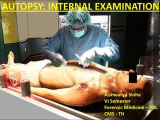
  • 1. AUTOPSY: INTERNAL EXAMINATION Aishwarya Sinha VI Semester Forensic Medicine – PBL CMS - TH
  • 2. INTERNAL EXAMINATION It consists of: 1. Incising the body 2. Inspecting the various organs 3. Examining the cavities systematically 4. Weighing and measuring the organs 5. Checking for any pathology 6. Putting all the organs back in and padding 7. Suturing the body
  • 3. 1. ‘I’ shaped Incision: It is a straight line incision extending from the chin to the symphysis pubis. 2. ‘Y’ Shaped Incision: This type of incision starts near the acromian process and progresses downwards towards the xiphoid process. The incision is then extended till the symphysis pubis. Also, a similar incision is made on the opposite side of the body. 3. Modified ‘Y’ Shaped: A ‘Y’ shaped incision is made from the suprasternal notch to symphysis pubis. It extends from the suprasternal notch over the clavicle to its center on both sides and passes upwards over the neck, behind the ear.
  • 4.
  • 5. REMOVAL OF ORGANS 1. Virchow’s Technique: Head  Thorax  Abdomen 2. Rokitansky’s Technique: in situ dissection in parts combined with en block removal. 3. Letulle’s Technique: Cervical  Thoracic  Abdominal  Pelvic Organs  Removed en masse and dissected as organ block.
  • 6. ORDER OF EXAMINATION VIRCHOW’S TECHNIQUE  Head Thoracic (Cervical) Abdominal Organs
  • 7. HEAD • The neck is extended by placing a wooden block under the shoulders. • Fix head using a head rest. • Intermastoidal incision is made i.e. from the mastoid process behind one ear to the vertex and again to the mastoid process of the other ear. • Scalp flap is reflected forward to the superciliary ridge and backwards to the occipital protuberance.
  • 8. HEAD • With the help of a saw, ‘V’ shaped cut is made so that the skull fits back correctly after autopsy. • This ‘V’ shaped cut passes through: the center of the forehead  base of the mastoid process  occipital protuberance  the base of the other mastoid process and back to the center of the forehead.
  • 9.
  • 10. CAUTION • Care must be taken to avoid any damage to the meninges or the brain during the process of skull removal. • Therefore, chisel and hammer should never be used. • Instead, a chisel should be inserted and twisted at various points in order to remove the skull flap. • By doing so, superior longitudinal sinus can be exposed.
  • 11. BRAIN • Dura is incised along the lines of the incision of the skull  folded to the midline  falx cerebri is freed from the cribriform plate  dura is pulled back. • 4 fingers of the left hand are inserted into the frontal lobe and the nerves and vessels are cut. • Tentorium is also cut along the posterior part of the petrous bone.
  • 12.
  • 13. BRAIN • A knife is then inserted into the occipital foramen and the cervical cord  cervical nerves and the vertebral arteries are cut as far as possible. • With the right hand the cerebellum is grasped and examined. • Any signs of hemorrhage, injury or any other disease should be carefully seen. • Vessels, especially the Circle of Willis, should be checked for any arteriosclerosis or aneurysm etc.
  • 14. FIXATION Brain will start to deteriorate as soon as the blood supply is interrupted. This deterioration is rapid and therefore, it is of utmost importance to arrest this procedure. This process of preservation of the brain is called fixation. There are 2 main methods of fixation: 1. Freezing 2. Prefusion
  • 15. FIXATION OF THE BRAIN Brain is fixed in 10% formalin for at least 1-2 week(s). To help with the penetration of the preservative, lateral fissures are made through, which tear open the pia and the arachnoid mater. A long sagittal cut is made through the corpus callosum so as to allow the formalin to pass into the ventricles.
  • 16. AFTER FIXATION  Brain is placed on a board and the cerebellum along with the pons and the medulla are removed.  Straight cut is made to expose the fourth ventricle.  Thereafter an oblique cut is made to expose the dentate nucleus.  Entire stem is sectioned at a few millimeters distance and examined for any pathology.  Cerebellum is examined by making a series of sagittal cuts beginning from the frontal pole passing backwards to the occiput.  Sequentially cuts are made and serially placed aside in the same order for later identification.
  • 17. SPINAL CORD • Not examined unless there is an indication. • In prone position, wooden block is placed under the chest and the head is bent downwards. • Mid-line incision from the occipital protuberance to the L4 vertebra is made. • Muscles are dissected all the way. • Atlas is disarticulated. • With the help of a double bladed saw, the laminae are sawed through the entire length and separated by a chisel.
  • 18. SPINAL CORD • Dura is cut open and checked for haemorrhage, inflammation, tumours, pus etc. • Nerves are cut from below as they pass though the spinal formaina. • And the cord is separated from the foramen magnum. • Cord is then sectioned transversely and serially. • Lastly, the vertebral column is examined for any fractures or dislocations.
  • 19. THORAX • Chest muscles are dissected away. • Chest is opened by cutting the costal cartilages with the help of a cartilage knife. • In case of elderly subjects, the costal cartilage may be calcified, hence, a bone saw or a rib shear is used to cut it out. • Thereafter, both the sternoclavicular joints are disarticulated and the chest is opened.
  • 20. HEART  Held at the apex and lifted upwards so that the pulmonary vessels, SVC & IVC and the ascending aorta can be examined.  The pulmonary artery is palpated and then the vessels are cut open. Pericardium is incised and examined for any blood or fluids. Heart, opened in the direction of the blood flow i.e. (Inflow-outflow method).
  • 21. HEART  The enterotome is inserted into the:  RA  Tricuspid valve  RV  Pulmonary Trunk  Pulmonary vein  LA  Mitral Valve  LV  Ascending aorta.  Both the auricular appendages are examined for thrombi.  Heart should eventually be weighed and the various measurements of the circumference of the valves or the thickness of the ventricles etc. are taken.
  • 22. CORONARY ARTERY  Examined by making serial cross sections to check for ante mortem clots.  To check for infarction and fibrosis, muscles of the right and the left ventricles are incised. SUBENDOCARDIAL HAEMORRHAGE:  Flame shaped, confluent, patchy, haemorrhages may be seen in the LV on the left side of the inter ventricular septum.  Seen in severe hypotension, intracranial damage, death due to ectopic pregnancies, arsenic poisoning etc.
  • 23. LUNGS • Anterior surface is faced upwards  PA is identified and cut as far as possible using small scissors  check for atherosclerosis, thrombi or emboli. • Steps are re-traced via PV  check for thrombi. • At hilum, a long bladed knife is placed with the blunt end facing upwards  pierced into the hilum and the knife is turned upwards so that the sharp end now faces upwards  Sawing motion  hilum is cut.
  • 24. LUNGS • Lungs are mounted on a board or held in the left hand incision is made from the apex to the base. • Hence, producing an antero-posterior slice. • Cut sections are then examined for consolidations, edema, atelectasis, cong estion, emphysema, Tardieu spots, emboli etc.
  • 25. If PENUMOTHORAX is suspected: • Pocket dissection on affected side between the chest wall and the skin  filled with water  wall is punctured under water. • If bubbles are present  Pneumothorax. • 16 gauge needle attached to a syringe without its plunger is inserted into the subcutaneous tissue over the ICS  water is filled in the syringe  needle is pierced into the pleural cavity if water bubbles are seen  Pneumothorax. • Thereafter, X-ray is done.
  • 26. If AIR EMBOLISM is suspected: • In such cases, before touching the thoracic organs, the pericardium and the heart should be examined. • The heart is lifted up and cut open at the apex. 1. Left ventricle is filled with frothy blood if air is present is good amount. 2. If right ventricle is filled with air, the heart will float in water.
  • 27. If AIR EMBOLISM is suspected: 1. Pericardial sac is filled with water  heart is punctured with a scalpel and twisted  if bubbles are present  AIR EMBOLISM. 2. A wide bore needle is attached to a syringe and water is filled in it  it is inserted in to right ventricle  if air is present bubbles are seen. 3. PYROGALLOL TEST: 4 mL of 2% freshly prepared pyrogallol is taken in two 10 mL syringes. In one of the syringes, 0.5 M NaOH is added. Gas is aspirated from the right heart and the needle is retracted and removed. A stopper is fixed to the syringe and the syringe is shaken. If the aspirate contains air, the colour of the solution would turn brown. 4. Air in the IVC can be demonstrated by puncturing it underwater. 5. Lastly, X-ray is done.
  • 28. NECK • Identify the oesophagus  incise it from the posterior aspect up till the cardiac end of the stomach  inspected for capsules, tablets or powders. • Larynx, trachea and the bronchi are also cut from the posterior aspect and checked for foreign bodies, blood, mucous, tumours, inflammation etc. • Thyroid is removed and examined. • The carotid arteries are examined for thrombi, esp. at their bifurcation. • Later, the hyoid bone, cricoid and the thyroid cartilages are examined.
  • 30. ABDOMEN • 5 cm above the symphysis pubis the rectus muscles are divided and a small cut is made. • Middle and the index fingers are then inserted and spread in a ‘V’ shape. • Sharp braded knife is inserted between them and the peritoneum is cut up to the xiphoid process.
  • 31. ABDOMEN • Firstly, inspection is done and if any damage, free fluid, perforations etc. are seen then these are noted. • Also, note should be made about the abnormalities, positions, abdominal organs, adhesions, pathology (if any), injuries etc.
  • 32. STOMACH After applying double ligatures, the stomach is opened along the greater curvature  from the cardiac to the pyloric end  size of the pyloric ring is noted. Contents are examined for any nature of food which might be present & its state of digestion, smell, colour, character, the presence of foreign bodies or any other suspicious matter etc. Mucous membrane is examined for congestion, haemorrhage, ulcerations or any other
  • 33. INTESTINES • Note the colour, consistency, adhesions, herniae, haem orrhage, serosal surface etc. • Small intestine is opened along its line of mesenteric attachments. • Large intestine is opened along the anterior taenia coli. • Examined for
  • 34. LIVER • Either liver is removed by itself or attached to the stomach and the duodenum. • It is examined for its weight, size, consistency and for the presence of any other pathology / injury. • It is cut along the long axis into 2 cm thick slices.
  • 35. SPLEEN • It is removed by itself. • Size, weight, consistency, capsule condition, rupture injuries or any pathology is noted. • This is also sectioned along its long axis and the character of the septa and the parenchyma are noted.
  • 36. PANCREAS • Along the long axis cuts are made at right angles. • Examined for fat necrosis.
  • 37. KIDNEYS • Size and weight are noted. • Capsules are excised and examined carefully. • Kidney is sectioned longitudinally the convex border of the hilum so that it splits into half and opens the pelvis. • Checked for calculi, inflammation etc. • Ureter is cut into and examined.
  • 38. BLADDER • Incision is made from the fundus and carried to the urethra. • Wall condition, amount and character of urine is noted.
  • 39. PROSTATE & TESTES • Examined for enlargement and malignancies. • Vertical cross-sections are made through the lateral and median lobes and the prostate is split open for examination. • Inguinal canal is incised from the peritoneal aspect and the loop of the vas deference is pulled to free it from the internal inguinal ring. • The testes is pushed with one hand and pulled out of the scrotum easily by the other. • These are cut longitudinally and checked for any clotted blood inside the scrotum and in the testis.
  • 40. FEMALE GENITALIA • Vagina and the uterus are cut either anteriorly or posteriorly upto the fundus. • Two short incisions are made at the fundus from the main incision towards each cornu so as to expose the endometrium. • Ovaries are sectioned longitudinally and the fallopian tubes are cut longitudinally. If the uterus contains a foetus, its age should be determined.
  • 41. AFTER COMPLETING INTERNAL EXAMINATIONS • Body cavities should be cleaned and made free from blood, fluids etc. • Organs are placed back in and excess space is packed with cotton/cloth etc. (esp. in the pelvis and the neck regions.) • Dissection flaps are closed and sutured with thin twine. • Skull is filled with cotton and absorbent material and the skull cap is placed back in and the scalp is stitched. • Body is washed with water, dried, covered with clothes and handed over to the police officials.