2. Traumatic spinal cord injury 179
Imaging Evaluation
The initial computed tomography (CT) scan obtained within
2 hours of the fall showed findings of early diffuse cerebral
edema, SDH, and SAH, including a prominent left parietal
focus of SAH and a focal right frontal SDH (Fig 1). A much
smaller SAH/SDH were also present along the convexities,
falx (ie, interhemispheric), and tentorium. There was no ev-
idence of brain hemorrhage. No scalp or skull abnormalities
were identified, although 1 observer could not rule out a
“healed” skull fracture along the sutures in the parieto-occip-
ital region. A CT scan performed 5 hours later showed pro-
gression of the cerebral edema and no change in the SAH or
SDH (Fig 2). A CT scan of the cervical spine was negative
(Fig 3). Magnetic resonance imaging was recommended but
never obtained. A skeletal survey showed anterior wedge-like
vertebral body deformities from T5 through T12 and inferior
L2. Some widening of the cranial sutures was present, but no
fractures were confirmed on the plain radiographs. The re-
mainder of the survey was negative. A postmortem CT scan of
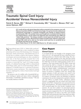
the entire spine (Fig 4), confirmed the vertebral deformities. Figure 2 A CT scan performed 5 hours after the initial CT scan shows
marked progression of cerebral edema with complete loss of gray/
Autopsy Findings by the Medical Examiner white matter differentiation, obliteration of sulci, and near complete
obliteration of the ventricular system. A SAH is seen within com-
Head and Brain
pressed sulci in the left parietal lobe.
A focus of hemorrhage was present in the left posterior
parietal scalp (ie, impact site). Microscopy showed acute
hemorrhage with acute vital reaction within the fibrous
connective tissue of the galea in that region. Brain weight axonal injury (TAI) observed microscopically on beta-
was 1,270 g with the spinal cord attached. No skull frac- amyloid precursor protein (B-APP) immunoperoxidase
ture was shown. The brain was extremely swollen with stains. There were bilateral, holohemispheric, thin-layer
generalized flattening and ablation of the normal gyral SDHs with no mass effect.
pattern. Histologically, a diffuse axonal injury pattern con-
sistent with HIE was present. There were no gross trau-
matic brain parenchymal injuries (eg, no contusion or
shear lesions) or any histological evidence of traumatic
Figure 1 A nonenhanced CT image of the brain preformed within 2
hours of the fall shows decreased differentiation of gray/white mat- Figure 3 Sagittal reconstructed CT image of the cervical spine shows
ter representing edema. normal alignment and no fractures.
3. 180 Barnes et al
Biomechanical Analysis
A court-approved, biomechanical evaluation was performed
including an investigation of the home setting where the
injury reportedly occurred. A number of potential accidental
and NAI (including SBS) scenarios were considered and an-
alyzed primarily to address the thoracic spinal injuries and
secondarily to address the cervical cord injury. The approach
to the biomechanical analysis was to assume that the care-
taker’s history was truthful and accurate and to then apply
the principle of mechanics to evaluate whether or not such a
history could be consistent with the subsequent injuries. This
approach was not intended to rule out other possibilities but
simply to evaluate the history provided.
In that light, the caretaker consistently reported to all au-
thorities that he had his back turned at the time of the inci-
dent but that the boy had been standing up on the seat of the
chair. He then reported hearing a noise and turning to find
the boy and the chair on the floor, with the chair lying on its
back. Using the caretaker’s consistent history as one scenario,
Figure 4 Sagittal reformation of the thoracic spine from a postmor- along with the imaging and clinical findings, the child was
tem CT scan shows multilevel anterior wedge compression fractures assumed to have fallen, rotating with the chair until a point of
of varying degrees. separation (Fig 6). From that point, it was further assumed
that he fell freely to strike the floor first with his head and
then with his dorsal neck and a shoulder, again based on the
Neck and Spinal Cord imaging and autopsy findings. This “impact” scenario would
Focal soft-tissue hemorrhages were present in relation to the
right posterior neck and shoulder regions as well as the pos-
terolateral transverse processes of the atlas and axis and at the
C1-C2 intervertebral junction. No vertebral artery abnormal-
ity was noted. The dura appeared tense and filled with blood-
stained fluid. Sagittal sections at the cervicomedullary junc-
tion showed partial transection and disruption of the central
cord immediately distal to the inferior medullary olives. The
tissue appeared elongated and physically separated suggest-
ing axial tension of the cord (Fig 5). The cellular response
consisted of round and polymorphonuclear cells with acute,
focal hemorrhage. Findings of ischemic neuronal degenera-
tion were most prominent in, and adjacent to, the dorsal and
ventral neurons and consisted of cytoplasmic swelling, de-
granulization, loss of fine detail, and nuclear pynknosis.
Eyes
On gross examination, the pigmented layer of the retina was
focally separated from the choroid. RHs were present primar-
ily in the ganglion cell layer anteriorly but extended posteri-
orly. An optic nerve sheath hemorrhage was also present
bilaterally. There was no mention of perimacular folds.
Vertebral Column
The vertebral bodies from T2 through L3 to 4 were removed
en bloc. A hemorrhage was seen in the anterior and lateral
perivertebral fibroconnective tissues. Microscopic sections
showed acute hemorrhage with fibrin deposition replacing
normal marrow of all vertebral bodies. Multiple areas of dis-
rupted cancellous bone with acute-phase osteogenic granu- Figure 5 A histological specimen through the cervicomedullary
lation tissue were especially prominent T7 through T10. No junction shows complete disruption of the central cord elements
callus or osteoblast activity was present. (circle). (Color version of figure is available online.)