Molecular marker technology in studies on plant genetic diversity

A molecular marker is a molecule contained within a sample taken from an organism (biological markers) or other matter. It can be used to reveal certain characteristics about the respective source. DNA, for example, is a molecular marker containing information about genetic disorders, genealogy and the evolutionary history of life. Specific regions of the DNA (genetic markers) are used to diagnose the autosomal recessive genetic disorder cystic fibrosis, taxonomic affinity (phylogenetics) and identity (DNA Barcoding). Further, life forms are known to shed unique chemicals, including DNA, into the environment as evidence of their presence in a particular location.Other biological markers, like proteins, are used in diagnostic tests for complex neurodegenerative disorders, such as Alzheimer's disease. Non-biological molecular markers are also used, for example, in environmental studies.

Recommandé
Contenu connexe
Tendances
Tendances (20)
En vedette
En vedette (8)
Similaire à Molecular marker technology in studies on plant genetic diversity
Similaire à Molecular marker technology in studies on plant genetic diversity (20)
Dernier
Dernier (20)
Molecular marker technology in studies on plant genetic diversity
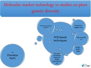
- 1. Molecular marker technology in studies on plant genetic diversity PCR based techniques PCR with arbitrary primers PCR with Amplified fragment length polymorphis ms Sequence- tagged sites Microsatellit es, SCARs, CAPS, ISSRsBy Chanakya Pachi
- 2. Molecular Markers • The development of molecular techniques for genetic analysis has led to a great augmentation in our knowledge of crop genetics and our understanding of the structure and behavior of various crop genomes. • These molecular techniques, in particular the applications of molecular markers, have been used to scrutinize DNA sequence and it’s variation(s) in and among the crop species and create new sources of genetic variation by introducing new and favorable traits from landraces and related crop species.
- 3. Markers can aid selection for target alleles that • Are not easily assayed in individual plants, • Minimize linkage drag around the target gene, and • Reduce the number of generations required to recover a very high percentage of the recurrent parent genetic background
- 4. Introduction • A genetic marker is a gene or DNA sequence with a known location on a chromosome and associated with a particular gene or trait. • It can be described as a variation, which may arise due to mutation or alteration in the genomic loci, that can be observed. • A genetic marker may be a short DNA sequence, such as a sequence surrounding a single base-pair change (single nucleotide polymorphism, SNP), or a long one, like minisatellites.
- 5. Some commonly used types of genetic markers RFLP (or Restriction fragment length polymorphism) AFLP (or Amplified fragment length polymorphism RAPD (or Random amplification of polymorphic DNA) VNTR (or Variable number tandem repeat) Microsatellite polymorphism SNP (or Single nucleotide polymorphism) STR (or Short tandem repeat) SFP (or Single feature polymorphism) DArT (or Diversity Arrays Technology)
- 6. DNA-based technologies • PCR based techniques • PCR with arbitrary primers • Amplified fragment length polymorphisms (AFLPs) • Sequence-tagged sites • Microsatellites • SCARs • CAPS • ISSRs
- 7. PCR BASICS • The polymerase chain reaction • PCR is a rapid, inexpensive and simple way of copying specific DNA fragments from minute quantities of source DNA material • It does not necessarily require the use of radioisotopes or toxic chemicals • It involves preparation the sample DNA • A master mix with primers, • Followed by detecting reaction products
- 8. PCR procedures: steps • Denaturation: • DNA fragments are heated at high temperatures, which reduce the DNA double helix to single strands. • These strands become accessible to primers • Annealing: • The reaction mixture is cooled down. • Primers anneal to the complementary regions in the DNA template strands, and double strands are formed again between primers and complementary sequences • Extension: • The DNA polymerase synthesises a complementary strand. • The enzyme reads the opposing strand sequence and extends the primers by adding nucleotides in the order in which they can pair.
- 9. • The DNA polymerase, known as 'Taq polymerase', is named after the hot-spring bacterium Thermus aquaticus from which it was originally isolated. • The enzyme can withstand the high temperatures needed for DNA-strand separation, and can be left in the reaction tube. • The cycle of heating and cooling is repeated over and over, stimulating the primers to bind to the original sequences and to newly synthesised sequences. • The enzyme will again extend primer sequences. • This cycling of temperatures results in copying and then copying of copies, and so on, leading to an exponential increase in the number of copies of specific sequences. • Because the amount of DNA placed in the tube at the beginning is very small, almost all the DNA at the end of the reaction cycles is copied sequences. The reaction products are separated by gel electrophoresis. • Depending on the quantity produced and the size of the amplified fragment, the reaction products can be visualized directly by staining with ethidium bromide or a silver-staining protocol, or by
- 10. PCR procedures: cycle 1
- 11. PCR procedures: cycle 2
- 12. PCR procedures: cycle 3
- 13. CONDITIONS Step Reaction Temperature (°C) Time (Seconds) 1 Initial Denaturation 94 120 2 Cycle Denaturation 94 15 3 Cycle Annealing 58 30 4 Cycle Extension 72 80 5 Repeat from Step 2 to 4 30 Cycles 6 Final extension 72 600 7 Infinite Hold 4
- 14. PCR with arbitrary primers • MAAP (multiple arbitrary amplicon profiling) is the acronym proposed to cover the three main technologies that fall in this category: • Random amplified polymorphic DNA (RAPD) • DNA amplification fingerprinting (DAF) • Arbitrarily primed polymerase chain reaction (AP-PCR)
- 15. RAPD • Random Amplified Polymorphic DNA • It is a type of PCR reaction, but the segments of DNA that gets amplified are random. • The researcher performing RAPD creates several arbitrary, short primers (8–12 nucleotides) • Then proceeds with the PCR using a large template of genomic DNA, to enable amplification of fragments. • By resolving the resulting patterns, a semi-unique profile can be gleaned from a RAPD reaction. • No knowledge of the DNA sequence for the targeted genome is required, as the primers will bind somewhere in the sequence, but it is not certain exactly where.
- 16. • This makes the method popular for comparing the DNA of biological systems that have not had the privileged attention of the scientific community, or in a system in which relatively few DNA sequences are compared (it is not suitable for forming a DNA databank). • Because it relies on a large, intact DNA template sequence, it has some limitations in the use of degraded DNA samples. • Its resolving power is much lower than targeted, species specific DNA comparison methods, such as short tandem repeats. • In recent years, RAPD has been used to characterize, and trace, the phylogeny of diverse plant and animal species.
- 17. How it Works? • Unlike traditional PCR analysis, RAPD does not require any specific knowledge of the DNA sequence of the target organism: • The identical 1somer primers may or may not amplify a segment of DNA, depending on positions that are complementary to the primers' sequence. • For example, no fragment is produced if primers annealed too far apart. • Therefore, if a mutation has occurred in the template DNA at the site that was previously complementary to the primer, a PCR product will not be produced, resulting in a different pattern of amplified DNA segments on the gel
- 19. • This picture shows an image of a very high quality RAPD gel. Both, presence and absence of most bands are very clear and the background is transparent. • The researcher would have no doubts while selecting bands and collecting data from this gel. • Consequently, the interpretation of results can be very confident.
- 20. Interpreting RAPD banding patterns • DNA polymorphism among individuals can be because of: • Mismatches at the primer site • The appearance of a new primer site • The length of the amplified region between primer sites • Because of the nature of RAPD markers, only the presence or absence of a particular band can be assessed. • Criteria for selecting scoring bands: • Reproducibility—need to repeat experiments • Thickness • Size
- 22. Amplified fragment length polymorphisms(AFLPs) • AFLP is a PCR-based tool used in genetics research, DNA fingerprinting, and in the practice of genetic engineering. • Developed in the early 1990s by Keygene • AFLP uses restriction enzymes to digest genomic DNA, followed by ligation of adaptors to the sticky ends of the restriction fragments. • A subset of the restriction fragments is then selected to be amplified. • This selection is achieved by using primers complementary to the adaptor sequence, the restriction site sequence and a few nucleotides inside the restriction site fragments. • The amplified fragments are separated and visualized on polyacrylamide gels, either through autoradiography or fluorescence methodologies, or via automated capillary
- 23. Digestion Amplificatio n Electrophore sis AFLP The procedure of this technique is divided into three steps: • Digestion of total cellular DNA with one or more restriction enzymes and ligation of restriction halfsite specific adaptors to all restriction fragments. • Selective amplification of some of these fragments with two PCR primers that have corresponding adaptor and restriction site specific sequences. • Electrophoretic separation of amplicons on a gel matrix, followed by visualisation of the band pattern.
- 24. Main features: • A combination of the RFLP and PCR technologies • Based on selective PCR amplification of restriction fragments from digested DNA • Highly sensitive method for fingerprinting DNA of any origin and complexity • Can be performed with total genomic DNA or with cDNA ('transcript profiling') • DNA is digested with two different restriction enzymes • Oligonucleotide adapters are ligated to the ends of the DNA fragments • Specific subsets of DNA digestion products are amplified, using combinations of selective primers • Polymorphism detection is possible with radioisotopes, fluorescent dyes or silver staining
- 25. DNA digestion and ligation • One restriction enzyme is a frequent cutter (four- base recognition site, e.g. MseI) • The second restriction enzyme is a rare cutter (six- base recognition site, e.g. EcoRI) • Specific synthetic double-stranded adapters for each restriction site are ligated to the DNA fragments generated
- 27. Interpreting AFLP bands • The AFLP technique detects polymorphisms arising from changes (presence or size) in the restriction sites or adjacent to these • Different restriction enzymes can be used, and different combinations of pre- and selective nucleotides will increase the probability of finding useful polymorphisms • The more selective bases, the less polymorphism will be detected • Bands are usually scored as either present or absent • Heterozygous versus homozygous bands may be detected, based on the thickness of the signal, although this can be tricky
- 28. Applications • Genetic diversity assessment • Genetic distance analysis • Genetic fingerprinting • Analysis of germplasm collections • Genome mapping • Monitoring diagnostic markers Examples • Limonium sp. • Stylosanthes sp. • Indian mustard
- 29. PCR-based technologies Sequences-tagged sites Microsatellites SCARs CAPS ISSRs
- 30. Unlike arbitrary primers, STS rely on some degree of sequence knowledge Markers based on STS are codominant They tend to be more reproducible because longer primer sequences are usedRequire the same basic laboratory protocols and equipment as standard PCR Sequence-tagged sites as markers
- 31. Microsatellites • Microsatellites are also called simple sequence repeats (SSRs) • Microsatellites are short tandem repeats (1-10 bp) • To be used as markers, their location in the genome of interest must first be identified • Polymorphisms in the repeat region can be detected by performing a PCR with primers designed from the DNA flanking region
- 33. Structure
- 35. Methodology and visualisation • Methodology: • DNA extraction • PCR with primers specific for microsatellite flanking regions • Separation of fragments • Visualization: • By agarose gel electrophoresis, using ethidium bromide staining and UV light, or • Acrylamide gels using silver staining or radioisotopes, or • Through automated sequencers, using primers prelabelled with fluorescence. • Data analysis
- 36. Staining with Ethidium bromide Staining with Silver Nitrate
- 37. • Advantages: • Require very little and not necessarily high quality DNA • Highly polymorphic • Evenly distributed throughout the genome • Simple interpretation of results • Easily automated, allowing multiplexing • Good analytical resolution and high reproducibility • Disadvantages: • Complex discovery procedure • Costly
- 38. Applications: Individual genotyping Germplasm evaluation Genetic diversity Genome mapping Phylogenetic studies Evolutionary studies
- 39. Sequence characterized amplified regions (SCARs) • SCARs take advantage of a band generated through a RAPD experiment • They use 16-24 bp primers designed from the ends of cloned RAPD markers • This technique converts a band—prone to difficulties in interpretation and/or reproducibility—into being a very reliable marker
- 40. Steps to obtain SCAR polymorphismsA potentially interesting band is identified in a RAPD gel The band is cut out of the gel The DNA fragment is cloned in a vector and sequenced Specific primers (16-24 bp long) for that DNA fragment are designed Re-amplification of the template DNA with the new primers will show a new and
- 41. Diagram of the SCAR procedure
- 43. Cleaved amplified polymorphic sequence (CAPS) • This method is based on • The design of specific primers, • Amplification of DNA fragments, and • Generation of smaller, possibly variable, fragments by means of a restriction enzyme • This technique aims to convert an amplified band that does not show variation into a polymorphic one
- 44. Steps for generating CAPS A band, DNA, gene sequence or other type of marker is identified as important Either the band is detected through PCR (and cut out of the gel, and the fragment cloned and sequenced) or the fragment sequence is already available Specific primers are designed from the fragment sequence The newly designed primers are used to amplify the template DNA The PCR product is subjected to digestion by a panel of restriction enzymes Polymorphism may be identified with some of the enzymes
- 45. Generating CAPS
- 47. Inter-simple sequence repeats (ISSRs) • They are regions found between microsatellite repeats • The technique is based on PCR amplification of intermicrosatellite sequences • Because of the known abundance of repeat sequences spread all over the genome, it targets multiple loci
- 48. Identifying ISSR polymorphisms • A typical PCR is performed in which primers have been designed, based on a microsatellite repeat sequence, and extended one to several bases into the flanking sequence as anchor points. • Different alternatives are possible: • Only one primer is used • Two primers of similar characteristics are used • Combinations of a microsatellite-sequence anchored primer with a random primer (i.e. those used for RAPD)
- 49. Designing primers for ISSR polymorphisms
- 50. • The diagram above presents three different items: • The original DNA sequence in which two different repeated sequences (CA), inversely oriented, are identified. Both repeated sections are, in addition, closely spaced. • If primers were designed from within the repeated region only, the interrepeat section would be amplified but locus- specificity might not be guaranteed. In the second row, a PCR product is shown as a result of amplification from a 3'- anchored primer (CA)n NN at each end of the interrepeat region. CA is the repeat sequence that was extended by NN, two nucleotides running into the interrepeat region. • Alternatively, anchors may be chosen from the 5' region. The PCR product in the third row is a result of using primers based on the CA repeat but extended at the 5' end by NNN and NN, respectively
- 51. • Do not require prior sequence information • Variation within unique regions of the genome may be found at several loci simultaneously • Tend to identify significant levels of variation • Microsatellite sequence-specific • Very useful for DNA profiling, especially for closely related species Disadvantages • Dominant markers • Polyacrylamide gel electrophoresis and detection with silver staining or radioisotopes may be needed
- 52. Latest strategies DNA sequencing Expressed sequence tags (ESTs) Microarray technology Diversity array technology (DArT) Single nucleotide polymorphisms (SNPs)
- 53. References… References Plant Biotechnology and Molecular Markers • P.S. Srivastava • Alka Narula • Sheela Srivastava Using molecular marker technology in studies on plant genetic diversity • M. Carmen de Vicente • Theresa Fulton Molecular Markers • Ashwani Kumar • Scientific Blogging 2.0
Notes de l'éditeur
- The aphanoplasmodium requires the presence of free water, forms a vein-like reticulate pattern that lacks distinct anteroposterior polarity, and exhibits erratic protoplasmic streaming. The characteristically thin and hyaline aphanoplasmodium is adapted to growing in the interstices of wood, flowing through the phloem and xylem (Gray and Alexopoulos, 1968). The protoplasmodium is microscopically rounded or irregular-shaped, does not develop the vein-like network characteristic of the other plasmodial types, and will give rise to only a single stalked sporangium. The trichiaceous plasmodium has characteristics of both the aphano- and phaneroplasmodium, requiring free water for development, having a generally appressed appearance and distinct antero-posterior polarity (Keller, 1971).
- The plasmodium phase of the acellular slime molds differs from the pseudoplasmodium of the cellular slime molds in that it is diploid. This mass gives rise to a fruiting body in which meiosis occurs and haploid spores are produced. The spores germinate to produce flagellated gametes. These gametes fuse to form a diploid zygote. The zygote grows and its nucleus divides mitotically, but the cytoplasm does not divide, resulting in another plasmodium.
- The life cycles of Dictyostelium discoideum. Most of its life, this haploid social amoeba undergoes the vegetative cycle, preying upon bacteria in the soil, and periodically dividing mitotically. When food is scarce, either the sexual cycle or the social cycle begins. Under the social cycle, amoebae aggregate to cAMP by the thousands, and form a motile slug, which moves towards light. Ultimately the slug forms a fruiting body in which about 20% of the cells die to lift the remaining cells up to a better place for sporulation and dispersal. Under the sexual cycle, amoebae aggregate to cAMP and sex pheromones, and two cells of opposite mating types fuse, and then begin consuming the other attracted cells. Before they are consumed, some of the prey cells form a cellulose wall around the entire group. When cannibalism is complete, the giant diploid cell is a hardy macrocyst which eventually undergoes recombination and meiosis, and hatches hundreds of recombinants. Not drawn to scale. CC Creative Commons Attribution – Share Alike 3.0, David Brown & Joan E. Strassmann http://dictybase.org/