Recommandé

Recommandé
Contenu connexe
Tendances
Tendances (20)
En vedette
En vedette (20)
Similaire à Thalamocortical tract ablation impacts motor performance and grip strength in rodents
Similaire à Thalamocortical tract ablation impacts motor performance and grip strength in rodents (20)
Thalamocortical tract ablation impacts motor performance and grip strength in rodents
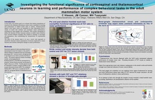
- 1. Introduction In this study, the primary goal is to evaluate how distinct populations of neurons in the thalamus and primary motor cortex contribute to learning and performance of skilled motor tasks in rodents. Thalamocortical inputs to the motor cortex are postulated to relay information from a variety of nuclei that mediate and coordinate motor performance, including the basal ganglia and cerebellum. Prior studies attempting to define the function of these neuronal systems in skilled motor behavior have involved ablation strategies that resulted in damage to non-target cell populations, thus confounding the interpretation of experimental findings. The goal of the present study is to use a novel viral approach to selectively eliminate distinct populations of neurons implicated in motor function and examine subsequent changes in motor performance. Methods Genetically engineered rabies pseudotype lentivirus expressing the human IL2-receptor α was used for selective ablation of thalamacortical tract. By fusing a pseudotype HIV-I Lentiviral vector with glycoprotein C type (Fug-C) we are able to produce the Neuret vector. The Lentivirus genome consists of envelope, transfer, and packaging plasmid. Envelope plasmid is controlled by cytomeg-alovirus enhancer/ chicken -β actin promoter and encodes for FugG-C (Inoue et al., 2012). Transfer plasmid encodes for Interleukin-2 receptor -α subunit (Il-2R )α as well as green fluorescent protein (GFP). The completed NeuRet-Il2R -α GFP vector is transferred into M1 region for retrograde infection of thethalamocortical tract neurons. Acknowledgments Supported by the NIH in part by a MARC U-STAR Award (T34GM087193), the Veterans Administration, and Academic enrichment program. Conclusions • Thalamacortical tract neurons dispersed within the Va/Vl nuclei necessitated adequate performance of complex motor task. Preceding disruption of TCT reveals an inability to initiate learnt motor behavior. •Apart from appropriate motor task initiation, inadequacies in the animal’s stride and forelimb strength were observed indicative of motor control deficits. •The basal ganglia and cerebellum are two significant brain regions fundamental for learning and motor performance whose input relies on TCT neurons for proper execution- the lack there of indicated by a decline in motor performance. •In an attempt to back tract through the complex circuitry involved in distal forelimb reach, succeeding basalthalamic and dentatethalimc ablations will be satisfied. Department of Neurosciences, UC San Diego; Veterans Affairs Med Ctr, San Diego, CA Investigating the functional significance of corticospinal and thalamocortical neurons in learning and performance of complex behavioral tasks in the adult mammalian motor system C Hissom, JM Conner, MH Tuszynski Pre and post ablation forelimb reach test exemplifies functional significance of TCT akin to motor performance Stride contact and stride intensity deviate from both control and CST for TCT lesion animals Figure 2: Eighteen rats were trained one week post injection in distal forelimb reach task to obtain a sugar pellet by extending the forelimb through panel opening. Performance is quantified by recording successful attempts; extend forelimb and retract sugar pellet, and unsuccessful attempt; reach but miss pellet or inability to retract pellet. Figure 3: A camera records the rat’s stride and the software is able to produce a multitude of stride assays. Animals that received the thalamocortical ablation (green) diverged from the rest of the group in both forelimb stride contact and intensity. This group showed greater stride contact with diminished stride intensity. Both control and corticospinal rats showed the opposite; greater stride intensity than stride contact . Animals with both CST and TCT ablations showed a decline in forelimb grip strength Figure 4: Post lesion rats are habituated to grip strength meter for 3 minutes prior to session. Each session consisted of 4 trials to obtain maximum grip strength for each animal. Figure 1: Rats are injected into primary motor cortex (M1) with Neuret viral vector, which retrogradely infects neurons projecting from thalamus to M1. Succeeding a one week period, the cells are infected and expressing Il2R receptor that will selectivelyα bind the selected immunotoxin. A second injection delivers the immunotoxin into the ventral anterior/ventral thalamic nuclei of thalamus insuring a selective thalamocotical track ablation. Figure 5: Primary motor cortex (M1) transmits several electrical impulses intracranially by way of the Basal ganglia and Cerebellum that are pivotal for motor execution and sensory processing. These signals coalesce onto the above mentioned thalamic nuclei. Both the Basal-ganglia thalamocortical circuit (back arrows) and corticopontine cerebellar loop (blue arrows) terminate on Va/Vl nuclei of thalamus (Cruikshank et al 2010). Basal ganglia facilitates initiation of motor commands by disinhibition of thalamic neurons and cerebellum coordinates and governs appropriate motor performance. Basal-ganglia thalamocortical circuit and corticopontine cerebellar loop impart motor command information by way of the Va/Vl nuclei of thalamus M1 Frontal Striatum GP SNr Dentate Red Nucleus Reticular NucleusPontine Thalamus Va/Vl Intermediate lobe of cerebellum References Cruikshank SJ., Urabe H., Nurmikko Av., and Connors BW.(2009) Pathway-Specific Feedforwardcircuits between Thalamus and Neocortex Revealed by Selective OpticalStimulation of Axons. Neuron 65.2: 230-45. Inoue KI., Koketsu D., Kato S., Kobayashi K., Nambu A., Takada M. (2012) Immunotoxin mediated tract targeting in the primate brain: selective elimination of the cortico subthalamic ‘‘hyperdirect’’ pathway. PLoS ONE 7.6: e39149.