

Recommandé
Contenu connexe
Tendances
Tendances (20)
En vedette
En vedette (20)
Similaire à Cardiac arrest
Similaire à Cardiac arrest (20)
Plus de Doha Rasheedy
Plus de Doha Rasheedy (20)
Dernier
Dernier (20)
Cardiac arrest
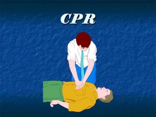
- 1. CPR
- 2. Pattern of cardiac arrest 1-Brady-asystole. 2-Pulseless electrical activity. (PEA) 3-Ventricular fibrillation, Pulseless Ventricular tachycardia.
- 4. Causes of reversible cardiac arrest H T Hypoxia. Toxin. Hypovolaemia. Thrombus Pulmonary Hypoglycemia. embolism. Hypothermia. Thrombus acute H+ions Acidosis. coronary syndrome. Hypo- hyperkalemia. Tension pneumothorax. Tamponade cardiac.
- 5. The primary survey Airway: Open the airway Breathing: Provide positive-pressure ventilation. Circulation: Give chest compressions. Defibrillation: Identify and shock ventricular fibrillation (VF) and ventricular tachycardia (VT)
- 6. Airway Assessment: 1- look for respiratory activity. 2-listen for breathing. 3- feel for air exchange at the patient’s nose and mouth. If these are present, assess the patient’s ability to protect the airway by asking them to speak. If the patient does not respond to questions, the absence of a strong gag reflex: confirms the inadequacy of protective airway mechanisms steps must be taken to provide airway support.
- 7. Steps to support airway 1- immediately call for assistance. 2- place the patient in a supine position. be careful in a patient with neck trauma in-line stabilization of the cervical spine. This is performed by keeping one hand behind the head and neck while the other hand rolls the patient toward you. 3- open airway: use the head tilt-chin lift maneuver or the jaw thrust maneuver. 4- Remove foreign material.
- 10. Breathing 1- bag-valve mask.(2 sec) if unsuccessful. 2-reposition the head and mask and try again. If unsuccessful, (obstructed airway). 3- Open the patient’s mouth by grasping both the tongue and the lower jaw between the thumb and fingers, and then lift the mandible. If you see obstructing material. 4- use a McGill forceps or clamp to remove it. If this equipment is not available, slide your index finger down the inside of the cheek to the base of the tongue and dislodge any foreign bodies using a hooking action. (if unsuccessful).
- 11. Breathing-2 5- Abdominal thrusts. 6- Reattempt ventilation. Rate 8-10 breaths min
- 12. Circulation check for a carotid pulse.( the most central of the peripheral arteries). If no pulse is present, chest compressions should be initiated and the patient should be placed on a cardiac monitor. To adequately perform chest compressions, the heel of one hand should be placed in the midline on the lower part of the sternum (just above the notch where the ribs meet the lower sternum).
- 13. The other hand is placed on top of the first hand and the fingers interlocked and kept off of the chest. Position your shoulders directly over your hands and lock your elbows. Depress the sternum about 1.5–2 inches approximately 100 times per minute. Properly performed compressions can produce a systolic blood pressure of 60mmHg.
- 14. Defibrillation When defibrillation can be successful performed within the first minute or two, as many as 90% of patients return to their pre-arrest neurologic status. The longer the patient remains in cardiac arrest, the more likely that defibrillation and resuscitation will be unsuccessful. Survival rates are 10% when defibrillation is delayed 10 minutes or more after a patient’s collapse.
- 15. Deliver an electric shock to convert the nonperfusing rhythm to a perfusing one. one paddle should be placed to the right of the sternum below the right clavicle and the other in the midaxillary line at the level of the nipple. Firm pressure of approximately 25 lb should be applied to each paddle. Alternatively, “hands off” defibrillator pads can be used that are placed on the chest and the back, sandwiching the heart.
- 16. Types of defibrillator Monophasic Biphasic Less energy needed with biphasic
- 17. Prepare patient Correct reversible causes Check lanoxin level 12 leads ECG before & after shock Iv line present Monitoring circulation and respiration Fasting 8 hr , sedation if elective cardioversion.
- 18. Size of paddle Adult debrillation, both handheld paddle electrodes and self-adhesive pad electrodes 8—12 cm in diameter are used and function well. Debrillation success may be higher with electrodes of 12-cm diameter compared with those of 8-cm.
- 19. Position of paddles the conventional sternal—apical position. The right (sternal) electrode is placed to the right of the sternum, below the clavicle. The apical paddle is placed in the mid axillary line, approximately level with the V6 ECG electrode . It does not matter which electrode (apex/sternum) is placed in either position.
- 20. Position of paddles-2 Other acceptable pad positions include: each electrode on the lateral chest wall, one on the right and the other on the left side (biaxillary); one electrode in the standard apical position and the other on the right or left upper back; one electrode anteriorly, over the left precordium, and the other electrode posterior to the heart just inferior to the left scapula.
- 21. Coupling agents Do not use medical gels or pastes of poor electrical conductivity (e.g., ultrasound gel). Pads versus paddles Self-adhesive debrillation pads are safe and effective and are preferable to standard debrillation paddles. Consideration should be given to use of self- adhesive pads in peri-arrest situations.
- 22. Successful defibrillation depends on the amount of current transmitted across the heart. (energy output of defibrillator transthoracic impedance). ↑ paddle size →↑ efficiency of shock current. Conductive gel (contain salt) so less energy is required, ↓ burn but not ↑ impedence.
- 23. The secondary survey Airway: Definitive airway management (tube). Breathing: Confirmation of adequate ventilation. Circulation: Intravenous access, ACLS medications, fluids. Defibrillation: Continued rhythm analysis and treatment.
- 24. Airway Endotracheal intubation is the most effective method of ensuring adequate ventilation, oxygenation, and airway protection against aspiration during cardiac arrest. In addition, it is an additional route of entry for some resuscitation medications, such as atropine, epinephrine, and lidocaine.
- 25. Breathing the adequacy of intubation should be checked by auscultating the chest for equal bilateral breath sounds, identifying fog in the endotracheal tube on exhalation, Monitoring end-tidal CO2 (using colorimetry or capnography). The presence of exhaled CO2 on a monitor indicates proper tracheal tube placement and can detect subsequent tube dislodgement. A chest X-ray can help determine the location of the tip of the endotracheal tube in relation to the carina.
- 26. Breathing-2 The patient should be placed on a ventilator for positive pressure ventilation. Continuous high flow oxygen and pulse oximetry should be maintained.
- 27. Circulation Intravenous (IV) access should be obtained, preferably with a central venous catheter in the internal jugular, subclavian, or femoral vein. Two large bore peripheral lines may be acceptable. And IV fluids should be infused. The patient’s rhythm should be identified and appropriate interventions instituted based on accepted ACLS guidelines.
- 31. Asystole and bradycardia Atropine has a vagolytic effect by antagonizing the parasympathetic system. Epinephrine improves myocardial and cerebral blood flow during CPR. Early transcutaneous pacing should be considered for bradycardia.transcutaneous pacing for asystole has not been shown to improve survival. As some
- 32. Somme patients with asystole are actually in fine VF, two or more cardiac leads should be checked before determining that the patient is truly in asystole. recent large randomized study from Europe comparing epinephrine with vasopressin for patients in asystole demonstrated that vasopressin was superior to epinephrine, suggesting that vasopressin followed by epinephrine may be more effective than epinephrine alone in the treatment of refractory cardiac arrest.
- 33. Pulseless electrical activity electromechanical dissociation Focus on determining and reversing the cause: The most common causes include severe hypovolemia (usually related to significant blood loss), hypoxia, acidosis, pericardial tamponade, tension pneumothorax, large pulmonary embolus, myocardial infarction, hypothermia, or drug overdose.
- 34. Patient should be intubated to provide adequate oxygenation and given a rapid IV infusion of crystalloid. If the patient has a treatable rhythm, appropriate rhythm-specific ACLS algorithms If the situation warrants, pericardiocentesis or needle thoracostomy should be performed. If no reversible cause can be determined, the patient should be given epinephrine every 3–5 minutes. If the PEA rate is slow, atropine can also be given. Unless a reversible cause is discovered, the prognosis of PEA is poor, with only 1–4% of patients surviving to hospital discharge.
- 35. PEA ASYSTOLE (monitor analysis) {nonshockable} CPR (5 cycles-30:2) 2 min Epinephrine 1mg (repeat 3-5 min) Vasopressin 40 IU then epinephrine. Atropine 1 mg (repeat 3-5 min) {max 3mg} (single dose) Then CPR 5 cycles Check pulse → CPR →E,A,V →CPR→ check pulse.
- 36. Ventricular fibrillation )VF( or pulseless )ventricular tachycardia )VT Attempt debrillation immediately (4 J kg-1 for all shocks). Resume CPR as soon as possible. After 2 min, check the cardiac rhythm on the monitor. Give second shock if still in VF/pulseless VT. Immediately resume CPR for 2 min and check monitor; if no change, give adrenaline followed immediately by a 3rd shock. CPR for 2 min. Give amiodarone if still in VF/pulseless VT followed immediately by a 4th shock.
- 37. Give adrenaline every 3—5 min during CPR. If remains in VF/pulseless VT, continue to alternate shocks with 2 min of CPR. If signs of life become evident, check the monitor for an organised rhythm; if this is present, check for a central pulse. Identify and treat any reversible causes (4Hs &4Ts). If debrillation was successful but VF/pulseless VT recurs, resume CPR, give amiodarone and debrillate again at the dose that was effective previously. Start a continuous infusion of amiodarone.
- 38. Precordial thump Consider giving a single precordial thump when cardiac arrest is confirmed rapidly after a witnessed, sudden collapse and a defibrillator is not immediately to hand. the technique: Using the ulnar edge of a tightly clenched fist, deliver a sharp impact to the lower half of the sternum from a height of about 20 cm, then retract the fist immediately to create an impulse-like stimulus. A precordial thump is most likely to be successful in converting VT to sinus rhythm.
- 39. Drug administration Routes IV access: central peripheral Drugs typically require 1 to 2 minutes to reach the central circulation when given via a peripheral vein but require less time when given via central venous access. If a resuscitation drug is administered by a peripheral venous route, administer the drug by bolus injection and follow with a 20-mL bolus of IV fluid. Elevate the extremity for 10 to 20 seconds to facilitate drug delivery to the central circulation.
- 40. Intraosseous (IO) cannulation: provides access to a noncollapsible venous plexus, enabling drug delivery similar to that achieved by central venous access. In the sternum , proximal tibia (2 cm below tt) , distal tibia (2 cm above mm) In endotracheal tube: Epinepherine , Atropine , Vasopressin , lidocaine , naloxone. Give at dose 2.5-3 times usual iv dose. Dilute in 5-10 ml saline.
- 41. Medications for Arrest Rhythms Vasopressors : at any stage during management of pulseless VT, VF, PEA, or asystole increases the rate of neurologically intact survival to hospital discharge. Epinepherine: It is appropriate to administer a 1- mg dose of epinephrine IV/IO every 3 to 5 minutes during adult cardiac arrest. Higher doses may be indicated to treat specific problems, such as В-blocker or calcium channel blocker overdose. may be given by the endotracheal route at a dose of 2 to 2.5 mg.
- 42. Vasopressin: Vasopressin is a nonadrenergic peripheral vasoconstrictor that also causes coronary and renal vasoconstriction.(40 U, with the dose repetition only once).
- 43. Atropine in asystole or slow PEA arrest. The recommended dose of atropine for cardiac arrest is 1 mg IV, whichcan be repeated every 3 to 5 minutes (maximum total of 3 doses or 3 mg) if asystole persists. Endotracheal route also used.
- 44. Antiarrhythmics Amiodarone: administered for VF or pulseless VT unresponsive to CPR, shock, and a vasopressor. An initial dose of 300 mg IV/IO can be followed by one dose of 150 mg IV/IO. Lidocaine: considered an alternative treatment to amiodarone.The initial dose is 1 to 1.5 mg/kg IV. If VF/ pulseless VT persists, additional doses of 0.5 to 0.75mg/kg IV push may be administered at 5- to 10 minute intervals, to a maximum dose of 3 mg/kg.
- 45. Magnesium: When VF/pulseless VT cardiac arrest is associated with torsades de pointes, magnesium sulfate at a dose of 1 to 2 g diluted in 10 mL D5WIV/IO push, typically over 5 to 20 minutes. When torsades is present in the patient with pulses, the same 1 to 2 g is mixed in 50 to 100 mL of D5Wand given as a loading dose. It can be given more slowly (eg, over 5 to 60 minutes IV)
- 46. Pacing in Arrest Several randomized controlled trials failed to show benefit from attempted pacing for asystole. At this time use of pacing for patients with asystolic cardiac arrest is not recommended.
- 47. Routine Administration of IV Fluids During Cardiac Arrest There were no published human studies evaluating the effect of routine fluid administration during normovolemic cardiac arrest There is insufficient evidence to recommend routine administration of fluids to treat cardiac arrest. Fluids should be infused if hypovolemia is suspected.
- 48. Monitoring Assessment During CPR: At present there are no reliable clinical criteria that clinicians can use to assess the efficacy of CPR. Although end-tidal CO2 serves as an indicator of cardiac output produced by chest compressions and may indicate return of spontaneous circulation
- 49. Assessment of Hemodynamics 1-Coronary Perfusion Pressure (CPP= aortic relaxation [diastolic] pressure minus right atrial relaxation phase blood pressure) during CPR correlates with both myocardial blood flow and ROSC. A CPP of 15 mm Hg is predictive of ROSC. Increased CPP correlate with improved 24-hour survival rates in animal studies. Rarely available
- 50. Pulses-2 Clinicians frequently try to palpate arterial pulses during chest compressions to assess the effectiveness of compressions. No studies have shown the validity or clinical utility of checking pulses during ongoing CPR. Because there are no valves in the inferior vena cava, retrograde blood flow into the venous system may produce femoral vein pulsations. Thus palpation of a pulse in the femoral triangle may indicate venous rather than arterial blood flow. Carotid pulsations during CPR do not indicate the efficacy of coronary blood flow or myocardial or cerebral perfusion during CPR.
- 51. Assessment of Respiratory Gases 1-Arterial Blood Gases not a reliable indicator of the severity of tissue hypoxemia, hypercarbia, or tissue acidosis. 2-Oximetry During cardiac arrest, pulse oximetry will not function because pulsatile blood flow is inadequate in peripheral tissue beds.
- 52. End-Tidal CO2 Monitoring useful as a noninvasive indicator of cardiac output generated during CPR. major determinant of CO2 excretion is its rate of delivery from the peripheral production sites to the lungs. In the low-flow state during CPR, ventilation is relatively high compared with blood flow, so that the end-tidal CO2 concentration is low. If ventilation is reasonably constant, then changes in end-tidal CO2 concentration reflect changes in cardiac output.
- 53. Duration of CPR Arrest time< 6 min→30 min CPR Arrest time> 6 min→15 min CPR
- 54. Post-CPR Management Induced hypothermia(32-34o)(12-24 hr) Glucose control Organ-Specific Evaluation and Support
- 55. Prognosis 1-strongly predict death or poor neurologic outcome, with 4 of the 5 predictors detectable at 24 hours after resuscitation: Absent corneal reflex at 24 hours Absent pupillary response at 24 hours Absent withdrawal response to pain at 24 hours No motor response at 24 hours No motor response at 72 hours
- 56. 2-An electroencephalogram performed 24 2- to 48 hours after resuscitation has also been shown to provide useful predictive value. 3-GCS: <5 ON 3rd day =no chance for neurological recovery. 4- Duration of coma: >4-6 hr =poor prognosis >24 hr =10% recovery >72 hr = 5% recovery > 2 wk = no recovery at all.