1. Forum
493 June 2012, Vol. 102, No. 6 SAMJ
Cervical cancer is the second most common cancer in women
worldwide and the leading gynaecological malignancy in women in
Africa.1
In 2008 the International Agency for Research on Cancer
estimated that 493 243 women are newly diagnosed with cervical
cancer annually. Of these, more than 273 000 die each year.1,2
It is
estimated that around 80 000 women, of whom 60 000 die each
year, live in Africa. Because there are inadequate cancer registries
in many African countries, it is likely that these figures are a gross
under-representation.
Infection of the cervix with high-risk human papillomavirus
(HPV) is regarded as the main causal factor in cervical cancer.3
There are more than 150 genotypes of HPV with around 40 known
to infect the anogenital tract, giving rise to genital warts or neoplastic
lesions.3
Recently, 2 prophylactic vaccines against the main high-
risk HPV variants, 16 and 18, have been introduced by Merck &
Company (Gardasil) and GlaxoSmithKline (Cervarix). These induce
an immune response that blocks initial HPV infection and confers
protection against cancer associated with HPV 16 and 18 and some
closely related variants. These have limited benefit for women already
infected with high-risk HPV and in addition are out of reach of the
majority of women in Africa due to the high costs involved. Although
HPV infection initiates disease, cervical cancer is a multi-step
process, with other contributing factors, including multiple sexual
partners, tobacco carcinogens, a weakened immune system and
sexually transmitted infection by human immunodeficiency virus
(HIV), Chlamydia trachomatis and Neisseria gonorrhoeae, thought to
contribute to the aetiology.
HPV initially infects basal keratinocytes and epithelial cells and
uses the host’s cellular machinery for replication and persistence.4
The HPV genome consists of 3 domains, a non-coding upstream
region, an early region containing open reading frames E1, E2, E4,
E5, E6 and E7, and a late region encoding the major and minor
capsid proteins.4
In the vast majority of women, infections and
HPV-induced lesions are transient and are naturally resolved.
However, approximately 10 - 20% of women fail to eliminate the
virus.4
In these cases, persistence of infection, viral integration and
activation of inflammatory pathways have been linked to neoplastic
transformation and malignant progression.4,5
In this review, we
highlight our findings relating to the activation of inflammatory
pathways in cervical cancers and address their role in disease
progression.
Persistent HPV infection and
inflammation
By broad definition, inflammation involves tissue remodelling
events brought about by alterations to epithelial, vascular and
immune cell function. These are orchestrated by specific molecular
pathways involving a host of cytokines, chemokines, growth factors
and lipid mediators.6
Compelling evidence has shown that the
majority of cancers arise from sites of chronic irritation, infection
and inflammation,7
solidifying the concept that chronic unabated
inflammation is critical for tumour progression.
Persistent HPV infection and integration of E6 and E7 oncogenes
into the host genome is considered key to development of cervical
cancer.4
The HPV E6 and E7 early genes encode oncoproteins
responsible for cervical neoplastic transformation3
by inactivating
tumour suppressors as well as promoting the accumulation of
genetic mutations.3,4
Although E6 and E7 oncogenes appear to be
the main HPV genes involved in transformation, recent studies have
highlighted an important role for E5 oncogene in tumorigenesis
and immune cell modulation8
and regulation of late viral functions
together with the E4 oncogene. In addition, E1 and E2 oncogenes
encode replication factors and are thought to play a role in HPV
persistence by allowing episomal copies of the virus to be maintained
in the nucleus and partitioned into daughter cells during mitosis.9
Immune evasion is an essential aspect of HPV persistence and
development of cervical cancer. Since there is no viraemia or
cytolysis associated with initial viral infection of the cervix, there is
no activation of the innate immune system and no inflammation.
ANALYSIS
Inflammatory pathways in cervical cancer – the University of
Cape Town’s contribution
Kurt J Sales, Arieh A Katz
Cervical cancer is the leading gynaecological malignancy in
southern Africa. The main causal factor for development of the
disease is infection of the cervix with human papillomavirus.
It is a multi-step disease with several contributing co-factors
including multiple sexual partners, a compromised immune system
and cervical inflammation caused by infections with Chlamydia
trachomatis or Neisseria gonorrhoeae. Inflammation involves
extensive tissue remodelling events which are orchestrated by
complex networks of cytokines, chemokines and bio-active lipids
working across multiple cellular compartments to maintain tissue
homeostasis. Many pathological disorders or diseases, including
cervical cancer, are characterised by the exacerbated activation and
maintenance of inflammatory pathways. In this review we highlight
our findings pertaining to activation of inflammatory pathways
in cervical cancers, addressing their potential role in pathological
changes of the cervix and the significance of these findings for
intervention strategies.
S Afr Med J 2012;102(6):493-496.
K J Sales and A A Katz are principal investigators at the MRC Research Group for
Receptor Biology, at the Institute of Infectious Disease and Molecular Medicine
and the Division of Medical Biochemistry, Faculty of Health Sciences, University
of Cape Town.
Corresponding author: K J Sales (kurt.sales@uct.ac.za)
2. Forum
494 June 2012, Vol. 102, No. 6 SAMJ
Despite this, the virus actively induces mechanisms to evade immune
detection and ensure its success by deregulating the interferon
pathway4
and via the down-regulation of pattern recognition receptors
such as Toll-like receptor 9, thereby allowing infection to proceed
undetected.4
The virus requires actively dividing cells and active host
cellular machinery for replication and persistence.4
Once established,
persistentinfectionspromotealterationsinthereleaseofinflammatory
cytokines which in turn can alter immune cell infiltration and
inflammation. Alterations in immune responsiveness and elevated
systemic levels of inflammatory cytokines have been observed in
older women (about 50 years of age) with persistent HPV infection.5
Since this is the age group most likely to present with cervical cancer
in the clinic, it is likely that sustained elevation in systemic cytokine
release contributes to HPV-mediated tumorigenesis.
Although the direct association between HPV infection and
inflammation is controversial, transgenic mouse models, expressing
the early genes from HPV 16 under the control of the human
keratin 14 promoter, have shown that HPV-induced lesions release
the chemokine CCL2 which enhances macrophage recruitment
into tumours via CCR2.10
In human neoplastic cervical epithelial
cells, HPV 16 E5, E6 and E7 oncogenes have been shown to induce
the inflammatory cyclo-oxygenase (COX)-prostaglandin axis, by
elevating expression of the immediate early oncogene COX-2.11
These
findings provide a direct link between HPV oncogenes and activation
of potent inflammatory cascades, with known roles in promoting
cancer. Thus, although HPV is not associated with inflammation
at the initial point of infection, it is likely that following integration
and transformation, persistent HPV infection drives inflammatory
pathways, such as the COX-prostaglandin pathway in neoplastic
epithelial cells, to promote immune cell infiltration, inflammation
and tumour progression.
The inflammatory cyclo-oxygenase-
prostaglandin pathway
COX enzymes, of which there are 2 isoforms in humans (COX-1
and COX-2), catalyse the rate-limiting conversion of arachidonic
acid to the unstable intermediate prostaglandin H2, which in turn
is converted by terminal prostaglandin synthase enzymes to specific
classes of prostaglandins.12
For many years COX-1 was considered
to be constitutively expressed in tissues at low levels, generating
prostaglandins for normal physiological functions, whereas
COX-2 was considered to be an immediate early gene involved in
pathology.12
Studying tissue biopsies, we showed that COX-1 and
COX-2 expression were both significantly elevated in the neoplastic
epithelial and vascular endothelial cells of cervical cancers of all
grades and stages.13,14
These findings highlighted a role for both COX
isoforms in pathology of the cervix. In order to elucidate the role of
COX-1 in cervical cancers, we used an in vitro model system where
we stably expressed the COX-1 gene in cervical adenocarcinoma
(HeLa) cells under the control of a tetracycline-inducible promoter
(HeLa COX-1 TET-OFF system).14
Induction of COX-1 expression
in HeLa cells caused a rapid and sustained elevation in the expression
of COX-2 and terminal PGE synthase (PTGES), resulting in the
biosynthesis of PGE2.14
Furthermore, the PGE2 was produced by both
COX-1 and COX-2, indicating that they can contribute equally, or
work synergistically, to promote cervical cancer.
The selectivity for prostaglandin production is determined by the
terminal prostaglandin synthase enzyme present in cells expressing
COX-1 and COX-2.12
Santin and colleagues15
showed that the terminal
PTGES enzyme, which converts PGH2 to PGE2, is significantly over-
represented in invasive cervical cancers. This is consistent with our
observations of elevated biosynthesis of PGE2 in cervical cancers,13
suggesting a dominance of this prostaglandin in cervical cancer.
PGE2 exerts its biological role via 4 subtypes of E-series prostanoid
G protein-coupled receptors (PTGER1-4).14
These receptors are
often co-expressed on the same cell. We found that cervical cancers
expressed elevated PTGER2 and PTGER4 in addition to elevated
expression of COX enzymes and biosynthesis of PGE2
. Until recently,
the molecular mechanisms regulating prostaglandin receptor
expression in cervical cancer cells were unknown. However, in vitro
studies have shown that HPV oncogenes and PGE2
can regulate the
expression of prostaglandin receptors. For example, the HPV 16 E5
oncogene has been shown to regulate expression of PTGER4 in a
cervical cancer cell line in a PGE2-cAMP-dependent manner.8
We
have shown that PGE2
, either directly16
or following induction of
COX-1 and COX-2 in HeLa cells, using the HeLa COX-1 TET-OFF
system, can regulate prostaglandin receptor (PTGER2/PTGER4)
expression.14
These findings suggest that in cervical cancers the
elevated PGE2 could regulate neoplastic cervical cell function in an
autocrine/paracrine manner via the elevated PTGER2 and PTGER4
receptors. Indeed, our studies using tissue biopsies showed that
cAMP levels were augmented in cervical cancer biopsies, relative to
normal cervix, treated ex vivo with PGE2.13
Taken together, our findings demonstrate that following HPV
infection and viral integration in cervical epithelial cells, activation of
viral oncogenes induces COX enzyme expression, PGE2 biosynthesis
and PTGER expression. In turn, PGE2 via PTGER can regulate
tumour cell function via cAMP signalling.
Regulation of vascular function and
immune cell recruitment by the COX-
prostaglandin pathway
In several in vitro and in vivo model systems employing cell lines
and rodents, overexpression of PGE2 as a consequence of elevated
COX enzyme expression has been shown to promote tumorigenesis.
This occurs by inducing tissue remodelling within the tumour by
inhibiting apoptosis, enhancing cellular proliferation, facilitating
tumour metastases and elevating angiogenesis.12
We have shown that
PGE2
, either directly, or biosynthesised following induction of COX-1
and COX-2 in HeLa cells, elevates the expression of potent pro-
angiogenic factors such as basic fibroblast growth factor 2, vascular
endothelial growth factor (VEGF) and angiopoietins.14,17
Following
their biosynthesis and release from neoplastic cervical epithelial cells,
angiogenic factors can then exert a paracrine activity on endothelial
cells to enhance blood supply to facilitate tumour growth, as well as
alter vascular permeability to allow extravasation of leucocytes and
macrophages into the surrounding tissues.6
Macrophage infiltration into cervical tumours has been positively
correlated with tumour vascularity18
and women with advanced-stage
invasive cancer have higher blood neutrophil counts than those with
early-stage disease.19
Although the precise mechanism for immune
cell recruitment into the cervix in humans has not been elucidated,
prostaglandins biosynthesised by COX enzymes in epithelial, stromal
and vascular cells have been shown to induce the expression of a host
of cytokines and chemokines. These can in turn act in an autocrine or
paracrine manner in the cervix to enhance inflammation by promoting
tissue remodelling and recruitment of immune cells via chemotaxis
and extravasation, which in turn can promote disease progression.6
In order to allow for leucocyte extravasation, changes in the
vasculature and angiogenesis are required. This involves tissue
remodelling of the extracellular matrix, a process facilitated by
matrix metalloproteinases (MMPs).6
Several studies have correlated
transcription of HPV E6 and E7 with transcription of MMPs,20
suggesting that HPV oncogenes can drive tissue and vascular
3. Forum
495 June 2012, Vol. 102, No. 6 SAMJ
remodelling. Indeed, micro-array analysis has shown that HPV 16 E6
oncoprotein regulates several genes involved in tissue differentiation
and remodelling, which are important for inflammation and tumour
progression.21
Whether HPV oncogenes directly regulate these genes
involved in tissue remodelling events, or drive their transcription
via intermediary pathways such as the COX-prostaglandin pathway,
remains to be determined.
Nonetheless, our studies, and others, highlight a mechanism
whereby activation of a chronic inflammatory pathway following
HPV infection and cellular transformation can induce tissue
remodelling events in cervical epithelial cells. Disease progression
is promoted by altered vascular function and angiogenesis via the
increased biosynthesis and signalling of PGE2.
Seminal fluid as a regulator of
cervical inflammation and cancer
The main route of HPV transmission is via exposure of the cervix
to virus present in seminal fluid and in the infected partner’s skin
during coitus. In addition to being a vehicle for the dissemination
of HPV, seminal fluid contains a diversity of molecules that include
cytokines, angiogenic factors, proteases, protein kinases, transporter
proteins, structural molecules and immune response proteins.22
Based on our research, we have proposed that the inflammatory
environment of cervical cancers can be further modulated by these
mediators present in seminal fluid.16,17
Deposition of seminal fluid
into the female reproductive tract elicits a wave of cytokine release
and recruitment and activation of leucocytes.23
Little is known about
the effect of seminal fluid on the neoplastic cervical epithelium.
However, it can promote the release of MMPs which can alter the
integrity of the epithelial barrier at the endocervical canal and
can enhance metastases24
and promote the release of local pro-
inflammatory mediators to regulate immune cell recruitment.23
We have shown that seminal plasma can induce expression
of COX-1 and COX-2 and the E-series prostaglandin receptors
(PTGER1, PTGER3 and PTGER4) in normal cervical tissue explants
(Fig. 1A) and neoplastic cervical epithelial cells.16
Furthermore, in
addition to the inflammatory COX-prostaglandin receptor axis,
seminal plasma induces the expression of inflammatory cytokine
interleukin (IL)-6, chemokines (IL-8 and growth-regulated oncogene
(GRO) alpha) and VEGF in cervical tissue explants (Fig. 1B). These
observations have been confirmed by Sharkey et al.,23
who have shown
that seminal fluid induces an inflammatory response in the cervix in
humans after coitus, characterised by the influx of leucocytes and
dendritic cells into the epithelium and stromal compartments and
an accompanying increase in inflammatory cytokines such as IL-6
and IL-8. These data provide robust evidence for a regulatory role
of seminal fluid on the cervical micro-environment in favour of
inflammation which might facilitate disease progression.
We earlier discussed the role of PGE2, produced by elevated COX
enzyme expression in cervical cancers. PGE2 is abundant in seminal
fluid, present at concentrations of up to 10 000-fold greater than at
the site of chronic inflammation. We have shown that the PGE2 in
seminal fluid can enhance the biosynthesis and release of VEGF from
cervical cancer cells via the PTGER4-mediated transactivation of the
epidermal growth factor receptor and extracellular signal-regulated
kinase signalling pathways.17
The elevated synthesis and release
of VEGF in turn can regulate vascular permeability to facilitate
extravasation of immune cells from the vasculature into the tumour,
as well as promote angiogenesis in cervical cancers.17
Taken together,
our observations, as outlined in Fig. 2, suggest that repeated exposure
of neoplastic cervical epithelial cells to seminal fluid can promote
tissue remodelling events associated with inflammation. These
exogenous inflammatory stimuli can act together with inflammatory
stimuli, regulated endogenously by HPV oncogenes and COX
enzymes, to augment cervical cancer progression.
Therapeutic management strategies
In Africa a large proportion of women have HPV infections; the
majority of women with cervical cancer present with advanced-
stage disease and poor prognosis. Treatment of early-stage cervical
cancer is generally surgical, often combined with radiation and/
or chemotherapy. However, radiation and chemotherapy are not
available in all African countries and it is evident that adequate
national screening programmes to detect HPV and early cervical
cancer precursors are needed.
In a number of tumour model systems, including colon cancer cells
implanted into nude mice and carcinogen-induced tumours in rats,
the application of non-steroidal anti-inflammatory drugs (NSAIDs)
and selective COX enzyme inhibitors exhibit dramatic anti-cancer
activity.12
This is mediated partially by reducing PGE synthesis in
the COX-2-overexpressing cells, which in turn down-regulates the
Fig. 1. Regulation of inflammatory pathways in cervical tissue explants by
seminal plasma. Human cervical explants were obtained with informed
written patient consent as described in our study.14
Tissue sections were
finely chopped and incubated with a 1:100 dilution of seminal plasma
or control for 24 hours.16
(A) The expression of COX-1, COX-2 and the
E-series prostaglandin receptors (PTGER1-4), and (B) inflammatory
cytokine interleukin (IL)-6 and chemokines IL-8 and growth-regulated
oncogene (GRO) alpha and vascular endothelial growth factor (VEGF) were
determined by Taqman quantitative RT-PCR analysis. Data shown is from
4 individual experiments using tissue taken from 4 different patients and are
expressed as mean ± SEM (* and ** denote statistical significance p<0.05
and p<0.01, respectively).
4. Forum
496 June 2012, Vol. 102, No. 6 SAMJ
survival, metastatic, and angiogenic potentials of the cancerous
tissue.12
Our observations of elevated biosynthesis and signalling
of PGE2 in cervical cancers prompt us to suggest that inhibition of
PGE2 secretion by the application of COX enzyme inhibitors may
suppress growth and invasiveness of cervical carcinomas. One of the
most widely available and cheapest NSAIDs is aspirin. Recent clinical
trials have shown that long-term aspirin treatment can be beneficial
in colorectal cancer.25
It is tempting to speculate that such anti-
inflammatory agents may similarly prove beneficial and cost-effective
for preventing progression of cervical cancer.
Our observations of the role of seminal plasma in regulating
potent inflammatory and angiogenic pathways in neoplastic cervical
epithelial cells suggest use of barrier contraceptives as a method of
preventing disease, not only as a barrier against HPV transmission,
but as a method of preventing the inflammatory actions of seminal
fluid on the neoplastic cervical micro-environment. In the absence
of barrier contraceptives, our research has highlighted the potential
advantages of using prostaglandin receptor antagonists to prevent
the activation and signalling of prostaglandin receptors by PGE2
present in seminal fluid. Potentially, these antagonists could be
utilised in combination therapy with COX enzyme inhibitors such
as aspirin to prevent endogenous production of PGE2 in the cervical
tumour as well as the exogenous actions of prostaglandin present in
the seminal fluid.
1. Arbyn M, Castellsague X, de Sanjose S,
et al. Worldwide burden of cervical cancer in
2008. Ann Oncol 2011;22(12):2675-2686.
2. Ferlay J, Shin HR, Bray F, et al.
Estimates of worldwide burden of cancer
in 2008: GLOBOCAN 2008. Int J Cancer
2010;127(12):2893-2917.
3. zur Hausen H. Papillomaviruses in the
causation of human cancers - a brief historical
account. Virology 2009;384:260-265.
4. Stanley MA, Pett MR, Coleman N. HPV:
from infection to cancer. Biochem Soc Trans
2007;35:1456-1460.
5. Kemp TJ, Hildesheim A, Garcia-
Pineres A, et al. Elevated systemic levels of
inflammatory cytokines in older women with
persistent cervical human papillomavirus
infection. Cancer Epidemiol Biomarkers Prev
2010;19:1954-1959.
6. Jabbour HN, Sales KJ, Catalano RD,
Norman JE. Inflammatory pathways in female
reproductive health and disease. Reproduction
2009;138:903-919.
7. Coussens LM, Werb Z. Inflammation
and cancer. Nature 2002;420:860-867.
8. Oh JM, Kim SH, Lee YI, et al. Human
papillomavirus E5 protein induces expression
of the EP4 subtype of prostaglandin E2
receptor in cyclic AMP response element-
dependent pathways in cervical cancer cells.
Carcinogenesis 2009;30:141-149.
9. Bodily J, Laimins LA. Persistence of
human papillomavirus infection: keys to
malignant progression. Trends Microbiol
2011;19:33-39.
10. Pahler JC, Tazzyman S, Erez N, et al.
Plasticity in tumor-promoting inflammation:
impairment of macrophage recruitment evokes
a compensatory neutrophil response. Neoplasia
2008;10:329-340.
11. Subbaramaiah K, Dannenberg AJ.
Cyclooxygenase-2 transcription is regulated
by human papillomavirus 16 E6 and E7
oncoproteins: evidence of a corepressor/
coactivator exchange. Cancer Res
2007;67:3976-3985.
12. Sales KJ, Jabbour HN. Cyclooxygenase
enzymes and prostaglandins in reproductive
tract physiology and pathology. Prostaglandins
Other Lipid Mediat 2003;71:97-117.
13. Sales KJ, Katz AA, Davis M, et
al. Cyclooxygenase-2 expression and
prostaglandin E(2) synthesis are up-regulated
in carcinomas of the cervix: a possible
autocrine/paracrine regulation of neoplastic cell function via EP2/EP4 receptors. J Clin Endocrinol
Metab 2001; 86:2243-2249.
14. Sales KJ, Katz AA, Howard B, et al. Cyclooxygenase-1 is up-regulated in cervical carcinomas:
autocrine/paracrine regulation of cyclooxygenase-2, prostaglandin e receptors, and angiogenic factors
by cyclooxygenase-1. Cancer Res 2002;62:424-432.
15. Santin AD, Zhan F, Bignotti E, et al. Gene expression profiles of primary HPV16- and HPV18-infected
early stage cervical cancers and normal cervical epithelium: identification of novel candidate molecular
markers for cervical cancer diagnosis and therapy. Virology 2005;331:269-291.
16. Sales KJ, Katz AA, Millar RP, Jabbour HN. Seminal plasma activates cyclooxygenase-2 and
prostaglandin E2 receptor expression and signalling in cervical adenocarcinoma cells. Mol Hum
Reprod 2002;8:1065-1070.
17. Muller M, Sales KJ, Katz AA, Jabbour HN. Seminal plasma promotes the expression of tumorigenic
and angiogenic genes in cervical adenocarcinoma cells via the E-series prostanoid 4 receptor.
Endocrinology 2006;147:3356-3365.
18. Mazibrada J, Ritta M, Mondini M, et al. Interaction between inflammation and angiogenesis during
different stages of cervical carcinogenesis. Gynecol Oncol 2008;108:112-120.
19. Tavares-Murta BM, Mendonca MA, Duarte NL, et al. Systemic leukocyte alterations are associated
with invasive uterine cervical cancer. Int J Gynecol Cancer 2010;20:1154-1159.
20. da Silva Cardeal LB, Brohem CA, Corrêa TC, et al. Higher expression and activity of metalloproteinases
in human cervical carcinoma cell lines is associated with HPV presence. Biochem Cell Biol
2006;84:713-719.
21. Duffy CL, Phillips SL, Klingelhutz AJ. Microarray analysis identifies differentiation-associated genes
regulated by human papillomavirus type 16 E6. Virology 2003;314:196-205.
22. Fung KY, Glode LM, Green S, Duncan MW. A comprehensive characterization of the peptide and
protein constituents of human seminal fluid. Prostate 2004;61:171-181.
23. Sharkey DJ, Tremellen KP, Jasper MJ, Gemzell-Danielsson, Robertson SA. Seminal fluid induces
leukocyte recruitment and cytokine and chemokine mRNA expression in the human cervix after
coitus. J Immunol 2012 (in press).
24. Jeremias J, Witkin SS. Effect of human seminal fluid on production of messenger ribonucleic acid
for metalloproteinase 2 and metalloproteinase 9 in cervical epithelial carcinoma cells. Am J Obstet
Gynecol 1999;181:591-595.
25. Burn J, Gerdes AM, Macrae F, et al. Long-term effect of aspirin on cancer risk in carriers of
hereditary colorectal cancer: an analysis from the CAPP2 randomised controlled trial. Lancet
2011;378(9809):2081-2087.
Accepted 23 March 2012.
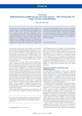
Fig. 2. A schematic diagram highlighting pathways involved in inflammation and cervical cancer progression. Seminal
plasma and prostaglandins elevate expression of COX-1 and COX-2 in cervical epithelial cells. This can occur via
prostaglandin G protein-coupled receptors (PTGER), causing the cells to release a host of local inflammatory mediators
including cytokines, interleukins, growth factors and prostaglandins. These mediators activate numerous pathways
which act synergistically to control tissue remodelling and tumour progression. For example, inflammatory mediators
are released to facilitate cellular proliferation and extravasation of immune cells into the tumour tissue by chemotaxis in
response to stimuli from local inflammatory mediators. In parallel, angiogenic factors and vascular permeability factors
promote the remodelling of the vasculature thereby facilitating angiogenesis.