The dental measurements showed few changes with growth in all groups. In terms of skeletal measurements from ages 9 to 18, similar growth changes were found between the sexes in most angular measurements, but males had larger values in linear measurements than females.
2. It has been shown that 60% to 70% of children have
Class I malocclusions.8,9
The dentofacial growth of
children with Class I malocclusions has always been
intriguing for many investigators. Many studies have
been made, but very few took into account the effect of
a high, average, or low MP-SN angle on facial growth.
Moreover, most reported studies have focused on the
facial growth of dental Class I subjects instead of
skeletal Class I. For example, Kerr10
examined the
longitudinal dentofacial growth of children from 5 to 15
years from the Belfast Growth Study. He divided the
subjects into different groups by sex and dental rela-
tionship (Angle classifications). He found that the
gonial and MP-SN angles decreased in all groups
(Classes I, II, and III) between 5 and 15 years. Also, the
SNA angle did not change significantly, and the SNB
angle increased slightly except for the Class II Division
2 female group. He did not divide the subjects accord-
ing to their MP-SN angles (high, average, or low).
Sinclair and Little11
studied longitudinal craniofacial
growth of untreated Class I male and female subjects
with good occlusions. They reported that from mixed
dentition (6.18 to 10.30 years) to adult dentition (17.98
to 21.83 years), both the SNA and SNB angles in-
creased and the ANB angle decreased and a forward
(bite closing) rotation of the mandible occurred. How-
ever, the mean MP-SN angles were 36.68° Ϯ 0.77° in
their male mixed dentition group and 34.93° Ϯ 0.86° in
female group; no higher or lower MP-SN angle subjects
were included in their study. Bishara and Jakobsen12
examined longitudinal growth of 20 male and 15
female untreated subjects with dental Class I relation-
ship from ages 5 to 25. The subjects of each sex were
categorized according to 3 facial types: relative long,
average, and relative short faces. They divided the
subjects into different groups using the ratio of poste-
rior to anterior face heights (S-Go/N-Me) and the
Frankfort horizontal-MP angle (FH-MP) of the adult
cephalograms. They reported that most subjects (77%)
had the same facial type at 5 years and 25.5 years of
age; there was a strong tendency to maintain the
original facial type with age. Also, the subjects in each
facial type had relatively large variations in the size and
relationships of the various dentofacial structures. They
suggested that longitudinal analysis of the data gave
more consistent and meaningful results than cross-
sectional comparisons when facial growth trends are
evaluated.
More recently, Chung and Wong13
incorporated
both the sagittal skeletal relationship and the degree of
MP-SN in their growth study. They examined the
craniofacial growth of skeletal Class II (ANBϾ4°)
untreated male and female subjects with low (Ͻ27°),
average (27°-36°), and high (Ͼ36°) MP-SN angles
from ages 9 to 18. They found that the SNA and SNB
angles increased, and the ANB angle decreased in all
groups with age. Also, all groups showed a mandibular
forward rotation with decreased gonial and MP-SN
angles. They also reported that the skeletal growth
changes in angular measurements were similar between
the male and female groups. Yet linear measurements
showed significant sex differences, especially in the
high-angle group. Craniofacial growth studies of skel-
etal Class I and Class III subjects with high, average, or
low MP-SN angles are not available in the literature.
The purpose of this study was to investigate the
longitudinal craniofacial growth changes in untreated
skeletal Class I subjects with low, average, and high
MP-SN angles.
MATERIAL AND METHODS
The sample consisted of 68 subjects—32 (14 males
and 18 females) from the Bolton-Brush Growth Study
at Case Western Reserve University in Cleveland,
Ohio, and 36 (22 males and 14 females) from the
Burlington Growth Center at the University of Toronto
in Canada. The subjects were selected according to the
following criteria: (1) lateral cephalograms available at
about ages 9 and 18, (2) skeletal Class I (0° ϽANB
Ͻ4° as determined from lateral cephalogram at age 9),
(3) skeletal age determined by hand-wrist radiographs
compared with standards by Greulich and Pyle14
(those
whose skeletal ages were greater than their chronolog-
ical ages by Ϯ 1 year were excluded), and (4) good
health with no orthodontic treatment.
The sample was divided into male (n ϭ 36) and
female (n ϭ 32) groups. For each subject, 2 lateral
cephalograms were traced by hand on acetate paper by
an examiner (V.D.M.). For the male group, the mean
ages were 8.64 years for the first tracing (T1) and 17.36
years for the second tracing (T2) (Table I). For the
female group, the mean ages were 8.66 years at T1 and
17.53 years at T2.
The sample was further divided into groups based
on the MP-SN angle at T1: (1) low angle (MP-SN Յ
27°), (2) average angle (MP-SN greater than 27° and
Table I. Age of subjects
n Mean age (y) Range (y)
Male
First tracing (T1) 36 8.64 8-10
Second tracing (T2) 36 17.36 16-18
Female
First tracing (T1) 32 8.66 8-9
Second tracing (T2) 32 17.53 16-18
American Journal of Orthodontics and Dentofacial Orthopedics
Volume 124, Number 6
Chung and Mongiovi 671
3. less than 37°), and (3) high angle (MP-SN Ն 37°). The
MP was drawn from menton (Me) to the inferior border
of the angular area of the mandible.1,2
These MP-SN
values represented about 1 SD from the mean MP-SN
angle of children ages 8 to 11 reported by Riedel.15
For
boys, the mean MP-SN angles at age 9 were 25.27° for
the low-angle group, 32.71° for the average-angle
group, and 40.68° for the high-angle group (Table II).
For girls, the mean MP-SN angles at age 9 were 26.08°
for the low-angle group, 33.13° for the average-angle
group, and 40.75° for the high-angle group.
The definitions of the landmarks used in this study
correspond to those of Riolo et al.16
All lateral cepha-
lometric tracings were digitized on a digitizer (Numon-
ics Corp, Montgomeryville, Pa) by an examiner
(V.D.M.) on a computer with Quick Ceph Orthodontic
Processing software, Version 2.6 (Quick Ceph Sys-
tems, San Diego, Calif). The computer software was
tested and confirmed for accuracy and reliability by
comparing values to the examiner’s hand measure-
ments.
Because subjects from 2 growth studies were ex-
amined, all linear measurements had to be converted
because of different enlargement factors for each
cephalostat. At the Burlington Growth Center, all
lateral cephalograms, regardless of the patient’s age,
were magnified by 9.84%.17
However, in the Bolton-
Brush Growth Study, magnification was regulated ac-
cording to the age of the patient (age 8, 5.5%; age 9-10,
5.6%; age 6-18, 5.9%).18
All linear measurements from
both studies were converted by eliminating the magni-
fication factor to allow the data to be analyzed.
Cross-sectional and longitudinal data were col-
lected and analyzed for each group. Statistical analysis
of the cephalometric measurements included descrip-
tive statistics at a 95% confidence interval and single
factor analysis of variance (AVOVA) calculated for
each group. Paired t tests were conducted, and statisti-
cal significance of compared measurements was de-
fined at P Յ .05.
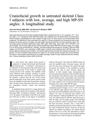
The following measurements were made and ana-
lyzed for each of the 136 lateral cephalograms (Fig).
Sagittal: SNA angle (in degrees), SNB angle (in de-
grees), ANB angle (in degrees), convexity (N-A-Pog,
in degrees), Pog-NB (effective chin, in millimeters),
ACB (anterior cranial base, N-S, in millimeters), man-
dibular body (Go intersection-Me, in millimeters);
vertical: MP-SN angle (in degrees), PP-SN (palatal
plane ANS-PNS to SN, in degrees), facial taper (N-Gn-
Go, in degrees), AFH (anterior facial height, N-Me, in
millimeters), PFH (posterior facial height, S-Go inter-
section, in millimeters), PFH:AFH, ANS-Me (lower
facial height, in millimeters), ANS-Me/N-Me (LFH:
AFH), saddle angle (N-S-Ar, in degrees), articular
angle (S-Ar-Go intersection, in degrees), gonial angle
(Ar-Go intersection to MP, in degrees), ramus height
(Ar-Go, in millimeters), PCB (posterior cranial base,
Table II. Group description at T1
Groups n Mean ANB Range Mean MP-SN Range
Male
Low-angle 13 1.94° 0-3.9° 25.27° 22-27°
Average-angle 12 2.93° 0.4-3.7° 32.71° 28-35.5°
High-angle 11 3.02° 1.1-4° 40.68° 37.5-45°
Female
Low-angle 6 2.62° 1.3-3.5° 26.08° 24-27°
Average-angle 12 2.94° 2.3-4° 33.13° 29-36°
High-angle 14 2.99° 1.6-4° 40.75° 37.5-51°
Fig. Cephalometric landmarks and planes.
American Journal of Orthodontics and Dentofacial Orthopedics
December 2003
672 Chung and Mongiovi
4. S-Ar, in millimeters), Y axis (FH to S-Gn, in degrees);
dental: 1/ to NA (maxillary incisor to NA, in millime-
ters), /1 to NB (mandibular incisor to NB, in millime-
ters), interincisal angle (in degrees), overbite (in milli-
meters), overjet (in millimeters), 1/ to NA (maxillary
incisor to NA, in degrees), /1 to NB (mandibular incisor
to NB, in degrees).
In addition, 15 randomly chosen lateral cephalo-
grams were traced twice by the same examiner
(V.D.M.) and measured separately on the Quick Ceph
computer software to determine whether an intraexam-
iner error resulted from landmark selection, tracing, and
measurement error. The same measurements were
made as in the subjects to be studied. Repeated mea-
sures ANOVA and paired Student t tests were carried
out for all linear and angular measurements to deter-
mine whether they were within acceptable limits. The
significance of differences was predetermined at P Յ
.05.
The mean and SD were calculated for each cepha-
lometic variable (measurement), and the differences of
each variable between the groups were tested with the
Student 2-tailed t test. The significance of differences
was predetermined at P Յ .05.
RESULTS
The assessment of intraexaminer error showed no
statistically significant difference between angular or
linear measurements (P ϭ 0.84). In addition, the mean
differences in replicate measures of the same cephalo-
grams showed a mean change of 0.3° between repeated
angular measurements and a 0.3 mm mean change
between linear measurements.
For the boys, the mean and SD of each measure-
ment at age 9 of the low-, average-, and high-angle
groups (cross-sectional data), and the statistical signif-
icance (P value) between the groups are given in Table
III. The growth changes (longitudinal data) of the
Table III. Cross-sectional data of boys at age 9 (T1)
Group I (low)
n ϭ 13
Group II (average)
n ϭ 12
Group III (high)
n ϭ 11 Significance (P value)
Mean SD Mean SD Mean SD I vs II II vs III I vs III
Sagittal
SNA (°) 80.90 2.75 80.67 2.39 78.46 2.68 .82 .05 .04
SNB (°) 78.96 2.29 77.74 2.54 75.44 2.45 .22 .04 .00
ANB (°) 1.94 1.19 2.93 0.96 3.03 0.98 .03 .80 .02
Convexity (°) 0.53 1.67 1.83 1.15 2.20 1.43 .03 .50 .02
Pog-NB (mm) 2.01 1.02 1.64 1.16 1.36 1.24 .42 .58 .18
ACB (mm) 63.43 2.00 64.42 2.43 64.22 3.24 .28 .87 .49
Mand. body (mm) 60.95 4.37 61.14 3.63 60.64 2.03 .90 .68 .82
Vertical
MP-SN (°) 25.27 1.75 32.71 2.68 40.68 2.40 .00 .00 .00
PP-SN (°) 7.08 1.62 7.11 1.77 8.16 1.88 .96 .18 .15
Facial taper (°) 72.95 2.88 68.60 2.57 63.47 1.48 .00 .00 .00
AFH (mm) 97.65 5.80 101.99 5.36 103.13 3.21 .06 .54 .01
PFH (mm) 68.98 5.81 66.97 4.43 60.72 2.86 .34 .00 .00
PFH:AFH (%) 70.58 3.07 65.70 2.99 58.88 2.19 .00 .00 .00
ANS-Me (mm) 54.06 3.64 57.77 3.02 58.54 2.90 .01 .54 .00
ANS-Me/N-Me (%) 55.37 1.89 56.67 1.54 56.76 1.84 .07 .89 .08
Saddle angle (°) 121.74 3.66 121.68 3.32 124.25 3.77 .96 .10 .11
Articular angle (°) 143.86 6.52 143.54 4.34 139.83 7.06 .89 .15 .16
Gonial angle (°) 121.16 5.38 127.43 3.35 136.16 4.95 .00 .00 .00
Ramus height (mm) 40.65 3.14 39.62 4.17 35.74 3.49 .50 .02 .00
PCB (mm) 31.94 3.48 30.86 2.72 28.97 1.95 .39 .07 .02
Y-axis (°) 57.48 4.55 58.66 3.49 61.54 2.23 .47 .03 .01
Dental
1/ to NA (mm) 4.53 2.37 4.44 2.04 4.72 1.58 .92 .72 .82
/1 to NB (mm) 3.50 1.50 4.81 1.36 4.91 1.65 .03 .87 .04
Interincisal angle (°) 127.67 11.28 127.94 8.57 125.98 8.17 .95 .58 .68
Overbite (mm) 1.77 2.04 1.70 1.38 1.99 1.36 .92 .62 .76
Overjet (mm) 3.36 1.09 3.36 0.90 3.87 0.91 1.00 .19 .22
1/ to NA (°) 25.65 9.02 24.66 5.95 25.60 4.53 .75 .67 .99
/1 to NB (°) 22.67 6.79 25.33 3.99 25.37 4.65 .24 .98 .26
American Journal of Orthodontics and Dentofacial Orthopedics
Volume 124, Number 6
Chung and Mongiovi 673
5. measurements in the low-, average- and high-angle
groups from ages 9 to 18 are shown in Table IV.
For the girls, Table V shows the mean and SD of
each measurement at age 9 of the low-, average-, and
high-angle groups (cross-sectional data), and the statis-
tical significance (P value) between the groups. Table
VI gives the longitudinal growth changes of the mea-
surements in the low-, average-, and high-angle groups
from ages 9 to 18.
The statistical significance data (P value) of each
measurement between the low-angle male and female
groups, the average-angle male and female groups, and
the high-angle male and female groups are listed in
Table VII. Similar patterns of skeletal growth were
found in most angular measurements of boys and girls,
but a significant sex difference was shown in some
linear measurements. Between male and female groups,
no significant differences in dental angular and linear
changes were found from ages 9 to 18.
DISCUSSION
The cross-sectional data of this study showed that,
at age 9, high-angle male and female groups had
smaller SNA and SNB values than did the low- and
average-angle groups (P Յ .05). Previous cross-sec-
tional studies by Isaacson et al4
and Bishara and
Augspurger19
had similar results (they did not divide
their subjects into skeletal Class I, II, or III). A recent
report by Chung and Wong,13
who studied the cranio-
facial growth in untreated skeletal Class II subjects with
low, average, and high MP-SN angles, also showed
similar findings. Bishara and Augspurger,19
in their
study of men, found that the ACB of high-angle
subjects was significantly smaller than the average- and
the low-angle subjects. In the present study, there was
no difference between male groups with regard to the
length of ACB at age 9. However, in the girls, the
high-angle group had a significantly smaller ACB than
Table IV. Male longitudinal growth changes from age 9 (T1) to age 18 (T2)
Group I (low)
n ϭ 13
Group II (average)
n ϭ 12
Group III (high)
n ϭ 11 Significance (P value)
Mean change SD Mean change SD Mean change SD I vs II II vs III I vs III
Sagittal
SNA (°) 2.32 1.38 2.73 3.71 0.98 3.56 .73 .26 .26
SNB (°) 2.79 1.17 4.16 3.27 2.89 2.98 .19 .34 .92
ANB (°) Ϫ0.47 1.05 Ϫ1.43 1.37 Ϫ1.91 1.38 .06 .42 .01
Convexity (°) Ϫ1.29 1.21 Ϫ2.08 1.66 Ϫ2.51 1.69 .19 .55 .06
Pog-NB (mm) 2.07 0.78 1.69 1.09 1.37 1.22 .34 .52 .13
ACB (mm) 6.30 1.34 5.85 2.51 6.38 2.91 .59 .65 .93
Mand. body (mm) 14.17 1.93 12.32 2.14 13.36 2.37 .03 .28 .38
Vertical
MP-SN (°) Ϫ2.42 2.36 Ϫ3.92 2.93 Ϫ3.18 2.19 .18 .50 .42
PP-SN (°) Ϫ0.18 2.44 Ϫ1.38 2.66 Ϫ1.07 3.21 .25 .80 .46
Facial taper (°) 0.17 3.06 Ϫ0.27 3.57 Ϫ0.24 2.26 .75 .98 .71
AFH (mm) 17.58 4.21 15.90 3.40 16.84 4.24 .28 .57 .67
PFH (mm) 17.53 3.51 16.09 3.63 13.41 3.07 .33 .07 .01
PFH:AFH (%) 4.52 3.24 5.02 3.93 2.93 1.68 .73 .11 .14
ANS-Me (mm) 9.23 2.83 9.46 1.63 9.84 2.18 .81 .64 .56
ANS-Me/N-Me (%) Ϫ0.46 1.20 0.39 0.95 0.18 1.56 .06 .71 .28
Saddle angle (°) 0.91 2.59 Ϫ1.58 3.20 0.08 4.11 .05 .30 .57
Articular angle (°) 0.47 5.39 0.95 3.61 2.23 6.01 .79 .55 .46
Gonial angle (°) Ϫ5.25 4.20 Ϫ4.05 5.11 Ϫ5.55 4.00 .53 .44 .86
Ramus height (mm) 11.74 3.40 10.41 4.04 8.57 3.20 .39 .24 .03
PCB (mm) 6.45 2.71 6.29 1.64 5.20 1.86 .86 .15 .19
Y-axis (°) Ϫ0.22 3.68 0.55 2.53 Ϫ1.95 2.30 .54 .02 .18
Dental
1/ to NA (mm) 0.62 1.37 2.10 1.73 2.73 3.00 .03 .55 .05
/1 to NB (mm) 0.25 0.96 0.67 2.12 1.26 1.62 .54 .46 .09
Interincisal angle (°) 1.62 6.96 Ϫ1.20 7.86 Ϫ3.35 10.31 .36 .58 .19
Overbite (mm) 1.01 2.62 0.69 1.33 0.03 0.50 .70 .13 .21
Overjet (mm) 0.19 1.62 Ϫ0.07 0.98 Ϫ0.85 1.09 .62 .09 .07
1/ to NA (°) 1.18 8.58 1.52 3.44 3.55 7.36 .90 .42 .47
/1 to NB (°) Ϫ0.23 5.36 0.35 6.56 1.72 4.63 .81 .57 .35
American Journal of Orthodontics and Dentofacial Orthopedics
December 2003
674 Chung and Mongiovi
6. the low-angle group at age 9. In terms of PCB, the male
high-angle group showed a significantly smaller value
than the male low-angle group at age 9.
Our cross-sectional data also showed that, at age 9,
for both boys and girls, the facial taper, PFH:AFH, and
ramus height were significantly greater in the low-angle
group than the high-angle group (P Յ .05), and the
AFH, LFH, and the gonial angle were significantly
greater in the high-angle group than the low-angle
group (P Յ .05). Isaacson et al4
and Bishara and
Augspurger19
also reported greater AFH and LFH in
the high MP-SN angle subjects than in the low MP-SN
angle subjects. We also found that there was no
significant difference in mandibular body length be-
tween groups of the same sex. Thus, we suggest that, in
the mandible, it is not the body that indicates diver-
gency, but the ramus height.
From ages 9 to 18, the mean SNA and SNB angles
of all groups were not constant, but instead they
increased. Similar findings were reported by Sinclair
and Little.11
Differently, Bishara and Jakobsen12
found
that from ages 5 to 25, the mean SNA angle of the
female subjects with average facial height decreased
slightly (Ϫ0.8°). Our data showed that as the SNA and
SNB angles increased, so did the ACB (SN). Therefore,
nasion (N) must have grown anteriorly less than Point
A or Point B. The commonly used Steiner20
normal
values, which do not change according to age, might, in
essence, not apply to younger subjects. Riolo et al16
reported the mean of each cephalometric measurement
on 47 boys and 36 girls yearly from ages 6 to 16. They
also found that the mean was not constant; it changed
with age. However, they did not separate their sample
according to skeletal Class I, II, or III. Thus, normal
cephalometric values for skeletal Class I subjects at
different ages are needed; this notion deserves further
attention and future research. Interestingly, our data
showed that the amount of SNB increase was greater
than the SNA increase with age in all groups. As a
result, the ANB angle became smaller. Of the 68
Table V. Cross-sectional data of girls at age 9 (T1)
Group I (low)
n ϭ 6
Group II (average)
n ϭ 12
Group III (high)
n ϭ 14 Significance (P value)
Mean SD Mean SD Mean SD I vs II II vs III I vs III
Sagittal
SNA (°) 81.70 1.85 81.09 2.13 78.52 2.76 .54 .01 .01
SNB (°) 79.08 1.77 78.14 1.89 75.53 2.80 .32 .01 .00
ANB (°) 2.62 0.86 2.94 0.50 2.99 0.92 .42 .86 .40
Convexity (°) 1.63 0.94 2.31 1.21 1.94 1.23 .22 .45 .55
Pog-NB (mm) 1.49 0.62 0.83 1.19 1.45 1.15 .14 .19 .91
ACB (mm) 64.69 3.14 61.87 2.32 60.94 2.32 .09 .32 .03
Mand. body (mm) 58.91 2.95 59.15 4.74 60.23 4.30 .90 .55 .44
Vertical
MP-SN (°) 26.08 1.11 33.13 2.59 40.75 4.27 .00 .00 .00
PP-SN (°) 9.00 1.94 8.82 2.56 10.53 3.79 .87 .18 .25
Facial taper (°) 72.83 2.29 69.02 2.67 63.78 2.89 .01 .00 .00
AFH (mm) 96.23 5.62 97.99 4.52 101.26 5.50 .52 .11 .10
PFH (mm) 67.58 4.78 64.37 3.48 60.70 4.67 .18 .03 .02
PFH:AFH (%) 70.20 1.56 65.72 3.36 60.00 3.98 .00 .00 .00
ANS-Me (mm) 52.20 4.40 54.94 4.21 56.84 3.28 .24 .22 .05
ANS-Me/N-Me (%) 54.22 2.36 53.50 8.17 56.14 1.64 .78 .29 .11
Saddle angle (°) 119.57 2.62 120.18 3.78 123.12 4.50 .69 .08 .04
Articular angle (°) 143.20 3.26 148.90 5.33 144.71 5.19 .01 .05 .44
Gonial angle (°) 124.28 3.11 123.39 5.57 131.91 4.02 .67 .00 .00
Ramus height (mm) 40.85 3.86 37.47 3.37 35.43 3.06 .10 .12 .02
PCB (mm) 30.28 1.81 29.39 1.57 28.32 3.01 .33 .26 .09
Y-axis (°) 56.53 3.02 58.15 4.50 59.19 3.84 .38 .54 .12
Dental
1/ to NA (mm) 3.66 2.33 4.38 1.63 4.67 1.73 .52 .66 .37
/1 to NB (mm) 3.87 2.38 5.27 1.65 4.83 1.50 .24 .49 .39
Interincisal angle (°) 128.82 10.90 122.47 7.63 128.38 6.95 .24 .05 .93
Overbite (mm) 1.92 1.16 1.56 1.43 0.93 1.76 .58 .32 .16
Overjet (mm) 3.49 0.69 2.89 1.03 3.59 1.40 .17 .16 .83
1/ to NA (°) 23.98 4.91 24.60 5.30 24.14 4.87 .81 .82 .95
/1 to NB (°) 24.30 7.98 29.83 5.32 24.46 3.39 .17 .01 .96
American Journal of Orthodontics and Dentofacial Orthopedics
Volume 124, Number 6
Chung and Mongiovi 675
7. subjects in our study, only 8 (11.7%) had an increase in
ANB angle from ages 9 to 18. Of the 8 subjects, there
were 5 with low angles, 2 with average angles, and 1
with a high angle with mean increases of 1.0°, 1.3°, and
1.1°, respectively, with no gender preference. Lande,21
Riolo et al,16
Sinclair and Little,11
Bishara and Jakob-
sen,12
and Chung and Wong13
also reported a decrease
in ANB angle with age in their subjects. Consequently,
new norms at different ages need to be developed for
the ANB angle as well, because it can be expected to be
somewhat larger for a 9-year-old than for an adult
whose norm is 2 Ϯ 2° according to Steiner.20
Another interesting finding of the present study was
that the mean MP-SN angle decreased from ages 9 to
18 in all groups. Of the 68 subjects, we observed only
7 (10.3%) who had an increase in MP-SN angle during
the growth period. Of the 7 subjects, there were 1 with
a low angle, 2 with average angles, and 4 with high
angles, with mean increases of 1.0°, 1.0°, and 2.0°,
respectively, with no sex preference. Our findings
agreed with those of Karlsen,6
who found that all 15
high-angle untreated males in his study had forward
rotation, and Riolo et al,16
who reported an MP-SN
angle decrease from ages 6 to 16 in their male and
female subjects. In Bjo¨rk and Skieller’s5
growth study,
19 of 21 (90.5%) subjects had decreased MP-SN angles
and only 2 (9.5%) had increased MP-SN angles from 3
years prepuberty to 3 years postpuberty. Chung and
Wong13
reported that, in their skeletal Class II subjects,
79 of 85 (93%) had decreased MP-SN angles, and only
6 (7%) had increased MP-SN angles from ages 9 to 18.
Therefore, our data suggest that, in the treatment of
skeletal Class I growing patients, the MP-SN angle
tends to decrease with age as long as orthodontic
mechanics do not extrude the posterior teeth.
Using the PHF:AFH ratio as an indicator of man-
dibular rotation as suggested by Bjo¨rk,22
we found that
PFH:AFH increased in all groups; this demonstrated a
Table VI. Female longitudinal growth changes from age 9 (T1) to age 18 (T2)
Group I (low)
n ϭ 6
Group II (average)
n ϭ 12
Group III (high)
n ϭ 14 Significance (P value)
Mean change SD Mean change SD Mean change SD I vs II II vs III I vs III
Sagittal
SNA (°) 1.73 2.31 1.48 1.54 0.91 2.04 .81 .43 .47
SNB (°) 1.77 1.14 2.19 1.64 2.02 2.04 .53 .82 .73
ANB (°) Ϫ0.05 1.58 Ϫ0.71 1.06 Ϫ1.11 1.27 .39 .39 .19
Convexity (°) Ϫ0.67 1.85 Ϫ1.24 1.32 Ϫ1.16 1.41 .52 .89 .57
Pog-NB (mm) 1.52 0.85 1.27 1.32 0.55 0.82 .64 .12 .04
ACB (mm) 4.71 1.40 4.02 0.71 3.66 1.33 .30 .39 .15
Mand. body (mm) 8.51 2.63 9.78 2.85 7.65 4.34 .37 .15 .60
Vertical
MP-SN (°) Ϫ0.83 1.29 Ϫ2.75 2.85 Ϫ1.39 2.75 .07 .23 .54
PP-SN (°) Ϫ0.53 2.32 Ϫ0.75 2.95 Ϫ0.60 1.73 .87 .88 .95
Facial taper (°) Ϫ0.43 1.09 0.36 1.57 Ϫ0.92 1.61 .23 .05 .44
AFH (mm) 12.62 3.04 11.89 1.53 11.99 3.43 .60 .92 .69
PFH (mm) 11.81 2.87 11.72 2.73 9.20 2.40 .95 .02 .08
PFH:AFH (%) 2.68 1.09 3.57 2.50 1.76 1.73 .31 .05 .17
ANS-Me (mm) 6.92 2.73 6.78 1.65 7.32 2.53 .91 .52 .77
ANS-Me/N-Me (%) Ϫ0.33 1.54 2.59 8.66 0.49 1.11 .28 .42 .27
Saddle angle (°) 2.17 3.90 2.64 3.56 Ϫ0.38 3.82 .81 .05 .21
Articular angle (°) Ϫ0.85 4.91 Ϫ3.48 4.37 0.75 3.79 .29 .02 .50
Gonial angle (°) Ϫ3.37 3.18 Ϫ2.55 3.59 Ϫ1.55 2.30 .63 .42 .24
Ramus height (mm) 9.41 2.90 8.68 2.86 6.08 2.78 .62 .03 .04
PCB (mm) 3.17 1.83 3.93 2.00 3.43 2.26 .44 .56 .79
Y-axis (°) 2.02 1.98 0.69 3.18 1.52 3.42 .30 .53 .69
Dental
1/ to NA (mm) 0.58 1.87 1.52 1.67 2.00 1.84 .32 .50 .15
/1 to NB (mm) 0.61 1.46 0.47 1.42 1.09 1.12 .85 .23 .49
Interincisal angle (°) Ϫ0.50 9.24 1.16 5.78 Ϫ4.90 6.98 .70 .02 .33
Overbite (mm) 0.20 0.84 0.59 1.54 0.35 1.77 .49 .71 .80
Overjet (mm) Ϫ0.09 0.88 0.45 0.89 Ϫ0.23 1.33 .25 .14 .79
1/ to NA (°) Ϫ0.67 4.95 1.52 5.06 3.31 3.76 .40 .32 .12
/1 to NB (°) 1.48 6.95 Ϫ1.83 2.55 2.73 4.25 .30 .00 .70
American Journal of Orthodontics and Dentofacial Orthopedics
December 2003
676 Chung and Mongiovi
8. forward mandibular rotation. Other indicators sug-
gested by Sinclair and Little11
were the MP-SN and
gonial angles. As stated above, in our study, the mean
MP-SN angle and gonial angle decreased in all groups;
this suggested a forward mandibular rotation. Sinclair
and Little11
also reported a forward (bite closing)
rotation of the mandible with growth in their Class I
normal occlusion subjects. However, they did not
separate their subjects with respect to skeletal vertical
pattern (high or low mandibular plane angle).
In agreement with Lande,21
Riolo et al,16
Bjo¨rk,23
Bishara and Jakobsen,12
and Chung and Wong,13
we
also recognized a decrease in convexity with growth in
all groups. Interestingly, the Pog-NB (effective chin)
increased in all groups with age; this might have been
due to the mandibular forward rotation or forward
growth of the chin. This might help to explain why the
face flattens with age. There was no difference ob-
served between groups in ACB (SN) for either sex from
a longitudinal outlook. However, between sexes, there
was a difference in magnitude. From ages 9 to 18,
males on average had an incremental growth change of
about 0.68 mm per year in ACB, and the females had an
increase of 0.45 mm per year. These values were
calculated without considering the magnification factor
and the growth spurt.
The dental measurements in this study showed few
changes from ages 9 to 18 in all groups. For overbite,
there was a net increase in all groups, but the value was
very small (Ͻ 1 mm). Overjet was also observed not to
worsen with age. Thus, it is suggested that overbite and
overjet remain relatively stable with growth regardless
of the person’s vertical pattern. Sinclair and Little11
showed similar results and reported that incisor angu-
lation appeared to be relatively stable.
Our results showed that there were some significant
growth differences between the low-, average-, and
high-angle groups from ages 9 to 18. For males, this
difference resulted in a significantly greater similarity
between the groups in ANB angle (low and average,
low and high), convexity (low and average, low and
high), AFH (low and high), and Y-axis (average and
high, low and high), and a significantly greater differ-
ence in Pog-NB (low and high), ANS-Me/N-Me (low
and average, low and high), saddle angle (average and
high), PCB (average and high), 1/ to NA in mm (low
and high), and /1 to NB in degrees (low and high). For
females, a significantly greater similarity between the
groups was seen in facial taper (low and average),
saddle angle (low and high), articular angle (low and
average, average and high), interincisal angle (average
and high), and /1 to NB in degrees (average and high),
but a significantly greater difference was noted in
Pog-NB (low and high), ANS-Me/N-Me (low and
high), ramus height (average and high), 1/ to NA in mm
(low and average, low and high), and 1/ to NA in
degrees (average and high). In general, the facial type
of each group was maintained with age; this agreed
with the previous report by Bishara and Jakobsen.12
In this study, similar growth changes were found
between male and female groups in most skeletal
angular measurements. However, marked sex differ-
ences were found in most skeletal linear measurements.
Males showed larger dimensions than females. Similar
findings were reported by Sinclair and Little11
and
Chung and Wong.13
CONCLUSIONS
The longitudinal growth changes from ages 9 to 18
of 68 skeletal Class I subjects with low, average, and
Table VII. Comparison of longitudinal changes from
age 9 to age 18 between groups
Significance (P value)
Low male vs
low female
Average male
vs average
female
High male vs
high female
Sagittal
SNA (°) .58 .30 .96
SNB (°) .10 .08 .42
ANB (°) .57 .16 .15
Convexity (°) .47 .18 .05
Pog-NB (mm) .21 .41 .07
ACB (mm) .04 .03 .01
Mand. body (mm) .00 .02 .00
Vertical
MP-SN (°) .08 .33 .08
PP-SN (°) .77 .59 .67
Facial taper (°) .54 .59 .41
AFH (mm) .01 .00 .01
PFH (mm) .00 .00 .00
PFH:AFH (%) .09 .29 .10
ANS-Me (mm) .12 .00 .01
ANS-Me/N-Me (%) .86 .40 .58
Saddle angle (°) .49 .01 .78
Articular angle (°) .61 .01 .49
Gonial angle (°) .30 .42 .01
Ramus height (mm) .15 .24 .05
PCB (mm) .01 .00 .04
Y-axis (°) .10 .91 .01
Dental
1/ to NA (mm) .96 .41 .49
/1 to NB (mm) .60 .79 .78
Interincisal angle (°) .63 .41 .68
Overbite (mm) .33 .87 .53
Overjet (mm) .62 .19 .21
1/ to NA (°) .56 1.00 .92
/1 to NB (°) .61 .30 .58
American Journal of Orthodontics and Dentofacial Orthopedics
Volume 124, Number 6
Chung and Mongiovi 677
9. high MP-SN angles were examined. Our conclusions
are as follows:
1. At age 9, for boys, significant differences were
found between the low- and the high-angle groups in
SNA, SNB, ANB angles, convexity, facial taper,
AFH, PFH, PFH:AFH, ANS-Me, gonial angle, ra-
mus height, PCB, Y-axis, and mandibular incisor to
NB (mm).
2. At age 9, for girls, significant differences were
found between the low- and the high-angle groups in
SNA and SNB angles, ACB, facial taper, PFH,
PFH:AFH, ANS-Me, saddle angle, gonial angle, and
ramus height.
3. From ages 9 to 18, the SNA and SNB angles
increased in all groups, and the ANB angle de-
creased in all groups. The male high-angle group
showed a greater decrease in ANB angle than did
the male low-angle group (P Յ .05). Among the
females, there was no difference between groups.
4. From ages 9 to 18, a mandibular forward rotation
(bite closing) was noted in all groups with a de-
crease in MP-SN and gonial angles and an increase
of PFH:AFH ratio.
5. From ages 9 to 18, few changes in the dental
measurements were found in all groups.
6. Similar growth changes were found between male
and female groups in most angular measurements,
but marked sex differences were found in most
linear measurements. Males had larger overall val-
ues in these linear measurements than females.
We thank Mrs Elizabeth Mongiovi and Drs Wallace
Wong, Solomon Katz, Jamie Ahl, and Stephen Tjoa for
their help.
REFERENCES
1. Schudy FF. Vertical growth versus anteroposterior growth as
related to function and treatment. Angle Orthod 1964;34:75-93.
2. Schudy FF. The rotation of the mandible resulting from growth:
its implications in orthodontic treatment. Angle Orthod 1965;35:
36-50.
3. Creekmore TD. Inhibition or stimulation of the vertical growth of
the facial complex, its significance to treatment. Angle Orthod
1967;37:285-97.
4. Isaacson JR, Isaacson RJ, Speidel TM, Worms FW. Extreme
variation in vertical facial growth and associated variation in
skeletal and dental relations. Angle Orthod 1971;41:219-29.
5. Bjo¨rk A, Skieller V. Facial development and tooth eruption: an
implant study at the age of puberty. Am J Orthod 1972;62:339-
83.
6. Karlsen AT. Craniofacial growth differences between low and
high MP-SN angle males: a longitudinal study. Angle Orthod
1995;65:341-50.
7. Karlsen AT. Association between facial height development and
mandibular growth rotation in low and high MP-SN angle faces:
a longitudinal study. Angle Orthod 1997;67:103-10.
8. Massler M, Frankel JM. Prevalence of malocclusion in children
aged 14-18 years. Am J Orthod 1951;37:751-68.
9. Ast DB, Carlos JP, Cons DC. Prevalence and characteristics of
malocclusion among senior high school students in upstate New
York. Am J Orthod 1965;51:437-45.
10. Kerr WJS. A longitudinal cephalometric study of dentofacial
growth from 5 to 15 years. Br J Orthod 1979;6:115-21.
11. Sinclair PM, Little RM. Dentofacial maturation of untreated
normals. Am J Orthod 1985;88:146-56.
12. Bishara SE, Jakobsen JR. Longitudinal changes in three normal
facial types. Am J Orthod 1985;88:466-502.
13. Chung C-H, Wong WW. Craniofacial growth in untreated Class
II subjects: a longitudinal study. Am J Orthod Dentofacial
Orthop 2002;122:619-26.
14. Greulich WW, Pyle SI. Radiographic atlas of skeletal develop-
ment of the hand and wrist. 2nd ed. Stanford (Calif): Stanford
University Press; 1959.
15. Riedel RA. The relation of maxillary structures to cranium in
malocclusion and normal occlusion. Angle Orthod 1952;22:
142-5.
16. Riolo ML, Moyers RE, McNamara JA, Hunter WS. An atlas of
craniofacial growth: cephalometric standards from the University
School Growth Study, monograph no. 2. Craniofacial Growth
Series. Ann Arbor: Center for Human Growth and Development;
University of Michigan; 1974.
17. Requirements and limitations of roentgenographic cephalometry.
Burlington Growth Center; Faculty of Dentistry, University of
Toronto.
18. Broadbent BH Sr, Broadbent BH Jr, Golden WY. Bolton
standards of dentofacial developmental growth. St. Louis: C. V.
Mosby; 1975.
19. Bishara SE, Augspurger EF. The role of mandibular plane
inclination in orthodontic diagnosis. Angle Orthod 1975;45:273-
81.
20. Steiner CC. The use of cephalometrics as an aid to planning and
assessing orthodontic treatment. Am J Orthod 1960;46:721-35.
21. Lande MJ. Growth behavior of the human bony facial profile as
revealed by serial cephalometric roentgenology. Angle Orthod
1952;22:78-90.
22. Bjo¨rk A. Prediction of mandibular growth rotation. Angle Orthod
1969;55:585-99.
23. Bjo¨rk A. The significance of growth changes in facial pattern and
their relationship to changes in occlusion. Dental Record 1951;
71:197-208.
American Journal of Orthodontics and Dentofacial Orthopedics
December 2003
678 Chung and Mongiovi