Ash Poster Final 120608
•
0 j'aime•633 vues

American Society of Hematology 75th Annual Conference. Accelerated Repair of Vascular Injury in Diabetes by TGF-B1 Modified Hematopoietic Stem Cells (HSC)
Recommandé

Recommandé
Contenu connexe
Similaire à Ash Poster Final 120608
Similaire à Ash Poster Final 120608 (19)
Dernier
Dernier (20)
Ash Poster Final 120608
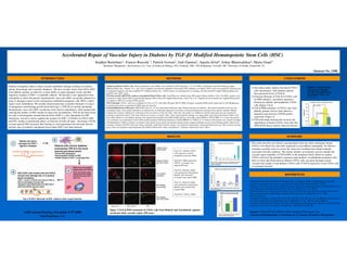
- 1. Accelerated Repair of Vascular Injury in Diabetes by TGF-β1 Modified Hematopoietic Stem Cells (HSC) Stephen Bartelmez1, Francis Ruscetti 2, Patrick Iversen3, Gail Gannon1, Aqeela Afzal4, Ashay Bhatwadekar4, Maria Grant4 1BetaStem Therapeutics, San Francisco CA, 2Lab. of Leukocyte Biology, NCI, Frederick, MD, 3AVI-Biopharma, Corvallis, OR, 4University of Florida, Gainesville, FL Abstract No. 1398 INTRODUCTION METHODS CONCLUSIONS Isolation of human CD34+ cells: Blood mononuclear cells (MNC) were enriched by F/H separation. 3.3 X 10⁷ MNCs and blocked with FcR blocking reagent Diabetic retinopathy induces retinal vascular endothelial damage resulting in ischemia, Figure 6: Vascular Repair 1) Our initial studies indicate that blood CD34+ (Miltenyi Biotec Inc., Auburn, CA) , and 33 µl of magnetic microbeads conjugated with an anti-CD34 antibody was added. CD34+ cells were positively selected using edema, hemorrhage and eventually blindness. We have recently shown that CD34+ /EPC is increased by Blocking cells (and plasma) from diabetic patients an automated magnetic selection autoMACS™ (Miltenyi Biotec Inc.). CD34+ purity was determined by co-staining with PE-anti-CD34 (high) (Miltenyi Biotec Inc.) from diabetic patients are defective in their ability to repair damaged vessels and their TGF-β in both Diabetic and and FITC-anti-CD45 (mid). have increased levels of TGF-β. Non-diabetic CD34+ cells migratory response to SDF-1 is markedly reduced. We describe a new approach to treat TGF-beta specific (qRT-PCR) Analyses of peripheral blood CD34+ cells: Total RNA was isolated using TRI reagent (Sigma-Aldrich, USA). The RNA samples were 2) Transient blockade of TGF-β in CD34+ cells (from Fig 2) retinopathy in which the patient’s hematopoietic stem cells (HSC) are greatly enhanced to converted to single strand cDNA using the high capacity DNA archive kit (Applied Biosystems, Foster City, CA). Taqman universal master mix (Applied Biosystems) by PMO enhances vasculature reparative was added to cDNA for real-time PCR analyses. home to damaged retinal vessels and generate endothelial progenitor cells (EPCs) which function by diabetic and nondiabetic CD34+ TGF-b blockade: CD34+ cells were incubated for 8 hrs at 37°C with either 40 ug/ml TGF-β PMO, 40 ug/ml scrambled PMO (both synthesized by AVI-Biopharma), repair vessel endothelium. We recently demonstrated that a transient blockade (2-4 days) cells (Figure 2 & 6). or medium alone then re-suspended in PBS alone for injection. of endogenous transforming growth factor-beta type 1 (TGF-β1) in murine and human Ischemia/Reperfusion (I/R) assay: NOD-SCID mice (n = 6) were kept under Isoflurane vapor during induction of ischemia. The anterior chamber of the eye was 3) TGF-β PMO treatment of CD34+ cells from hematopoietic stem cells (HSC) accelerates bone marrow engraftment, while dramatically cannulated using a 30-ga needle attached to an infusion line of saline and subjected to two hours of increased hydrostatic pressure in the anterior chamber. Retinal diabetic patients restores their defective reducing the number of HSC needed for long-term reconstitution. TGF-β1 has been shown ischemia then ensued, confirmed by whitening of the iris and loss of the red reflux. After 2hrs. the needle was withdrawn and the intraocular pressure normalized, migration and increases CXCR4 protein resulting in reperfusion injury. The contra lateral eye served as a control. After 7 days retinal capillary damage was appreciable, and at that point human CD34+ cells not only to downregulate stromal derived factor (SDF)-1, a key chemokine for EPC expression (Figure 4). from either diabetic or non-diabetic patients were injected intravitreally with 5000-20,000 cells/eye with either control PMO or TGF-β PMO. At 2-4 days the animals chemotaxis, but also to down regulate the receptor for SDF-1 (CXCR4) on CD34+ HSC 4) TGF-β blockade dramatically increases the were sacrificed, eyes enucleated, fixed in PFA and flat mounted. Following blocking in 10mM HEPES, 0.2 %BSA, 0.2 % Triton X, flat mounts were reacted overnight and EPC, leading to detrimental effects on function of both cell types. Increasing CXCR4 with Rhodamine 1:1000 to visualize vasculature. The mounts were treated with primary human nuclear antigen antibodies in goat serum blocking buffer, followed by engraftment of human CD34+ stem cells into on bone marrow HSC increases their homing to, and engraftment into the bone marrow, secondary staining with FITC-Goat-anti-mouse antibodies. Individual confocal images were analyzed using Image J and green fluorescence per 100 sq micron area per NOD-SCID mouse marrow (data not shown). and into sites of ischemic and injured tissue where SDF-1 has been released. group. Data was analyzed using Graph pad with ANOVA followed by Tukey and plotted as Arbitrary Fluorescent Units” (AFU). RESULTS SUMMARY This study describes pre-clinical experimentation that may allow autologous human CD34+ to be effectively and safely employed to treat diabetic retinopathy. No effective treatment currently exists to reverse the vision loss resulting from retinal ischemia (Fig A-C), Diabetic CD34+ associated with this condition. The chronic diabetic environment severely disrupts the cells exposed to control vascular repair capability of CD34+/EPC in the peripheral blood. However, patient morpholino antisense PMO CD34+ cells have the potential to generate large numbers of endothelial progenitor cells. Here we show that blood derived diabetic CD34+ cells can repair damaged retinal (Fig D-F), PMO targeted to Figure 3: TGF-β, a pleiotropic autocrine and paracrine regulator of TGF-β1, overnight, and vascular beds similar to non-diabetic CD34+ cells if TGF-β expression in the CD34+ cells all stages of hematopoiesis is increased in HSC and serum of patients with diabetes. injected I/R mice is transiently blocked. Migration of human BM CD34+ Cells to SDF-1 (Fig G-I), Identical studies were performed using human Figure 2 80 REFERENCES diabetic cells untreated Migrating cells (RFU) 60 or treated with control PMO 40 20 1 Bartelmez SH, Iversen P, and Ruscetti FW. Blockade of endogenous TGFβ1 in hematopoietic stem cells (Fig J-L), Identical studies 0 accelerates engraftment and enhances repopulating efficiency. Submitted to BLOOD 12/2008 Negative Medium PMO control PMO anti- were performed using human -20 control alone TGF-β1 2 Ruscetti, F.W. & Bartelmez, S.H. Transforming growth factor beta, pleiotropic regulator of hematopoietic stem diabetic cells treated with Figure 4: Migration of CD34+ cells toward SDF-1 increases after cells: potential physiological and clinical relevance. Int J Hematol 74, 18-25. 2001. control PMO TGF-β PMO treatment. 3 Harris JR, Brown GA, Jorgensen M, Kaushal S, Ellis EA, Grant MB, Scott EW. Bone marrow-derived cells home to and regenerate retinal pigment epithelium after injury. Invest Ophthalmol Vis Sci. 2006 RPM I Scrambled TGF-be ta M AS (Fig M-O), TGF-β1- PMO May;47(5):2108-13. 200 180 4 Sengupta N, Caballero S, Mames RN, Butler JM, Scott EW, Grant MB. The role of adult bone marrow-derived Fig 1:TGFß-1 blockade in HSC enhances their repair function 160 stem cells in choroidal neovascularization. Invest Ophthalmol Vis Sci. 2003 Nov;44(11):4908-13. 140 CXCR4 protein 120 5 Lanning DA, Nwomeh BC, Montante SJ, Yager DR, Diegelmann RF, Haynes JH. TGF-beta1 alters the healing 100 80 of cutaneous fetal excisional wounds. J Pediatr Surg. 1999 May;34(5):695-700. Blood Vessels HSC in Murine Both 60 40 Figure 2 TGF-β PMO treatment of CD34+ cells from Diabetic and Non-diabetic patients 20 ASH Annual Meeting, December 6-9th 2008 Support: NIH EY07739, NIH EY12601, BetaStem Therapeutics Inc. 0 accelerates their vascular repair (I/R assay) Commercial Relationship: S. Bartelmez & G. Gannon, BetaStem Therapeutics Figure 5: CXCR4 Protein Expression Increases in San Francisco, CA CD34+ after TGF-β PMO treatment