
Recommandé
Recommandé
Contenu connexe
Dernier
Dernier (20)
En vedette
En vedette (20)
Equi thermo power point english
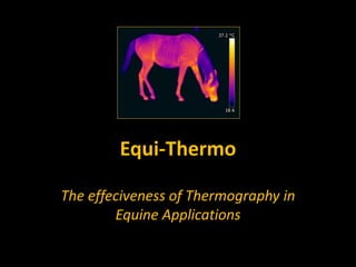
- 1. Equi-Thermo The effeciveness of Thermography in Equine Applications
- 2. This image was taken on the 14/08/11 after 3 months of rest showing clearly where she injured herself. Maissa du Pech
- 3. This image was taken on the 02/11/11 after 6 months rest and show clearly that both legs are now the same temperature. Maissa du Pech
- 4. This horse fell in his trailer. This image taken 2 weeks after the accident – His near hind was shaved to better treat his open wounds. His off hind looked normal but showed thermal heat which probably indicates bruising.
- 5. This horse had infiltrations to the knee on the 08/11/12 – Two weeks before this image was taken. He had two infiltrations to the front of the knee and one to the The treatment was for a tendon problem just below the back of the knee. We can see where the infiltrations were carried out and note that his whole knee is hotter than the near side knee. Image taken 24/11/11 Two weeks after this horse had Infiltrations
- 6. Image taken 03/01/12 8 weeks after the Infiltrations The sites of Infiltration still visiable Two months later and the knee is still warmer.
- 7. Image taken 20/01/12 10 weeks after the Infiltrations The sites of Infiltration are still visiable, but seem to be cooler. The near front hoof is showing hot. I was normal in the first image dated 24/11/11. Seemed slightly warmer in the image of the 03/01/11 and is now showing much hotter.
- 8. The same horse We are now looking at his off hind, this hot spot was apparent in the first images I took on the 24/11/11
- 9. The off hind from a different angle. Same horse
- 10. The same horse 10 weeks later and The hot spot is much Cooler with just a slight Trace.
- 11. This horse showed this hot spot to it’s near fore on the 03/01/12 and it was still hot the day after 04/01/12, however images taken on the 20/01/12 showes nothing. I assume it was a knock of some sort. The same horse again
- 12. He had an infection which showed up on these images 4 days before they became apparent to the naked eye. This horse was slightly lame.
- 13. This horse is slightly lame This image shows heat to the off front heal area of the hoof. To be diagnosed.
- 14. Horse with hot hocks. This horse shows a hot hock with an unusual heat pattern. To be diagnosed.
- 15. The same horse.
- 16. A horse with back problems. To be diagnosed
- 17. This horse is lame To be diagnosed.
- 18. The same horse from a different angle.
- 19. Horse with a susspected hoof abcess. We can see the heat up as far as his knee. This photo was taken in a sand school and this is not ideal as it does not allow for the hooves to be placed flat.
- 20. This horse has been treated with a Blister for a tendon problem – Nothing was visiable to the naked eye but here is quite clear. Horse treated with a Blister product
- 21. This horse would not / could not lower his head to eat. Nothing of note was observed appart from the fact that he has occular heat around his near side eye. This was apparent in other images and I have never seen this before. The vet attended 4 days later but was unable to diagnose as by then the horse was eating normally. te
- 22. Fin