Super Resolution Microscopy publications 2012
•
1 j'aime•697 vues

Localization Microscopy: Molecular Galaxies of protein transport in human blood-brain-barrier, tight junctions networks, gene transcription, single molecules, nuclear histones, viruses

Recommandé
Contenu connexe
Tendances
Tendances (20)
Similaire à Super Resolution Microscopy publications 2012
Similaire à Super Resolution Microscopy publications 2012 (20)
Dernier
Dernier (20)
Super Resolution Microscopy publications 2012
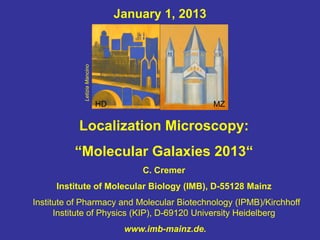
- 1. January 1, 2013 Letizia Mancino HD MZ Localization Microscopy: “Molecular Galaxies 2013“ C. Cremer Institute of Molecular Biology (IMB), D-55128 Mainz Institute of Pharmacy and Molecular Biotechnology (IPMB)/Kirchhoff Institute of Physics (KIP), D-69120 University Heidelberg www.imb-mainz.de.
- 2. Molecular Galaxies Conventional Localization of Protein Transport Microscopy in a Human Blood- Brain Barrier Model O. Huber, A. Brunner, P. Maier, R. Kaufmann, P.-O. Couraud, C. Cremer, G. Fricker (2012)
- 3. Molecular Galaxies of Tight Junction Networks formed by individual Claudin Molecules in Human Cells Conventional Microscopy Localization Microcopy R. Kaufmann, J.Piontek, F. Grüll, M. Kirchgessner, J. Rossa, H. Wolburg, I. E. Blasig, C. Cremer (2012)
- 4. Molecular Galaxies of Gene Transcription Conventional Microscopy Localization Microscopy Positions R. Kaufmann , C.Cremer, J.Gall (2012)
- 5. Deep Insights into the Interior of Molecular Galaxies: Single Molecules Single Molecules Nuclear Histones a) on Surface b) Inside Cell 300 nm Nucleus c),d) in Virus Single Viruses C. Cremer (2012)
- 6. Principle of Spectrally Assigned Localization Microscopy (SPDM, Spatial Position Determination Microscopy): Optical Isolation of Diffraction Images of individual Molecules: Determination of Gravity Centers 3 point-like objects in x,y plane with next neigbour distances 50 nm a) Labelling with same spectral signature b) Labeling with different unique spectral signatures B,G,R Computation (scalar Theory): NA = 1.4, λexc = 488 nm Optical Isolation by the “Supernova“ Mode: Induction of stochastically distributed short flashes of light emission by Cremer et al. 1996, 1999, 2010, 2012 individual molecules
- 7. Present State of Localization Microscopy (wide field) realized @ C. Cremer Lab (January 1, 2013) - Best Optical Resolution OR (resolvable distance): OR ~ 5 nm (~ 1/100 λexc, from localization precision) - Structural Resolution (imaging capability): ● Mean (2D) distance between individual molecules actually detected: ~ 6 nm ( ~ 1/80 λexc) ● Density of individually detected molecules : ~ 2,8 · 104/µm2 ● 3D single molecule resolution inside cells: ca. 20 nm laterally, 30 – 50 nm axially ● Optical Sectioning: ~ 15 nm
- 8. Publications Cremer-Lab 2012 part I C. Cremer, Optics far Beyond the Diffraction Limit: From Focused Nanoscopy to Spectrally Assigned Localization Microscopy (2012). In: Springer Handbook of Lasers and Optics, 2nd edition (F. Träger, Edit.), pp. 1351 – 1389. T. Cremer, Y. Markaki, B. Hübner, A. Zunhammer, H. Strickfaden, S. Beichmanis, M. Heß, L. Schermelleh, M. Cremer, C. Cremer (2012) Chromosome Territory Organization within the Nucleus. In: Encyclopedia of Molecular Cell Biology and Molecular Medicine: Epigenetic Regulation and Epigenomics, 2nd Edition (R.A. Meyers, Edit.), pp. 1-30.Wiley-VCH. R. Kaufmann, C. Cremer, J. G. Gall (2012a) Superresolution imaging of transcription units on newt lampbrush chromosomes. Chromosome Research, DOI 10.1007/s10577-012-9306-z-
- 9. Publications Cremer-Lab 2012 part II R. Kaufmann, J. Piontek, F. Grüll, M. Kirchgessner, J.Rossa, H. Wolburg, I. E. Blasig, C. Cremer (2012b) Visualization and quantitative analysis of reconstituted tight junctions using localization microscopy. PLoS One 7 (2 ) e31128: 1 – 9. O. Huber, A. Brunner, P. Maier, R. Kaufmann, P.-O. Couraud, C. Cremer, G. Fricker (2012) Localization microscopy (SPDM) reveals clustered formations of P-Glycoprotein in a human blood- brain barrier model. PLoS ONE 7 (9) e44776: 1-10. T. Ach, G. Best, S. Rossberger, R. Heintzmann, C. Cremer, S. Dithmar (2012) Autofluorescence imaging of human RPE cell granules using structured illumination microscopy. Br. J. Ophthalmology, DOI 10.1136/bjophthalmol-2012-301547.