
Recommended
Recommended
More Related Content
What's hot
What's hot (20)
Similar to Core Stability Measures as Risk Factors for Lower Extremity Injuries
Similar to Core Stability Measures as Risk Factors for Lower Extremity Injuries (20)
More from GrandFinalTechnologies
More from GrandFinalTechnologies (20)
Recently uploaded
Recently uploaded (20)
Core Stability Measures as Risk Factors for Lower Extremity Injuries
- 1. Core Stability Measures as Risk Factors for Lower Extremity Injury in Athletes DARIN T. LEETUN1 , MARY LLOYD IRELAND1 , JOHN D. WILLSON2,3 , BRYON T. BALLANTYNE2 , and IRENE MCCLAY DAVIS2,3 1 Kentucky Sports Medicine Clinic, Lexington, KY; 2 Joyner Sportsmedicine Institute, Lexington, KY; and 3 University of Delaware, Department of Physical Therapy, Newark, DE ABSTRACT LEETUN, D. T., M. L. IRELAND, J. D. WILLSON, B. T. BALLANTYNE, and I. M. DAVIS. Core Stability Measures as Risk Factors for Lower Extremity Injury in Athletes. Med. Sci. Sports Exerc., Vol. 36, No. 6, pp. 926–934, 2004. Introduction/Purpose: Decreased lumbo-pelvic (or core) stability has been suggested to contribute to the etiology of lower extremity injuries, particularly in females. This prospective study compares core stability measures between genders and between athletes who reported an injury during their season versus those who did not. Finally, we looked for one or a combination of these strength measures that could be used to identify athletes at risk for lower extremity injury. Methods: Before their season, 80 female (mean age ϭ 19.1 Ϯ 1.37 yr, mean weight 65.1 Ϯ 10.0 kg) and 60 male (mean age ϭ 19.0 Ϯ 0.90 yr, mean weight 78.8 Ϯ 13.3 kg) intercollegiate basketball and track athletes were studied. Hip abduction and external rotation strength, abdominal muscle function, and back extensor and quadratus lumborum endurance was tested for each athlete. Results: Males produced greater hip abduction (males ϭ 32.6 Ϯ 7.3%BW, females ϭ 29.2 Ϯ 6.1%BW), hip external rotation (males ϭ 21.6 Ϯ 4.3%BW, females ϭ 18.4 Ϯ 4.1%BW), and quadratus lumborum measures (males ϭ 84.3 Ϯ 32.5 s, females ϭ 58.9 Ϯ 26.0 s). Athletes who did not sustain an injury were significantly stronger in hip abduction (males ϭ 31.6 Ϯ 7.1%BW, females ϭ 28.6 Ϯ 5.5%BW) and external rotation (males ϭ 20.6 Ϯ 4.2%BW, females ϭ 17.9 Ϯ 4.4%BW). Logistic regression analysis revealed that hip external rotation strength was the only useful predictor of injury status (OR ϭ 0.86, 95% CI ϭ 0.77, 0.097). Conclusion: Core stability has an important role in injury prevention. Future study may reveal that differences in postural stability partially explain the gender bias among female athletes. Key Words: GENDER, HIP STRENGTH, TRUNK ENDURANCE, BASKETBALL, TRACK N umerous reports indicate that females who partici- pate in athletics experience particular injuries at a disproportionate rate versus males (14,26,38). Such injuries include traumatic anterior cruciate ligament (ACL) ruptures to overuse injuries such as patellofemoral pain syndrome, iliotibial band friction syndrome, and femoral, pubic, tibial, and metatarsal stress fracture (14,26,36,38). The identification of risk factors for these lower extremity injuries continues to interest researchers, health care profes- sionals, and athletes alike. Recent studies suggest that structural differences between males and females (18,24) may lead to altered movement patterns that may, in turn, contribute to this gender bias (12). In a study of gender differences in runners, female subjects demonstrated greater hip adduction, knee abduction, hip inter- nal rotation, and tibial external rotation during the stance phase of running (12). The authors felt that these kinematic differ- ences placed greater demands on female lumbo-pelvic muscu- lature, commonly referred to as the core. Increasingly, scientists are widening their focus to include assessment of joint mechanics proximal and distal to the sites where injuries tend to occur. This is largely due to the closed chain nature of athletic activities. When the distal ends of a segment are relatively fixed, motion at one seg- ment will influence that of all other segments in the chain. The influence of foot mechanics on proximal structures has been studied extensively (35,39). However, the influence of proximal stability on lower extremity structure and pathol- ogy remains largely unknown. Bouisset (7) initially pro- posed that stabilization of the pelvis and trunk is necessary for all movements of the extremities. Hodges and Richard- son (17) later identified trunk muscle activity before the activ- ity of the lower extremities, which he felt served to stiffen the spine to provide a foundation for functional movements. Considering the wide variety of movements associated with athletics, athletes must possess sufficient strength in hip and trunk muscles that provide stability in all three planes of motion. Indeed, recent research demonstrates that the contribution of different muscle groups to lumbar spine stability depends on the direction and magnitude of trunk loading (10). The abdominal muscles control external forces that may cause the spine to extend, laterally flex, or rotate (2). The abdominals have also been reported to increase the stability of the spine through co-contraction with the lumbar extensors (2). Ireland (19) further suggests that the abdomi- Address for correspondence: John D. Willson, MSPT, University of Del- aware, Department of Physical Therapy, 305 McKinly Lab, Newark, DE 19716; E-mail: jdwillson@yahoo.com. Submitted for publication November 2003. Accepted for publication January 2004. 0195-9131/04/3606-0926 MEDICINE & SCIENCE IN SPORTS & EXERCISE® Copyright © 2004 by the American College of Sports Medicine DOI: 10.1249/01.MSS.0000128145.75199.C3 926
- 2. nals also control excessive anterior pelvic tilt, which is believed to be coupled with femoral internal rotation and adduction. Due to its architectural features and location, the quadratus lumborum is also reported to be a major stabilizer of the lumbar spine (11,30). In addition to production of lateral trunk flexion, this important muscle has been shown to be active for most tasks that require lumbar flexion and extension moment development. Hip abductors and external rotators also play an important role lower extremity alignment. They assist in the mainte- nance of a level pelvis and in the prevention of movement into hip adduction and internal rotation during single limb support (40). Further, recent biomechanical studies indicate that hip muscle activation significantly affects the ability of the quadriceps and hamstrings to generate force or resist forces experienced by the entire leg during jumping (6). These findings in addition to years of empirical evidence have led some authors to suggest that the knee may be a “victim of core instability” with respect to lower extremity stability and alignment during athletic movements (6,28). In particular, in reference to injuries of the anterior cruciate ligament, Ireland (19) describes a “position of no return,” which is characterized by hip adduction and internal rota- tion, leading to knee valgus and tibial external rotation. Interestingly, the same alignment tendency has also been linked to repetitive injuries such as iliotibial band friction syndrome (13) and patellofemoral pain syndrome (20). Gender differences in the performance of core stabilizing muscles have been identified. Nadler et al. (33) reported that female athletes who reported an injury to their lower ex- tremity or low back demonstrated a greater difference in side-to-side hip extension strength symmetry than their male counterparts. McGill et al. (30) reported that males demon- strated significantly greater endurance in the side bridge exercise (a measure of quadratus lumborum function) than females. However, he found no difference in trunk flexor muscle endurance and found that females generally demon- strated greater trunk extension endurance. Although gender differences were not the focus of the study, Bohannon (5) identified greater isometric strength in males versus females in hip abduction by 19% after strength was normalized to body weight. Similarly, Cahalan et al. (8) reported a 39% greater hip external rotation torque in males versus females, although this measure was not normalized to body weight. These noted decreases in proximal strength measures suggest that females may have a less stable foundation upon which to develop or resist force in the lower extremities. This tendency for core instability has been suggested to predispose females to lower extremity injury (15,20). How- ever, despite this commonly held belief, there is no pro- spective evidence supporting this theory. Therefore, the purpose of this study was to prospectively examine the difference in core stability strength measures between males and females. Additionally, we intended to evaluate the re- lationship between these measures and the incidence of lower extremity injury. Based on previous literature and current thought, we hypothesized that males would demon- strate greater core strength measures versus females when normalized to body weight. Second, we hypothesized that those individuals who remain uninjured over the course of a sport season would demonstrate significantly greater core strength measures than those who reported an injury. Fi- nally, we felt that one or a combination of these strength measures could be used to identify those individuals at risk for lower extremity injury. METHODS Subjects. We estimated that 37% of the subjects would experience a back or lower extremity injury over the course of one season (32). Therefore, using a medium effect size of 0.60 and estimates of sample variability from previous lit- erature (8,21,30), we estimated that 140 subjects (51 inju- ries) were required to identify strength differences between groups using ␣ ϭ 0.05 and  ϭ 0.20. Data collection took place over a 2-yr period using athletes from six local uni- versities that employ full-time athletic trainers. Any athlete who presented with complaints of pain in their lower ex- tremities, low back, or abdominal region at the time of testing was excluded from participation, and only 10% of the athletes reported pain in a hip or their low back within the previous year. The first year, 44 male and 60 female varsity intercollegiate basketball athletes were recruited to participate. The second year, 16 male and 20 female varsity intercollegiate cross-country athletes were added to the study. Therefore, a total of 140 athletes, 80 females (mean age ϭ 19.1 Ϯ 1.37 yr, mean weight 65.1 Ϯ 10.0 kg) and 60 males (mean age ϭ 19.0 Ϯ 0.90 yr, mean weight 78.8 Ϯ 13.3 kg), were tested. Each athlete was tested within 2 wk of the beginning of organized practice in their respective sports and was followed for the length of one athletic season. One female athlete was later excluded from the analysis because of extended illness that caused her to miss most of the season. Thus, 139 athletes were followed throughout their competitive seasons. Procedure. The study protocol was approved by Essex Institutional Review Board, Inc. Each athlete read and signed a written informed consent before testing and com- pleted a detailed injury history questionnaire to identify previous injuries and surgeries. Foot dominance for each athlete was determined by asking which leg they would choose if they were to kick a ball as hard as possible. Body weight was recorded using a standard scale. To test the strength of the anterior, posterior, and lateral muscles that contribute to core stability, four testing stations were organized around the training room at each university. Athletes were assigned to begin testing at one of the stations and preceded to each of the other stations in a randomized fashion until testing was completed. Testing for each subject took approximately 45 min and was performed by the same two examiners throughout the study. Each tester performed the same tests at every data collection. All tests were based on those previously demonstrated to be reliable in peer reviewed literature for a comparable age group (8,21,23,30). CORE STABILITY IN ATHLETES Medicine & Science in Sports & Exerciseா 927
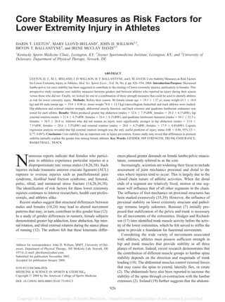
- 3. Hip abduction isometric strength testing was performed with subjects positioned in sidelying on a treatment table (Fig. 1). A pillow was placed between the subjects’ legs, using additional toweling as needed, such that the hip of the leg to be tested was abducted approximately 10° as mea- sured with respect to a line connecting the anterior superior iliac spines. A strap placed just proximal to the iliac crest and secured firmly around the underside of the table was used to stabilize the subjects’ trunk. The center of the force pad of a Nicholas hand-held dynamometer (Lafayette In- struments, Lafayette, IN) was then placed directly over a mark located 5 cm proximal to the lateral knee joint line. This dynamometer uses a load cell force detecting system to measure static force ranging from 0 to 199.9 kg with accu- racy to 0.1 kg Ϯ 2%. The dynamometer was secured be- tween the leg and a second strap that was wrapped around the leg and the underside of the table. The strap eliminated the effect of tester strength on this measure which has been reported to be a limitation of hand-held dynamometry (4). After zeroing the dynamometer, the subject was instructed to push the leg upward with maximal effort for 5 s. The force value displayed on the dynamometer was recorded and the device was re-zeroed. One practice trial and three ex- perimental trials were performed, with 15 s of rest between trials. The peak value from the three experimental trials was recorded. The athlete was then repositioned on their oppo- site side to test the hip strength of the contralateral limb using the same procedures. Hip external rotation (ER) isometric strength testing was performed with subjects positioned on a padded chair with the hips and knees flexed to 90° (Fig. 2). To limit the contri- bution of the hip adductors to force production in rotation, a strap was used to stabilize the thigh of the involved leg and a towel roll was placed between the subjects’ knees. The dyna- mometer was then placed such that the center of the force pad was directly over a mark that was 5 cm proximal to the medial malleolus. A strap around the leg and around the base of a stationary object held the dynamometer in place during con- tractions. Collection of peak hip external rotation isometric strength for each leg then proceeded in the same manner as that for hip abduction strength. Muscle capacity of the posterior core was measured using the modified Biering-Sorensen test (30) (Fig. 3). The athlete was positioned in prone with the pelvis at the edge of a treatment table. Straps were used to secure the athletes’ pelvis and legs to the table. The athlete supported their torso with their hands on a bench in front of the table until they FIGURE 2—Isometric testing of hip external rotation strength using hand-held dynamometry and strap stabilization. FIGURE 1—Isometric testing of hip abduction strength using hand- held dynamometry and strap stabilization. 928 Official Journal of the American College of Sports Medicine http://www.acsm-msse.org
- 4. were instructed to cross their arms and assume a horizontal position. The athlete was required to maintain the body in a horizontal position for as long as possible. The total time that the athlete was able to maintain the horizontal position until they touched down on the bench in front of them with their hands was recorded in seconds using a stopwatch. Athletes performed the side bridge test as described by McGill et al. (30) as a measure of lateral core muscle capacity, particularly the quadratus lumborum (Fig. 4). The athletes were positioned in right sidelying with their top foot in front of their bottom foot and their hips in zero degrees of flexion. The athletes were asked to lift their hips off the treatment table, using only their feet and right elbow for support. The left arm was held across their chest with their hand placed on the right shoulder. The total time the athlete was able lift their bottom hip from the table was recorded using a stopwatch. McGill (30) previously documented no significant difference between right and left side bridge endurance times. Therefore, the measure for the right lateral core muscles was used for data analysis. Anterior core muscle testing was performed using the straight leg lowering test for the first year of testing (23). This test was performed with the patient supine on the treatment table with their hips flexed to 90° and their knees fully extended. Patients were asked to steadily lower their legs back to the table over a 10-s period while they main- tained contact with the examiner’s hand at their L4–L5 interspace. A large board was placed behind the athlete during this test with marks indicating 10° increments of hip flexion. The angle at which the athlete’s low back raised from the examiner’s hand was recorded. Lower angles of hip flexion indicate a better performance on the test. After 1 yr of testing, we questioned the sensitivity of the straight leg lowering test for this population of subjects. There was very little variability in the measurement as nearly 70% of the athletes raised from the examiner’s hand between 50° and 60° of hip flexion, making the effect size small and increasing our likelihood of Type II error. There- fore, subjects enrolled in the second year of testing per- formed the flexor endurance test as described by McGill et al. (30) This test is performed seated on a treatment table with the athlete’s back supported on a 60° wedge (measured from horizontal). The athlete’s hands were crossed over their chest and their toes were placed under a stabilization strap. The athletes were then asked to maintain the position as the supporting wedge was pulled 10 cm away from the athlete. The time the athlete was able to maintain the 60° angle was recorded using a stopwatch. The test ended when the angle of the athlete’s upper body fell below the 60° threshold. Based on a larger range of evenly distrib- uted values, we found this test to be a more sensitive indicator of anterior core muscle capacity than the straight leg lowering test. Injuries. The head athletic trainers for each of the teams participating in the study recorded all back and lower ex- tremity injuries that occur during organized practices or games throughout the season. An injury was defined as an event that occurred during athletic participation and re- quired treatment or attention from the athletic trainer, team doctor, or other medical staff. Further, the event must have resulted in at least one full missed day of practice or sport participation. Trainers were given identical forms to record the details of each injury including the date, conditions (practice or game environment), mechanism of injury (con- tact with another player or object vs no contact), body part involved, and the type of injury that occurred. Finally, the number of whole days lost due to injury was recorded for each injury. Data analysis. Core stability measurements were com- pared between genders and between athletes who reported and injury and those who did not using two analysis of variance tests (SPSS 11.5.1, Chicago, IL). A significance level of 0.05 was used for all comparisons. The results of abdominal muscle performance for both tests are presented descriptively but were not included in the statistical analysis due to the previously described change in methods and associated lack of power for comparison. Logistic regres- sion was used to analyze the relationship between injury status and postural muscle strength measurements. The pro- cess began with simultaneous entry of the independent con- FIGURE 4—Endurance testing of the lateral trunk using the side bridge test. Left side test position shown here. FIGURE 3—Endurance testing of lumbar extensors using the modi- fied Beiring-Sorensen test. CORE STABILITY IN ATHLETES Medicine & Science in Sports & Exerciseா 929
- 5. tinuous variables into the model and was followed by back- ward, stepwise elimination of those variables with P values greater than 0.20. The continuous variables included in the model included strength measurements of hip abduction, hip external rotation, back extension, and side bridging. The response variable was injury status (1 ϭ injury reported, 0 ϭ no injury reported). RESULTS A total of 41 (28 females, 13 males) of the 139 athletes (29%) sustained 48 back or lower extremity injuries during a single competitive season. Thirty-five percent of the fe- males sustained an injury, compared with 22% for the males. Season-ending injuries occurred in one female (ACL tear) and one male (metatarsal stress fracture). For athletes who were able to return to participation, the average amount of time lost due to injury was 6.8 Ϯ 7.2 d. Twenty seven percent of the injuries occurred during a competitive event, and only 17% of the injuries were a result of direct contact to the player. A summary of the injuries by gender for situation, mechanism, and severity is presented in Table 1. The total number of injuries is larger than the number of injured athletes because six females and one male sustained more than one injury during the season. Although injury distribution was not a focus of this study, injuries were grouped into three body regions for general comparison (Table 2). The foot and ankle were the most frequently injured body region, accounting for 65% of all injuries. The knee was the primary complaint in 23% of the injuries, whereas injuries to the back, hip, or thigh occurred in 13% of the cases. For athletes who were able to return to competition, injuries to the knee required a 41% longer recovery than injuries to the hip, back, or thigh and 131% longer recovery than injuries to the ankle. There was no apparent difference in the location of injury between gen- ders. Basketball players, however, tended to experience injuries to the foot and ankle more frequently than the track athletes, who sustained a greater percentage of injuries to more proximal structures. Male athletes generally demonstrated greater core stabil- ity measures than female athletes (Table 3). These differ- ences were consistent whether the athlete participated in basketball or track (Table 4). Significant differences were noted between males and females for hip abduction, external rotation, and side bridging measures. Additionally, average abdominal muscle performance was slightly better for males than females for both the straight leg lowering test (average Ϯ SD: males ϭ 49° Ϯ 10°, females ϭ 59° Ϯ 9°) and the flexor endurance test (males ϭ 218 s Ϯ 146, females ϭ 204 s Ϯ 121). Athletes who experienced an injury over the course of the season generally demonstrated lower core stability measures than those who did not (Table 5). These strength differences were statistically significant for hip abduction and hip ex- ternal rotation. With respect to abdominal testing, athletes who experienced an injury generally demonstrated lower mean abdominal performance during the straight leg low- ering test (injured ϭ 59° Ϯ 8°, uninjured ϭ 54° Ϯ 11°) and the flexor endurance test (injured ϭ 199 s Ϯ 91, uninjured ϭ 217 s Ϯ 149). One female athlete experienced a season-ending injury to her ACL. Although it was not the purpose of this study to specifically analyze ACL injuries, it is interesting to note that this athlete demonstrated preseason deficiencies in each core stability test. Although certain females performed equally poorly on individual tests, this individual was unique in that she was well below the average performance of females who reported an injury as well as to those who did not (Table 6). The core stability measures included in this study gener- ally demonstrated moderate, but significant, correlation (Ta- ble 7). Side bridge scores were significantly correlated with the performance of all other postural muscle tests included in this study. Back extension, on the other hand demon- strated a very low correlation with hip abduction and exter- nal rotation isometric strength measurements. Results of the logistic regression analysis indicate that the regression equation fits our data reasonably well (likelihood ratio ϭ 12.72, P ϭ 0.013) (Table 8). Using this equation, the model predicted 62.6% of the injuries correctly. Hip exter- nal rotation accounted for the majority of the variability in the model and represented the only true significant risk factor (OR ϭ 0.86, 95% CI ϭ 0.77, 0.097). Indeed, back- ward stepwise logistic regression of the independent vari- TABLE 2. Injuries by gender and sport by body part injured. Back/Hip/Thigh Knee Ankle/ Foot Male (N ϭ 14) 7% 21% 71% Female (N ϭ 34) 15% 24% 62% Basketball (N ϭ 34) 6% 15% 79% Track (N ϭ 14) 29% 43% 29% Average time lost (days per injury) 8.0 11.3 4.9 TABLE 3. Comparison of core stability measures by gender. Hip Abduction (% Body Weight) Hip External Rotation (% Body Weight) Side Bridge (s) Back Extension (s) Average (SD) Average (SD) Average (SD) Average (SD) Male (N ϭ 60) 32.6 (7.3) 21.6 (4.3) 84.3 (32.5) 130.4 (40.0) Female (N ϭ 79) 29.2 (6.1) 18.4 (4.1) 58.9 (26.0) 123.4 (48.4) P 0.04 Ͻ0.001 Ͻ0.001 0.37 TABLE 1. Injuries by gender for situation, mechanism, and severity. Situation Mechanism Severity (days lost) Event Practice Contact Noncontact Average (SD) Male (N ϭ 14) 14% 86% 14% 86% 8.5 (11.0) Female (N ϭ 34) 32% 68% 18% 72% 6.0 (4.9) Combined (N ϭ 48) 27% 73% 17% 83% 6.8 (7.2) 930 Official Journal of the American College of Sports Medicine http://www.acsm-msse.org
- 6. ables established that hip external rotation was the only useful predictor of the likelihood of sustaining an injury over the course of a season (coefficient ϭ Ϫ0.154, t-statistic ϭ Ϫ3.15, P ϭ 0.002). However, the relatively low coeffi- cient of determination suggests that other factors not in- cluded in this study significantly contribute to injury status over the course of an athletic season. DISCUSSION The purpose of this study was to prospectively examine differences in core stability measures between males and females as well as between those athletes who became injured and those who did not. We also hoped to identify one or a combination of strength measures that could be used to identify those individuals at risk for lower extremity injury. Females in this study demonstrated significantly reduced side bridge endurance and hip abduction and external rota- tion isometric strength. Whereas weakness in females has been previously documented in these muscles groups (5,8,30), the consequence of this weakness is not well un- derstood. We suggest that hip and trunk weakness reduces the ability to of females stabilize the hip and trunk. There- fore, females may be more vulnerable to the large external forces experienced by these segments during athletics, es- pecially those forces in the transverse and frontal planes. As a result, females may be predisposed to excessive motion in the hip or trunk versus males, potentially permitting their entire lower extremity to move into positions frequently associated with noncontact injuries such as femoral adduc- tion and internal rotation. Indeed, recent literature verifies that females tend to display greater hip internal rotation and adduction during athletic tasks (12,22,25). Athletes who sustained an injury in this study displayed significantly less hip abduction and external rotation strength than uninjured athletes. To our knowledge, this is the first prospective study to demonstrate a relationship between these variables. However, several retrospective and cross-sectional studies have been performed that previously indicated that such a relationship may exist for a variety of injuries (1,13,20,21). For example, Ireland et al. (20) iden- tified significant weakness among young female athletes with patellofemoral pain in hip abduction and external ro- tation strength versus a healthy, age-matched control group. These authors further explained that the mechanism for this pain may be excessive femoral adduction and internal rota- tion during weight bearing activities. Citing cadaveric stud- ies, they reported that this alignment promotes lateral pa- tellar tracking and increases lateral retropatellar contact pressure (20). This study finds that hip external rotation strength weak- ness most closely predicts injury status over the course of one athletic season. However, hip external rotation strength is only one element of core stability, and other elements of core stability not included in this study may add to the predictive value of the regression equation. Core stability is the product of motor control and muscular capacity of the lumbo-pelvic-hip complex. Hewett et al. (16) has previously demonstrated the value of motor control on knee injury prevention. Females who participated in a general strength, flexibility, and neuromuscular training program experienced a 62% decrease in serious knee ligament injuries. Although the strengthening component of his intervention program included abdominal curls and back hyperextension exer- cises, our results suggest that these muscle groups may not have significantly contributed to his positive results. Rather, the benefit of his program may be a reduction in knee adduction and abduction moments due to advanced postural adaptations of the hip abductors and external rotators before landing from a jump. The other component of core stability, muscle capacity, is represented by the athlete’s ability to generate force or maintain force (endurance) in the lumbo-pelvic-hip complex. McGill et al. (29) suggest that the value of trunk muscle endurance is greater than the ability of these muscles to gen- erate force in the prevention of low back pain. Indeed, the endurance of the trunk extensors has been found to predict the occurrence of low back pain among 30- to 60-yr-old adults (3). However, in a more athletic population, this study suggests that isometric hip strength measures, particularly in external rota- tion, are more accurate predictors of back and lower extremity injury than trunk endurance measures. These results may reflect the significance of strength versus endurance for individuals who participate in high TABLE 4. Comparison of core stability measures by sport. Hip Abduction (% Body Weight) Hip External Rotation (% Body Weight) Side Bridge (s) Back Extension (s) Average (SD) Average (SD) Average (SD) Average (SD) Male BB (N ϭ 44) 32.9 (7.8) 21.7 (4.3) 82.7 (30.6) 131.4 (42.0) Male XC (N ϭ 17) 32.4 (6.2) 21.7 (4.3) 87.6 (37.1) 122.9 (38.7) Female BB (N ϭ 60) 29.3 (5.8) 18.0 (3.5) 57.8 (24.7) 115.7 (43.5) Female XC (N ϭ 18) 27.8 (7.0) 19.5 (5.3) 60.9 (30.5) 151.4 (52.5) TABLE 5. Comparison of core stability measures by injury status. Hip Abduction (% Body Weight) Hip External Rotation (% Body Weight) Side Bridge (s) Back Extension (s) Average (SD) Average (SD) Average (SD) Average (SD) Uninjured (N ϭ 99) 31.6 (7.1) 20.6 (4.2) 72.0 (32.4) 128.3 (43.6) Injured (N ϭ 41) 28.6 (5.5) 17.9 (4.4) 64.7 (28.8) 121.6 (48.9) P 0.02 0.001 0.22 0.43 CORE STABILITY IN ATHLETES Medicine & Science in Sports & Exerciseா 931
- 7. speed events. Cholewicki et al. (9) suggest that the kine- matic response of the trunk during sudden events depends on both the mechanical stability level of the spine before loading, as well as the reflex response of the trunk muscles immediately after loading. Considering that the endurance times between the injured and uninjured athletes were very similar, it appears that all athletes possessed the capacity for mechanical stability of the lumbar spine. However, as Cholewicki et al. (9) suggest, the injured athletes may lack the ability to generate sufficient force or resist external forces during high-speed events. Perhaps future study will find that isometric strength testing of the abdominals, back extensors, and quadratus lumborum is more closely associ- ated with the ability of individuals to sufficiently recruit the muscles of the trunk during high speed events, stabilize the lumbar spine, and prevent lower extremity injuries. The athletes in this study experienced an injury incidence of 0.35 (48 injuries/139 athletes). This incidence is very similar to the results of Messina et al. (32), who analyzed injuries sustained by male and female Texas high school basketball players. After adjustment to include only back and lower extremity injuries, the injury incidence for their study becomes 0.37 injuries/athlete. Meeuwisse et al. (31) reported that the incidence of back and lower extremity injuries was 0.50 injuries/athlete for their male intercolle- giate basketball players. Although the males in our study ex- perienced a much lower injury incidence of 0.23 injuries/ athlete, we only included injuries that resulted in at least one full day of missed participation. Meeuwisse et al. also included injuries that resulted in days of partially missed participation. The injury patterns in this investigation mirror those found in other, large-scale epidemiological studies. For ex- ample, a greater proportion of female athletes experienced an injury versus males (36,37,42). Additionally, the ankle was the most commonly injured structure of the lower extremity (31,32,42). Finally, a greater total number of injuries occurred during practice than during games (31,34,36). These results suggest that our sample of injuries represent a similar distribution of injuries for this population of athletes. A potential limitation of this study is that hip strength measurements were made in units of force instead of torque. Therefore, if injured athletes were systematically taller than the uninjured athletes, the difference in hip torque measure- ments may have been less significant than the force mea- surements found in this study. However, considering that females tend to be shorter than males and a greater propor- tion of females reported injuries, we believe we would have found even greater differences between groups with respect to gender and injury status if torque were used. A second potential limitation of this study is that the two examiners were not tested for intratester reliability before data collection. However, as noted above, each test was based on those previously described to be reliable in a similar group of subjects. Further, the use of straps for stabilization during isometric testing eliminated the variabil- ity of tester strength in these measures. Finally, the testers were aware of the potential influence of verbal feedback on the motivation of the subjects and used consistent verbal cues for all endurance tests. Intertester reliability was not a concern because each tester performed the same tests throughout data collection. The results of this investigation generate numerous ques- tions for further studies. For example, future studies may test the athlete’s ability to demonstrate core stability in more physiologic positions. The tests positions used in this study are those most commonly used for manual muscle testing and are conducive to methods for preparticipation screening. Although these tests gauge the capacity of each athlete to generate force or maintain force in core muscle groups, they do not necessarily reflect how these muscles function during closed chain activities. Further, these tests may not reflect the degree to which the muscles are recruited by the athletes during athletic participation. Considering these facts, future studies should consider the addition of a dynamic test of lower extremity alignment during a closed kinetic chain activity such as the single leg step down test (41). Future studies should also seek to understand the relationship be- tween these core strength measures and the result of this dynamic test. Future studies on the potential of core stability programs to prevent serious knee ligament injuries also seem justified. TABLE 6. Description of the core stability measurements for a female subject prior to an ACL injury. Hip Abduction (% Body Weight) Hip External Rotation (% Body Weight) Side Bridge (s) Back Extension (s) Average (SD) Average (SD) Average (SD) Average (SD) Uninjured females 29.4 (6.2) 19.0 (3.8) 59.0 (23.3) 124.3 (46.1) Injured females 28.9 (6.1) 17.4 (4.6) 58.8 (30.1) 121.7 (54.2) Female with ACL injury 23.0 16.5 25.0 38.0 TABLE 7. Pearson correlation matrix for core stability measures. Hip abduction Hip Ext Rotation Side Bridge Hip ext rotation 0.525* Side bridge 0.383* 0.440* Back extension 0.165 0.087 0.564* * Significant at P Ͻ 0.05. TABLE 8. Logistic regression results (dependent variable ϭ injury during the season). Variable Coefficient t P Odds Ratio (95% CI OR) Constant 2.931 2.37 0.018 Hip abduction Ϫ0.031 Ϫ0.85 0.40 0.97 (0.90, 1.04) Hip external rotation Ϫ0.146 Ϫ2.49 0.013 0.86 (0.77, 0.97) Side bridge 0.007 0.73 0.46 1.01 (0.99, 1.02) Back extension Ϫ0.004 Ϫ0.77 0.44 1.00 (0.99, 1.01) Likelihood ratio [df] 12.72 [4] 0.013 % correct prediction 62.6% McFadden’s-R2 0.076 932 Official Journal of the American College of Sports Medicine http://www.acsm-msse.org
- 8. The addition of an external valgus moment to a flexed knee has been documented to increase the force on the ACL (27). By increasing the strength of muscles that resist this moment, athletes may decrease the incidence of injury to this important ligament. Further, improving the strength of the hip external rotators and abductors may diminish the tendency for femoral internal rotation and adduction frequently observed in athletes with patellofemoral pain. Future studies in this area should also include exposure time in order to establish the injury rate for each group of athletes and use survival analysis statistics. Finally, the relationship between core strength and lower extremity mechanics needs to be examined. CONCLUSIONS Female athletes displayed significantly decreased hip external rotation and side bridge measures versus their male counterparts. Additionally, athletes who experi- enced an injury over the course of a season displayed significant weakness in hip abduction and external rota- tion. Backward, logistic regression analysis of the core stability measurements reveals that hip external rotation strength was the sole significant predictor of injury status for the athletes in this study. These results highlight the importance of proximal stabilization for lower extremity injury prevention. REFERENCES 1. BECKMAN, S. M., and T. S. BUCHANAN. Ankle inversion injury and hypermobility: effect on hip and ankle muscle electromyography onset latency. Arch. Phys. Med. Rehabil. 76:1138–1143, 1995. 2. BERGMARK, A. Stability of the lumbar spine: a study in mechanical engineering. Acta Orthop. Scand. Suppl. 230:1–54, 1989. 3. BIERING-SORENSEN, F. Physical measurements as risk indicators for low-back trouble over a one-year period. Spine 9:106–119, 1984. 4. BOHANNON, R. W. Intertester reliability of hand-held dynamome- try: a concise summary of published research. Percept. Mot. Skills 88:899–902, 1999. 5. BOHANNON, R. W. Reference values for extremity muscle strength obtained by hand-held dynamometry from adults aged 20 to 79 years. Arch. Phys. Med. Rehabil. 78:26–32, 1997. 6. BOBBERT, M. F., and J. P. VAN ZANDWIJK. Dynamics of force and muscle stimulation in human vertical jumping. Med. Sci. Sports Exerc. 31:303–310, 1999. 7. BOUISSET, S. Relationship between postural support and intentional movement: biomechanical approach. Arch. Int. Physiol. Biochim. Biophys. 99:77–92, 1991. 8. CAHALAN, T. D., M. E. JOHNSON, S. LIU, and E. Y. CHAO. Quanti- tative measurements of hip strength in different age groups. Clin. Orthop. 246:136–145, 1989. 9. CHOLEWICKI, J., A. P. D. SIMONS, and A. RADEBOLD. Effects of external trunk loads on lumbar spine stability. J. Biomech. 33: 1377–1385, 2000. 10. CHOLEWICKI, J., and J. J. VAN VLIET IV. Relative contribution of trunk muscles to the stability of the lumbar spine during isometric exertions. Clin. Biomech. 17:99–105, 2002. 11. CHOLEWICKI, J., and S. M. MCGILL. Mechanical stability of the in vivo lumbar spine: implications for injury and chronic low back pain. Clin. Biomech. 11:1–15, 1996. 12. FERBER, R., I. MCCLAY DAVIS, and D. S. WILLIAMS. Gender differ- ences in lower extremity mechanics during running. Clin. Bio- mech. 18:350–357, 2003. 13. FREDERICSON, M., C. COOKINGHAM, A. M. CHAUDHARI, B. C. DOWDELL, N. OESTREICHER, and S. A. SAHRMANN. Hip abductor weakness in distance runners with iliotibial band syndrome. Clin. J. Sport Med. 10:169–175, 2000. 14. GEMMELL, I. M. M. Injuries among female army recruits: a conflict of legislation. J. R. Soc. Med. 95:23–27, 2002. 15. GRIFFIN, L. Y., J. AGEL, M. J. ALBOHM, et al. Noncontact anterior cruciate ligament injuries: risk factors and prevention strategies. J. Am. Acad. Orthop. Surg. 8:141–150, 2000. 16. HEWETT, T., T. LINDENFELD, J. RICCOBENE, and F. NOYES. The effect of neuromuscular training on the incidence of knee injury in female athletes. A prospective study. Am. J. Sports Med. 27:699– 706, 1999. 17. HODGES, P. W., and C. A. RICHARDSON. Contraction of the abdom- inal muscles associated with movement of the lower limb. Phys. Ther. 77:132–144, 1997. 18. HORTON, M. G., and T. L. HALL. Quadriceps femoris muscle angle: normal values and relationships with gender and selected skeletal measures. Phys. Ther. 69:897–901, 1989. 19. IRELAND, M. L. The female ACL: why is it more prone to injury? Orthop. Clin. North Am. 33:637–651, 2002. 20. IRELAND, M. L., J. D. WILLSON, B. T. BALLANTYNE, and I. MCCLAY- DAVIS. Hip strength in females with and without patellofemoral pain. J. Orthop. Sports Phys. Ther. 33:671–676, 2003. 21. JARAMILLO, J., T. W. WORRELL, and C. D. INGERSOLL. Hip isometric strength following knee surgery. J. Orthop. Sports Phys. Ther. 20:160–165, 1994. 22. LEPHART, S. M., C. M. FERRIS, B. L RIEMANN, J. B. MYERS, and F. H. FU. Gender differences in strength and lower extremity kinematics during landing. Clin. Orthop. 401:162–169, 2002. 23. LINDSTROM, I., C. OHLUND, C. EEK, L. WALLIN, L. PETERSON, and A. NACHEMSON. Mobility, strength, and fitness after a graded activity program for patients with subacute low back pain. Spine 17:641– 652, 1992. 24. LIVINGSTON, L. A. The quadriceps angle: a review of the literature. J. Orthop. Sports Phys. Ther. 28:105–109, 1998. 25. MALINZAK, R. A., S. M. COLBY, D. T. KIRKENDALL, B. YU, and W. E. GARRETT. A comparison of knee joint motion patterns between men and women in selected athletic tasks. Clin. Biomech. 16:438–445, 2001. 26. MALONE, T. R., W. T. HARDAKER, W. E. GARRETT, et al. Relation- ship of gender to anterior cruciate ligament injuries in intercolle- giate basketball players. J. Southern Orthop. Assoc. 2:36–39, 1993. 27. MARKOLF, K. L., D. M. BURCHFIELD, M. M. SHAPIRO, M. F. SHEP- ARD, G. A. FINERMAN, and J. L. SLAUTERBECK. Combined knee loading states that generate high anterior cruciate ligament forces. J. Orthop. Res. 13:930–935, 1995. 28. MCCLAY DAVIS, I., and M. L. IRELAND. ACL research retreat: the gender bias, April 6–7, 2001. Clin. Biomech. 16:937–939, 2001. 29. MCGILL, S. M., S. GRENIER, N. KAVCIC, and J. CHOLEWICKI. Coor- dination of muscle activity to assure stability of the lumbar spine. J. Electromyogr. Kinesiol. 13:353–359, 2003. 30. MCGILL, S. M., A. CHILDS, and C. LIEBERMAN. Endurance times for low back stabilization exercises: clinical targets for testing and training from a normal database. Arch. Phys. Med. Rehabil. 80: 941–944, 1999. 31. MEEUWISSE, W. H., R. SELLMER, and B. E. HAGEL. Rates and risks of injury during intercollegiate basketball. Am. J. Sports Med. 31:379–385, 2003. 32. MESSINA, D. F., W. C. FARNEY, and J. C. DELEE. The incidence of injury in Texas high school basketball: a prospective study among male and female athletes. Am. J. Sports Med. 27:294–299, 1999. 33. NADLER, S. F., G. A. MALANGA, M. DEPRINCE, T. P. STITIK, and J. H. FEINBERG. The relationship between lower extremity injury, low back pain, and hip muscle strength in male and female collegiate athletes. Clin. J. Sport Med. 10:89–97, 2000. 34. POWELL, J. W., and K. D. BARBER-FOSS. Sex-related injury patterns among selected high school sports. Am. J. Sports Med. 28:385– 391, 2000. CORE STABILITY IN ATHLETES Medicine & Science in Sports & Exerciseா 933
- 9. 35. POWERS, C. M., P. Y. CHEN, S. F. REISCHL, and J. PERRY. Compar- ison of foot pronation and lower extremity rotation in persons with and without patellofemoral pain. Foot Ankle Int. 23:634–640, 2002. 36. RAUH, M. J., A. J. MARGHERITA, S. G. RICE, T. D. KOEPSELL, and F. P. RIVARA. High school cross country running injuries: a lon- gitudinal study. Clin. J. Sport Med. 10:110–116, 2000. 37. SALLIS, R. E., K. JONES, S. SUNSHINE, G. SMITH, and L. SIMON. Comparing sports injuries in men and women. Int. J. Sports Med. 22:420–423, 2001. 38. TAUNTON, J. E., M. B. RYAN, D. B. CLEMENT, D. C. MCKENZIE, D. R. LLOYD-SMITH, and B. D. ZUMBO. A retrospective case-control analysis of 2002 running injuries. Br. J. Sports Med. 36:95–101, 2002. 39. TIBERIO, D. The effect of excessive subtalar joint pronation on patellofemoral mechanics: a theoretical model. J. Orthop. Sports Phys. Ther. 9:160–165, 1987. 40. WINTER, D. A., J. J. ENG, and M. G. ISHAC. A review of kinetic parameters in human walking. In: Gait Analysis: Theory and Application, R. L. Craik and C. A. Oatis (Eds.). St. Louis: Mosby, 1995, pp. 252–270. 41. ZELLER, B. L., J. L. MCCRORY, W. B. KIBLER, and T. L. UHL. Differences in kinematics and electromyographic activity between men and women during the single-legged squat. Am. J. Sports Med. 31:449–456, 2003. 42. ZELISKO, J. A., H. B. NOBLE, and M. A. PORTER. A comparison of men’s and women’s professional basketball injuries. Am. J. Sports Med. 10:297–299, 1982. 934 Official Journal of the American College of Sports Medicine http://www.acsm-msse.org