Curación acelerada de tendón de aquiles en ratas en resp uesta a acs 2009 ajsm - majewski - en
•
1 like•425 views


Recommended
Recommended
More Related Content
What's hot
What's hot (20)
Viewers also liked
Viewers also liked (18)
Similar to Curación acelerada de tendón de aquiles en ratas en resp uesta a acs 2009 ajsm - majewski - en
Similar to Curación acelerada de tendón de aquiles en ratas en resp uesta a acs 2009 ajsm - majewski - en (20)
More from Dr. Manuel Concepción
More from Dr. Manuel Concepción (20)
Recently uploaded
Recently uploaded (20)
Curación acelerada de tendón de aquiles en ratas en resp uesta a acs 2009 ajsm - majewski - en
- 1. Accelerated Healing of the Rat Achilles Tendon in Response to Autologous Conditioned Serum ¨ Martin Majewski,*y MD, Peter E. Ochsner,z MD, Fanjun Liu,§ PhD, Rudolf Fluckiger,§ PhD, and Christopher H. Evans,§ PhD From the yOrthopaedic Clinic, University of Basel, Basel, Switzerland, the z Orthopaedic Clinic, St. Anna Clinic, Lucerne, Switzerland, and the §Center for Molecular Orthopaedics, Harvard Medical School, Boston, Massachusetts Background: Despite advances in the treatment of ruptured Achilles tendon, imperfections of endogenous repair often leave patients symptomatic. Local administration of autologous conditioned serum (ACS) in patients with inflammatory, degenerative conditions has shown beneficial effects. Purpose: Because ACS also contains growth factors that should accelerate tendon healing, we studied the effect of ACS on the healing of transected rat Achilles tendon. Study Design: Controlled laboratory study. Methods: In preliminary in vitro experiments, rat tendons were incubated with ACS and the effect on the expression of Col1A1 and Col3A1 was assessed by real-time quantitative polymerase chain reaction. To test its effect in vivo, the Achilles tendons of 80 Sprague Dawley rats were transected and sutured back together. Ten rats from each group (ACS group, n 5 40; control group, n 5 40) were euthanized at 1, 2, 4, and 8 weeks postoperatively for biomechanical (n 5 7) and histologic (n 5 3) testing. Lysyl oxidase activity was assayed by a flurometric assay. The organization of repair tissue was assessed histologically with hematoxylin and eosin- and with Sirius red-stained sections, and with immunohistochemistry. Results: Tendons exposed to ACS in vitro showed a greatly enhanced expression of the Col1A1 gene. The ACS-treated tendons were thicker, had more type I collagen, and an accelerated recovery of tendon stiffness and histologic maturity of the repair tissue. However, there were no differences in the maximum load to failure between groups up to week 8, perhaps because lysyl oxidase activities were unchanged. Conclusion and Clinical Relevance: Overall, our study demonstrates that treatment with ACS has the potential to improve Achilles tendon healing and should be considered as a treatment modality in man. However, as strength was not shown to be increased within the parameters of this study, the clinical importance of the observed changes in humans still needs to be defined. Keywords: growth factors; autologous conditioned serum; animal model; biological repair suturing21,31 or functional treatment,22 recovery times are still long and rerupture rates are substantial.23 Further major improvements in Achilles tendon rupture healing are most likely to result from the implementation of novel, biological approaches. In particular, numerous studies have documented the beneficial effects of individual growth factors on tendon healing in animal models. Such growth factors include vascular endothelial growth factor (VEGF),40 transforming growth factor-beta (TGF-b1),17 platelet-derived growth factor (PDGF),35 growth and differentiation factors,28 insulin-like growth factor-1,6 basic fibroblast growth factor (FGF-2, also bFGF),5 and bone morphogenetic proteins (BMP-12 and BMP-13).8 Exposure in vitro of patellar tendon fibroblasts to bFGF was found to affect cell proliferation and collagen type III expression, both early events in tendon healing.5 Additionally, an angiogenic effect was observed with bFGF application.10 Once a rare occurrence, rupture of the Achilles tendon has become the most common type of sports-related tendon rupture, with an estimated annual incidence of 10 ruptures per 100 000 people.30 Despite advances in the treatment of Achilles tendon rupture such as percutaneous ¨ *Address correspondence to Dr. med. Martin Majewski, Orthopadi¨ sche Universitatsklinik Basel, Spitalstrasse 26, CH-4031 Basel, Switzerland (e-mail: majewski01@yahoo.de). One or more authors has declared a potential conflict of interest: Christopher H. Evans is on the Supervisory Board of Orthogen and owns stock in the company. The authors gratefully acknowledge the support of the Swiss Society of Orthopedic Surgery. The American Journal of Sports Medicine, Vol. 37, No. 11 DOI: 10.1177/0363546509348047 Ó 2009 The Author(s) 2117 Downloaded from ajs.sagepub.com by guest on December 14, 2009
- 2. 2118 Majewski et al The American Journal of Sports Medicine However, of all growth factors studied, VEGF appears to have the strongest angiogenic activity.40 During embryogenesis of tendon tissue, BMPs—in particular, BMP-12 and BMP-13—affect the expression of elastin and collagen type I. Additionally it was found in animal experiments that BMP-12 stimulated the healing of the patellar tendon.7 Among the other growth factors that play critical roles in tendon healing is TGF-b1, which has been reported to induce the expression of collagen type I and fibronectin mRNA in experiments in tissue culture.11 Upregulation of mRNAs expressing the a-chains of type I and III collagens has been reported to occur in tenocytes isolated from sheep cultured with and without serum.4 It appears that optimal tendon repair requires high expression of collagen type I. As healing progresses, there is a switch from collagen type III, which is expressed early in the healing process, to collagen type I, which provides greater mechanical strength. As newly synthesized collagen is laid down, the enzyme lysyl oxidase converts certain lysine and hydroxylysine groups into aldehydes, which spontaneously form cross-links that stabilize collagen fibers.19 Although the effect of growth factors on tendon healing is impressive, it has become increasingly clear that tendon repair is not triggered by a single growth factor but requires the interplay of various such factors. Collectively, they exert powerful and comprehensive effects on healing. Consistent with this hypothesis, injections of platelet concentrates that contain a rich mixture of growth factors into the hematoma of severed Achilles tendons increased tendon callus strength and stiffness in the rat, but no such improvement was apparent when individual growth factors TGF-b1 or FGF-2 were injected.2 Autologous conditioned serum (ACS) preparations are a natural source of multiple growth factors. By exposing whole blood to glass beads, Meijer and coworkers26 obtained serum enriched in anti-inflammatory cytokines, including interleukins (IL-4, IL-10, IL-13) and interleukin-1 receptor antagonist but which also contained elevated concentrations of growth factors FGF-2, hepatocyte growth factor, and TGF-b1. In clinical trials, locally injected ACS was found to be beneficial in the treatment of equine osteoarthritis,9 lumbar radicular compression,3 and muscle injuries.36,38,39 We tested the hypothesis that the growth factors in ACS would improve the healing of ruptured and sutured Achilles tendons in rats. MATERIALS AND METHODS Autologous Conditioned Serum Serum was conditioned by incubating pooled rat blood for 9 hours at 37°C in Orthokine syringes (Orthogen AG, Dussel¨ dorf, Germany) containing glass beads (surface area 21 mm2). After incubation, samples were centrifuged and the conditioned serum collected and frozen in 0.2-mL aliquots at 220°C. For injection, aliquots were thawed on ice, allowed to reach room temperature, and 170 lL of ACS injected directly into the area of the sutured Achilles tendon using a 1-mL insulin syringe. The dose used was calculated from a study in mice,39 based on mean body weight. The exact location of the injection site was defined by the position of the stitches closing the skin. The gap was closed with 4 stitches and injections were made between stitches 2 and 3. Rat Achilles Tendon Healing Model Eighty adult male Sprague Dawley rats, weighing 400 to 425 g (Charles River Laboratories, Boston, Massachusetts) were used. The Institutional Animal Care and Use Committee approved this study. Animals were randomly assigned to an experimental and a control group of 40 rats each. The experimental group received ACS injections 24, 48, and 72 hours postoperatively directly into the sutured tendon area, while the control group did not receive any such injections. Injections of phosphate-buffered saline (PBS) alone were not considered as this has previously been shown to have no effect.17 Similarly, injection of unconditioned serum is of questionable value because platelet activation during its preparation cannot be avoided. Rats were placed under general anesthesia with isoflurane and received buprenorphine (0.06 mg/kg) intramuscularly into the left thigh. The right hind leg was shaved and disinfected with Betadine-Scrub (Betadine, Stamford, Connecticut) and rinsed with 70% alcohol. Animals were placed in a sterile field on a heated surgery table and covered with a sterile surgical drape such that only the prepared limb remained exposed. A 3-cm incision was made above the right Achilles tendon, the superficial tendon was exposed, and the peritendon opened. The tendon was transected with a no. 15 scalpel blade perpendicular to the collagen fibers, creating a gap 5 mm proximal to the calcaneal insertion of the tendons. The plantar tendon was transected to prevent internal splinting. After transsection, the Achilles tendon was sutured back together with PDS II 0-2 (Ethicon, Norderstedt, Germany) with a Kessler–Kirchmayr stitch. (In our experience from a pilot study, use of thinner thread increases rerupture rates and, according to the literature, decreases tendon strength.40) The subcutaneous layer and skin were closed by a continuous suture with Prolene 0-4 (Ethicon). After regaining consciousness the rats received twice-daily buprenorphine (0.06 mg/kg) for 2 days. To simulate early functional therapy, operated legs were left without cast immobilization At weeks 1, 2, 4, and 8, 10 animals from each group were euthanized with CO2 after isoflurane anesthesia, and whole bone-tendon units were removed for testing (n 5 7 for biomechanical testing, n 5 3 for histology). To harvest the whole muscle-Achilles tendon-bone units, a 5-cm incision was made from below the knee down to the plantar fascia of the foot. The muscle-Achilles tendon-bone units were exposed, dissecting the subcutaneous layer from muscle, tendon, and calcaneus. Muscle-Achilles tendon-bone units were harvested by transection of the middle part of the gastrocnemius muscle and of the calcaneus. Enzyme-Linked Immunoabsorbent Assay Tests Cytokine levels were determined with Quantikine ELISA (enzyme-linked immunoabsorbent assay) kits (R&D Downloaded from ajs.sagepub.com by guest on December 14, 2009
- 3. Vol. 37, No. 11, 2009 Effect of ACS on Healing of Achilles Tendon Rupture 2119 Systems, Minneapolis, Minneapolis): mouse/rat PDGF-BB (catalog No. MBB00), rat VEGF (catalog No. RRV00), and TGF-b1 (catalog No. DB100B), and used according to manufacturerÕs instructions. Real-Time Quantitative Polymerase Chain Reaction Eight Achilles tendons of the left, unoperated leg were collected at week 8. Tendons were collected and cultured for 7 days in 1 mL of DelbuccoÕs Modified Eagle Medium (DMEM) 1 0.2% fetal bovine serum (FBS) with and without 10% ACS for 7 days (4 tendons per group) without change of media. Ribonucleic acid was extracted from tendons for RTPCR analysis of Col1A1, Col3A1, and GAPDH expression. One milliliter of TRI reagent (Ambion, Austin, Texas) was added, incubated for 15 minutes at room temperature, centrifuged (12 000 3 g, 10 minutes), and supernatants collected for RNA purification according to the manufacturerÕs instructions. The RNA was dissolved in diethylpyrocarbonate-treated water at a concentration of 0.1 lg/lL. The RNA was reverse-transcribed using Super Script FirstStrand Synthesis Kits for RT-PCR (Invitrogen, Carlsbad, California) according to the manufacturerÕs instructions. The complementary DNA (cDNA) product was amplified on an Applied Biosystems 7500 Real-Time PCR System using Platinum SYBR Green pPCR SuperMix-UDG (Invitrogen) for detection. The primers used were for collagen type I (alpha 1 chain; Col1A1): cactggtgctaagggagagc (forward), aggagaaccacgttcaccag (reverse); for collagen type III (alpha 1 chain; Col3A1): tacggcaatcctgaacttcc (forward), caccttcatttgaccccatc (reverse); for glyceraldehyde 3-phosphate dehydrogenase (GAPDH): acccagaagactgtggatgg (forward), tctagacggcaggtcaggtc (reverse). The PCR conditions were 50°C, 2 minutes, 1 cycle; 95°C, 10 minutes, 1 cycle; 95°C, 30 seconds, 58°C, 30 seconds, 72°C, 30 seconds, 40 cycles; and 72°C, 10 minutes, 1 cycle. Relative gene expression was calculated and normalized to the internal standard GAPDH, using instrument software. Histology Three specimens per group were examined histologically at each time point. The tendons were immediately fixed in 4% buffered formalin (pH 7.4) for 24 hours, dehydrated in graded alcohols, and embedded in paraffin wax. Longitudinal sections (5 lm) at the midsubstance of the tendon were stained with hematoxylin and eosin. Slides were viewed under light microscopy. Five regions were evaluated for each sample (center of tendon, left border, right border, 2 mm above tendon center, and 2 mm below tendon center). Slides were evaluated in a blinded manner by visual inspection. Immunohistochemistry Collagen III was specifically detected using the primary rabbit antirat collagen type III polyclonal antibody (1:300, Batch 24042951, AbD Serotec, Dusseldorf, Ger¨ many) and visualized with conjugated secondary goat antirabbit immunoglobulin G antibodies (1:50, B6648, E8386, Sigma Aldrich, Hamburg, Germany). For staining, endogenous peroxidase was blocked by 30-minute incubation in 1% hydrogen peroxide in PBS. After washing in PBS, sections were incubated with 1:10 diluted rat serum to block nonspecific protein binding. Sections were incubated with the primary antibody for 1 hour at room temperature, followed by three 5-minute washes. For amplified detection with biotin-streptavidin, sections were incubated with biotinylated secondary antibody for 1 hour at room temperature, washed 3 times with PBS, and incubated with fluorescein 5-isothiocyanate–conjugated streptavidin for 20 minutes at room temperature. This was followed by three 5-minute washes with PBS. To detect visualized areas of collagen III, 5 regions were evaluated for each sample (center of tendon, left border, right border, 2 mm above tendon center, and 2 mm below tendon center). Images (magnification 3400, area evaluated 100 lm 3 100 lm) were captured with a digital camera (Olympus C-3030, Hamburg, Germany). The ratios were obtained by automated measurements of the area of visualized areas of collagen III versus nonvisualized areas of the image taken using digital imaging analysis software (Image-Pro Plus, Media Cybernetics, Silver Spring, Maryland). Results are expressed as the ratio of area of collagen. Slides were also evaluated in a blinded manner by visual inspection. There was excellent agreement between the scores obtained this way and the results of automated image analysis. Sirius Red Staining/Cross-Polarization Microscopy For cross-polarization microscopy, 5-lm sections were stained for 1 hour in picrosirius solution (Sirius red F3BA [0.1%] in saturated aqueous picric acid, pH 2) according to the method of Junqueira et al.15 Sections were washed for 2 minutes in 0.01 N HCl, dehydrated, cleared, and mounted in synthetic resin. Slides were viewed under polarized light microscopy. Thicker, mature collagen fibers, including type I collagen fibers, appeared red-orange, whereas thinner collagen fibers, including type III collagen, appeared pale green.16 To determine thicker, mature red-orange versus thinner pale green collagen ratios, 5 regions were evaluated for each sample (center of tendon, left border, right border, 2 mm above tendon center, and 2 mm below tendon center). Images (magnification 3400, area evaluated 100 lm 3 100 lm) were captured with a digital camera (Olympus C-3030). Collagen ratios were obtained by automated measurements of the area of thicker mature red-orange versus thinner pale green using digital imaging analysis software (Image-Pro Plus, Media Cybernetics). Results are expressed as the ratio of area of collagen. Slides were also evaluated in a blinded manner by visual inspection. There was excellent agreement between the scores obtained this way and the results of automated image analysis. Biomechanical Testing The muscle-tendon-bone units were kept wrapped in cotton gauze soaked in lactated RingerÕs solution and stored at Downloaded from ajs.sagepub.com by guest on December 14, 2009
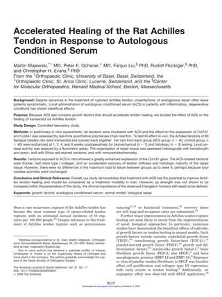
- 4. 2120 Majewski et al The American Journal of Sports Medicine 220°C until testing. On the day of testing the specimens were thawed in RingerÕs solution for 4 hours. Before testing, tendon thickness was measured with a precision caliper (Digital Caliper, Tesa, Lausanne, Switzerland) at the former site of tendon repair. The muscle-tendon-bone units were fastened in the clamping device by freezing the muscular segment between the cryojaws and by fixing the bony segment between the copper clamp.37 The clamping device was attached to an electrohydraulic materials testing machine (Fa. Zwick, Einsingen-Ulm, Germany). The tendons were not preconditioned or cyclically stretched before tensile testing. The room temperature was kept constant at 25°C. To prevent the specimens from dehydrating they were kept moist with RingerÕs solution. Both reservoirs of the cryojaws were filled with liquid nitrogen. The experiment was started as soon as the expansion of the freezing zone reached the border of the metal clamp but did not extend into the tendon substance or into the repair site (registered manually with a metal needle). The displacement rate was set at 1000 mm/min. Force-displacement curves were recorded and transferred to an IBM-compatible computer (Hewlett Packard, Berlin, Germany) for subsequent data analysis. Load to failure (N) and stiffness (N/mm) were measured. Lysyl Oxidase Activity Twelve Achilles tendons of the left, unoperated leg were collected for the determination of lysyl oxidase activity. Tendons were explanted (6 per group) and cultured for 7 days in 1 mL of DMEM 1 0.2% FBS with and without 10% ACS for 7 days. The activity of lysyl oxidase was determined according to the peroxidase-coupled fluorometric assay of Trackman et al.32 Tendon samples were homogenized in extraction buffer consisting of 0.15 M NaCl and 16 mM potassium phosphate, pH 7.8. The samples were centrifuged at 10 000 3 g for 20 minutes. The pellets were extracted with 3 volumes of 4 M urea and 16 mM potassium phosphate, pH 7.8. After centrifugation as noted, lysyl oxidase was batch-extracted with diethylaminoethyl cellulose. Samples were allowed to adsorb to the resin for 30 minutes with gentle mixing. The resin was then washed with 4 M urea and 16 mM potassium phosphate buffer, pH 7.8. The bound lysyl oxidase was eluted with 0.3 mL of 0.5 M NaCl, 4 M urea, and 16 mM potassium phosphate buffer. The fluorometric assay was conducted on an Aminco Bowman Series 2 Luminescence spectrophotometer (PerkinElmer, Waltham, Massachusetts) at 55°C with constant stirring. To the assay solution containing 0.25 mg of sodium homovanillate, 40 lg of horseradish peroxidase, 1.2 M urea, in 1.9 mL of 0.05 M sodium borate buffer, pH 8.2, a 100-lL test sample was added. Reactions were started by addition of 0.1 mL of a 50 mM solution of diaminopentane substrate solution. Fluorescence changes were monitored continuously for 10 minutes at excitation and emission wavelengths of 315 and 425 nm, respectively. The slope of the fluorescence change is proportional to the lysyl oxidase activity in the test solution. Fluorescence changes were completely inhibited by the lysyl oxidase inhibitor b-aminopropionitrile fumarate (2 3 1024 M). Statistical Analysis One-tailed Student t tests with Kolmogorov-Smirnov test for assessing normal distribution of values was performed using GraphPad InStat version 3.05 (GraphPad Software, San Diego, California). The level of significance was set to P .05 RESULTS In Vitro Data Effect of Conditioning on Growth Factor Content in Rat Conditioned Serum. Conditioning of rat blood in Orthokine syringes for 9 hours significantly increased TGF-b1 levels 1.4-fold from 23 030 to 32 450 pg/mL (P 5 .0122). The PDGF-BB (7550 pg/mL, basal), 7980 pg/mL in ACS were essentially unaffected, and increases in VEGF (21 pg/mL basal to 28 pg/mL, P 5 .0476) were small. Effect of Conditioned Serum on Collagen mRNA Expression. Incubation of Achilles tendons in vitro in ACS for 7 days caused substantial increases in Col1A1 and Col3A1 mRNA expression over levels in untreated tendons. The expression of Col1A1 mRNA was more than 5 times higher than that of Col 3A1 (Figure 1). In Vivo Data—Effect of ACS on Achilles Tendon Healing Histology and Immunohistochemistry. Sirius red staining did not reveal differences in collagen type deposition at weeks 1 and 4, but a distinct increase of thicker mature red-orange fibers was apparent at weeks 2 and 8 in tendons from animals with sutured tendons receiving ACS injections (Figures 2A and 3B). This finding is supported by the semiquantitative immunohistochemical analysis with a collagen type IIIspecific antibody. In ACS-treated animals, the collagen III content at weeks 1, 2, and 8 was approximately onethird that of animals that did not receive ACS injections (Figure 2B). Staining with hematoxylin and eosin revealed a much more rapid recovery of the normal histologic appearance of the tendon in response to ACS (Figure 3A). In the control group at week 1, cells were abundant and interlaced with adipose deposits. Occasional macrophages and fibroblastic elements were also apparent. Capillarization was distinct and some collagen fibrils were already bundled and properly aligned. At week 2, collagen deposition was markedly increased but tension fibers were not yet apparent. In addition to occasional fibrocytes, many fibroblasts were apparent. At week 4, thick fibers that often ran parallel to each other were present. Macrophages in close proximity to the sutures were also observed. At week 8, collagen crimp started to synchronize within big collagen bundles. In comparison with normal tendons, however, there were still more fibrocytes between the collagen fibers. Downloaded from ajs.sagepub.com by guest on December 14, 2009
- 5. Vol. 37, No. 11, 2009 Effect of ACS on Healing of Achilles Tendon Rupture 2121 Relative Quantity (Log10) 2.5 Control ACS 2.0 1.5 1.0 Ratio: red-orange / pale green A 3.0 0.5 10 8 6 4 Suture Suture with ACS 2 0 0 2 4 6 8 Time (weeks) 0.0 COL III Figure 1. Effect of autologous conditioned serum (ACS) on Col1A1 and Col3A1 mRNA expression in rat tendons. Tendon explants (4 per group) were cultured for 7 days in 1 mL of DelbuccoÕs Modified Eagle Medium 1 0.2% fetal bovine serum with and without 10% ACS for 7 days. The RNA was extracted for real-time polymerase chain reaction analysis of collagen and GAPDH expression. Marked increases of Col1A1 and Col3A1 mRNA levels in ACS exposed (filled bars) versus control (open bars) tendons were found. The mRNA quantities relative to GAPDH are depicted in log10 units. In tendons of ACS-treated rats, collagen fibers were apparent as early as week 1 postoperatively. These collagen fibers were visibly thicker and more bundled than in tendons of animals that did not receive ACS injections. This trend toward better organized collagen continued in week 2, at which time bundled collagen became apparent. Numerous fibrocytes but also some fibroblasts were found between the collagen fibrils. Four weeks postoperatively, there was a distinct overall tendonlike appearance with thick collagen bundles and cells oriented along the tension fibers, but occasional vascular conglomerates and fibrocytes were still present and around the sutures some macrophages could still be seen. At 8 weeks postoperatively, the tendons had a smooth appearance resembling very much normal tendons with small fibrocartilaginous areas in the center of the stress areas (Figure 3A). Tendon Thickness and Biomechanical Properties. The ACS treatment caused consistent increases in tendon thickness at the site of healing (Figure 4). Tendons of rats treated with ACS reached stiffness values 4 weeks earlier than the group that was not receiving ACS injections (Figure 5A). On the other hand, there were no differences in maximum load to failure between groups up to week 8 (Figure 5B). As judged from the continued increased thickness of ACS-treated tendons, remodeling of repair tissue is delayed with ACS treatment. Activity of Lysyl Oxidase. The activity of lysyl oxidase in Achilles tendons obtained from control and ACS-treated tendon in vitro as measured by the slopes of fluorescence B Collagen III (%) COL I 60 40 Suture Suture with ACS 20 0 0 2 4 6 8 Time (weeks) Figure 2. Autologous conditioned serum (ACS) treatment increases collagen I content in the healing Achilles tendon. Sirius red staining and polarization microscopy (A) and staining with collagen III-specific antibodies (B). s, mean 6 standard deviation of values in animals with sutured tendon and no ACS injections; , sutured tendon with ACS injections. development was not significantly different (control, 0.04115 per minute; ACS, 0.0368 per minute; P 5 .7579). DISCUSSION Patients suffering from complications after Achilles tendon rupture and suturing are common in clinical practice. The imperfections of endogenous repair often leave patients symptomatic and delay the time for return to work and sports activities. Because surgical treatment and posttreatment regimens are already refined, further major improvement of Achilles tendon healing is likely to come from the use of biological approaches, such as the administration of appropriate growth factors. Downloaded from ajs.sagepub.com by guest on December 14, 2009
- 6. 2122 Majewski et al The American Journal of Sports Medicine Figure 3. Effect of autologous conditioned serum (ACS) treatment on the histologic appearance of the repair site in the rat Achilles tendon. A, sections were made from tendons recovered 1, 2, 4, and 8 weeks after surgery (week 1 at top, week 8 at bottom) and stained with hematoxylin and eosin. Repair tissue formed with ACS injections (right) appears more structured, with a more developed crimp pattern, bigger collagen bundles than without (left). Sutured-only tissue shows more fibroblasts, fat cells, and blood vessels (left). Magnification 3200. B, sections were made from tendons recovered 1, 2, 4, and 8 weeks after surgery (week 1 at top, week 8 at bottom) and stained with Sirius red. Sirius red staining did not reveal differences in collagen type deposition at weeks 1 and 4, but a distinct increase of thicker mature red-orange fibers was apparent at weeks 2 and 8 in tendons from animals with sutured tendons receiving ACS injections (right) than those without ACS (left). Magnification 3400. Disturbances of the tendon structure lead to considerable loss of function in the lower extremities. To fulfill its function, the Achilles tendon has special morphologic properties: the helical Achilles tendon is relatively thick and collagenrich with a distinct parallel orientation of the collagen fibrils. This arrangement renders the tendon one of the most stress-resistant tissues in the human body. It is thus significant that, in our in vitro study, ACS supplementation of tendons increased markedly the expression of mRNA coding for collagen type I and collagen type III. The expression of type I collagen was increased to a greater degree than that of type III collagen, which is consistent with the in vivo findings. This explains the improved stiffness at 4 weeks produced by ACS, because type I collagen contributes to greater tensile strength. Measurements at a later time point showed that remodeling is delayed as postulated. We speculate that this increased expression of mRNA encoding the a chains of collagen types I and III may result from conditioning of blood, which increased the concentration of TGF-b1 1.4-fold. Wright-Carpenter et al38,39 and Meijer et al26 reported a comparable 1.8-fold increase in TGF-b1 after conditioning of mouse serum. These authors documented an even larger 5.6-fold increase in FGF-2, a growth factor that we did not measure. The FGF injections have been shown to affect the initial events, cell proliferation and collagen type III expression, in a rat patellar tendon model without having a significant effect on ultimate stress.5 It is quite possible that the elevated concentration of TGF-b1 in ACS may have mediated part of the observed effects on tendon healing. This cytokine has pleiotropic effects on fibroblasts and has emerged as a pivotal Downloaded from ajs.sagepub.com by guest on December 14, 2009
- 7. Vol. 37, No. 11, 2009 Effect of ACS on Healing of Achilles Tendon Rupture 2123 Figure 4. Autologous conditioned serum (ACS) application causes consistent increases in tendon thickness of the healing Achilles tendon. Significance was reached at week 1, P 5 .0417; week 4, P 5 .0162; and week 8, P 5 .0125. s, mean 6 standard error of values in sutured-only animals; , values in sutured animals receiving ACS injections. mediator in tissue repair.27 Transforming growth factor-b1 stimulates production of matrix molecules such as collagen and fibronectin from several fibroblast cell lines11,12,33,34 and plays important roles in the healing process of injured tendon or ligament by directing fibroblast migration and secretion of extracellular matrix proteins.17,18,24,29,36 Stimulation of type I collagen mRNA expression by TGF-b1 was also documented by the studies in tissue culture of Ignotz et al.11 Our analysis of collagen types in tendons from ACS-treated animals showed a marked increase of collagen type I and a decrease of collagen type III. An increased content of type III collagen in the fibers would tend to reduce their tensile strength.13,14,20,25 Our investigation of collagen maturation and collagen type III deposition also showed differences between the control and treatment groups. The ACS-treated animals showed markedly more mature, thick reddish-orange collagen fibers at weeks 2 and 8 postoperatively as compared with controls. The quantitative evaluation of collagen type III showed a 3-fold reduced level of this collagen at weeks 1, 2, and 8 in ACS-treated animals compared with controls. This finding is in agreement with the reports of Aspenberg and Virchenko,2 who also documented a beneficial effect of platelet concentrate administration on the histologic appearance of the healing tendon, with a concomitant increased tendon callus strength and stiffness by about 30% after week 1, an improvement which persisted for as long as 3 weeks after the injection.39 Despite the improved collagen synthesis, which is apparent from the significantly greater tendon thickness at weeks 1, 4, and 8 (Figure 4), stiffness was significantly (P 5 .038) increased at week 4 only. Figure 5. Effect of autologous conditioned serum (ACS) injections on Achilles tendon stiffness (A) and maximum load to failure (B). Shown are median values, 95% confidence interval, as well as the full range of individual values. A significant difference in tendon stiffness was apparent 4 weeks postoperatively (P 5 .038). Maximum load to failure was comparable in control and ACS-treated animals at all time points. In summary, it is clearly evident that ACS injections accelerate the rate of organization of repair tissue (Figure 3); they increase collagen mRNA expression (Figure 1), collagen deposition as reflected by tendon thickness (Figure 4), and accelerate collagen fiber maturation (Figure 2). Tendon stiffness does not improve significantly over control until week 4 (Figure 5), likely because of the time required for collagen cross-linking and remodeling of the repair site to occur. The fact that repair tissue remodeling Downloaded from ajs.sagepub.com by guest on December 14, 2009
- 8. 2124 Majewski et al The American Journal of Sports Medicine is a slow process is evidenced by the finding that tendon thickness remained elevated even at week 8 (Figure 4). Tendon thickness should decrease with longer healing times, an aspect we did not address with this study. Clinically, this could mean that ACS treatment shortens functional recovery times. One would not expect that new tendon tissue, which is still undergoing remodeling, should be stronger than the original normal tendon.1 The fact that ACS-aided tendon repair fills in well-organized, strong collagen fibers suggests that the healed tendon should contain fewer imperfections than in repair tissue formed following spontaneous healing or tendon suture. Such tissue would be expected to be less prone to rerupture. The biologically active component(s) in ACS that are responsible for the observed accelerated Achilles tendon healing remain to be defined. The ACS injections apparently do not critically limit inflammatory signals of the early healing phase but supplement critical growth factors that are present in serum in limiting concentrations. Overall, our study documents a beneficial effect of ACS administration on the collagen composition, histologic appearance, and mechanical strength of the tendon regenerates. In view of a possible clinical application of ACS in ruptured tendon healing in man, studies on the identity of growth factors and the dose-response relationship causing the observed accelerated healing as well as studies showing clinically important benefits in humans, such as early increase in strength, are indicated. ACKNOWLEDGMENT We are grateful to Mrs. H. Schaller and Mrs. C. Pilapil for histology preparation, Dr. H. Clahsen and Mrs. E. Krott for assistance during histology examination, and Associate Professor Dr. L. Durselen for help during biomechanical ¨ testing. We thank Dr. H. M. Kagan for his help with the lysyl oxidase activity determinations. This work was supported by a grant from the Swiss Society of Orthopaedic Surgery. REFERENCES 1. Anderson DD, Campbell PG, Guanche CA. The use of biological agents to accelerate recovery from rotator cuff repair: path to clinical application. Oper Tech Sports Med. 2002;10:58-63. 2. Aspenberg P, Virchenko O. Platelet concentrate injection improves Achilles tendon repair in rats. Acta Orthop Scand. 2004;75:93-99. 3. Becker C, Heidersdorf S, Drewlo S, de Rodriguez SZ, Kramer J, Willburger RE. Efficacy of epidural perineural injections with autologous conditioned serum for lumbar radicular compression: an investigatorinitiated, prospective, double-blind, reference-controlled study. Spine. 2007;32:1803-1808. 4. Brink HE, Miller GJ, Beredjiklian PK, Nicoll SB. Serum-dependent effects on adult and fetal tendon fibroblast migration and collagen expression. Wound Repair Regen. 2006;14:179-186. 5. Chan BP, Fu S, Qin L, Lee K, Rolf CG, Chan K. Effects of basic fibroblast growth factor (bFGF) on early stages of tendon healing: a rat patellar tendon model. Acta Orthop Scand. 2000;71:513-518. 6. Drissi H, Lomri A, Lasmoles F, Holy X, Zerath E, Marie PJ. Skeletal unloading induces biphasic changes in insulin-like growth factor-I mRNA levels and osteoblast activity. Exp Cell Res. 1999;251:275-284. 7. Enzura Y, Rosen V, Nifuji A. Introduction of hypertrophy in healing patella tendon by implantation of human recombinant BMP12. J Bone Min Res. 1996;11:401. 8. Forslund C, Rueger D, Aspenberg P. A comparative dose-response study of cartilage-derived morphogenetic protein (CDMP)-1, -2 and 3 for tendon healing in rats. J Orthop Res. 2003;21:617-621. 9. Frisbie DD, Kawcak CE, Werpy NM, Park RD, McIlwraith CW. Clinical, biochemical, and histologic effects of intra-articular administration of autologous conditioned serum in horses with experimentally induced osteoarthritis. Am J Vet Res. 2007;68:290-296. 10. Gabra N, Khayat A, Calabresi P, Khayat A. Detection of elevated basic fibroblast growth factor during early hours of in vitro angiogenesis using a fast ELISA immunoassay. Biochem Biophys Res Commun. 1994;205:1423-1430. 11. Ignotz RA, Endo T, Massague J. Regulation of fibronectin and type I collagen mRNA levels by transforming growth factor-beta. J Biol Chem. 1987;262:6443-6446. 12. Ignotz RA, Massague J. Transforming growth factor-beta stimulates the expression of fibronectin and collagen and their incorporation into the extracellular matrix. J Biol Chem. 1986;261:4337-4345. 13. Jozsa L, Balint BJ, Reffy A, Demel Z. Fine structural alterations of collagen fibers in degenerative tendinopathy. Arch Orthop Trauma Surg. 1984;103:47-51. 14. Jozsa L, Reffy A, Kannus P, Demel S, Elek E. Pathological alterations in human tendons. Arch Orthop Trauma Surg. 1990;110:15-21. 15. Junqueira LC, Bignolas G, Brentani RR. Picrosirius staining plus polarization microscopy, a specific method for collagen detection in tissue sections. Histochem J. 1979;11:447-455. 16. Junqueira LC, Cossermelli W, Brentani R. Differential staining of collagens type I, II and III by Sirius Red and polarization microscopy. Arch Histol Jpn. 1978;41:267-274. 17. Kashiwagi K, Mochizuki Y, Yasunaga Y, Ishida O, Deie M, Ochi M. Effects of transforming growth factor-beta 1 on the early stages of healing of the Achilles tendon in a rat model. Scand J Plast Reconstr Surg Hand Surg. 2004;38:193-197. 18. Leadbetter WB. Cell-matrix response in tendon injury. Clin Sports Med. 1992;11:533-578. 19. Liu SH, Yang RS, al Shaikh R, Lane JM. Collagen in tendon, ligament, and bone healing: a current review. Clin Orthop Relat Res. 1995; 318:265-278. 20. Maffulli N, Ewen SW, Waterston SW, Reaper J, Barrass V. Tenocytes from ruptured and tendinopathic Achilles tendons produce greater quantities of type III collagen than tenocytes from normal Achilles tendons: an in vitro model of human tendon healing. Am J Sports Med. 2000;28:499-505. 21. Majewski M, Rohrbach M, Czaja S, Ochsner P. Avoiding sural nerve injuries using percutaneous Achilles tendon repair. Am J Sports Med. 2006;34:793-798. 22. Majewski M, Schaeren S, Kohlhaas U, Ochsner P. Postoperative rehabilitation after percutaneous Achilles tendon repair: early functional therapy versus cast immobilization. Disabil Rehabil. 2008; 30:1726-1732. ¨ 23. Majewski M, Widmer KH, Steinbruck K. Achilles tendon ruptures: 25 yearsÕ experience in sport-orthopedic treatment [in German]. Sportverletz Sportschaden. 2002;16:167-173. 24. Marui T, Niyibizi C, Georgescu HI, et al. Effect of growth factors on matrix synthesis by ligament fibroblasts. J Orthop Res. 1997;15: 18-23. 25. Matthew CA, Moore MJ. Regeneration of rat extensor digitorum longus tendon: the effect of a sequential partial tenotomy on collagen fibril formation. Matrix. 1991;11:259-268. 26. Meijer H, Reinecke J, Becker C, Tholen G, Wehling P. The production of anti-inflammatory cytokines in whole blood by physico-chemical induction. Inflamm Res. 2003;52:404-407. 27. OÕKane S, Ferguson MW. Transforming growth factor beta and wound healing. Int J Biochem Cell Biol. 1997;29:63-78. Downloaded from ajs.sagepub.com by guest on December 14, 2009
- 9. Vol. 37, No. 11, 2009 Effect of ACS on Healing of Achilles Tendon Rupture 2125 28. Rickert M, Jung M, Adiyaman M, Richter W, Simank HG. A growth and differentiation factor-5 (GDF-5)-coated suture stimulates tendon healing in an Achilles tendon model in rats. Growth Factors. 2001;19:115-126. 29. Roberts AB, Flanders KC, Kondaiah P, et al. Transforming growth factor beta: biochemistry and roles in embryogenesis, tissue repair and remodeling, and carcinogenesis. Recent Prog Horm Res. 1988;44:157-197. 30. Suchak AA, Bostick G, Reid D, Blitz S, Jomha N. The incidence of Achilles tendon ruptures in Edmonton, Canada. Foot Ankle Int. 2005;26:932-936. 31. Tomak SL, Fleming LL. Achilles tendon rupture: an alternative treatment. Am J Orthop. 2004;33:9-12. 32. Trackman PC, Zoski CG, Kagan HM. Development of a peroxidasecoupled fluorometric assay for lysyl oxidase. Anal Biochem. 1981; 113:336-342. 33. Varga J, Jimenez SA. Stimulation of normal human fibroblast collagen production and processing by transforming growth factor-beta. Biochem Biophys Res Commun. 1986;138:974-980. 34. Varga J, Rosenbloom J, Jimenez SA. Transforming growth factor beta (TGF beta) causes a persistent increase in steady-state amounts of type I and type III collagen and fibronectin mRNAs in normal human dermal fibroblasts. Biochem J. 1987;247:597-604. 35. Wang XT, Liu PY, Tang JB. Tendon healing in vitro: genetic modification of tenocytes with exogenous PDGF gene and promotion of collagen gene expression. J Hand Surg Am. 2004;29: 884-890. 36. Wehling P, Moser C, Frisbie D, et al. Autologous conditioned serum in the treatment of orthopedic diseases: the OrthokineÒ therapy. BioDrugs. 2007;21:323-332. 37. Wieloch P, Buchmann G, Roth W, Rickert M. A cryo-jaw designed for in vitro tensile testing of the healing Achilles tendons in rats. J Biomech. 2004;37:1719-1722. 38. Wright-Carpenter T, Klein P, Schaferhoff P, Appell HJ, Mir LM, Wehling P. Treatment of muscle injuries by local administration of autologous conditioned serum: a pilot study on sportsmen with muscle strains. Int J Sports Med. 2004;25:588-593. 39. Wright-Carpenter T, Opolon P, Appell HJ, Meijer H, Wehling P, Mir LM. Treatment of muscle injuries by local administration of autologous conditioned serum: animal experiments using a muscle contusion model. Int J Sports Med. 2004;25:582-587. 40. Zhang F, Liu H, Stile F, et al. Effect of vascular endothelial growth factor on rat Achilles tendon healing. Plast Reconstr Surg. 2003;112: 1613-1619. For reprints and permission queries, please visit SAGE’s Web site at http://www.sagepub.com/journalsPermissions.nav Downloaded from ajs.sagepub.com by guest on December 14, 2009