
Recommended
Recommended
More Related Content
What's hot
What's hot (20)
Viewers also liked
Viewers also liked (20)
Similar to Epistaxis new
Similar to Epistaxis new (20)
Recently uploaded
Recently uploaded (20)
Epistaxis new
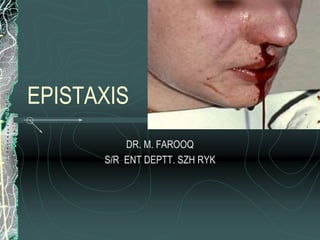
- 1. EPISTAXIS DR. M. FAROOQ S/R ENT DEPTT. SZH RYK
- 3. EPISTAXIS Definition Bleeding from nose Greek word- Epistazo ( Epi + Stazo) Epi - Over / above Stazo- To drip (from nostrils)
- 7. BLOOD SUPPLY Arterial supply Ext. Carotid A Int. Carotid A Venous Drainage Facial V Pterygoid Plexus Ant. & Post. Ethmoidal Veins
- 8. External Carotid Artery -Sphenopalatine artery -Greater palatine artery -Ascending pharyngeal artery -Posterior nasal artery -Superior Labial artery Internal Carotid Artery -Anterior Ethmoid artery -Posterior Ethmoid artery
- 9. BLOOD SUPPLY
- 10. BLOOD SUPPLY
- 11. BLOOD SUPPLY
- 12. BLOOD SUPPLY
- 13. EPIDEMIOLOGY 30% of ENT Admission Age Sex Season Area / Region
- 14. CLASSIFICATION On the basis of Etiology - Primary /Secondary Age - Children / Adult Site - Ant. / Post.
- 15. Anterior vs. Posterior Maxillary sinus ostium Anterior: younger, usually septal vs. anterior ethmoid, most common (>90%), typically less severe Posterior: older population, usually from Woodruff’s plexus, more serious.
- 16. CAUSES OF EPISTAXIS Local Causes General / systemic Causes Idiopathic Causes
- 19. GENERAL CAUSES Cardiovascular System HTN, Mitral stenosis, Pregnancy. Disorders of Blood & blood Vessels Aplastic Anaemia, Leukaemia, Thrombocytopaenias, Vascular Purpura, Haemophilia, Scurvy, Vit K Defficiency.
- 20. GENERAL CAUSES Liver Disease - Cirrhosis Kidney Disease- Ch. Nephritis Drugs- NSAIDS, Anticoagulants (Warfarin) Mediastinal Compression Accute General infections- Measles, Chicken pox.
- 21. MANAGEMENT Aims of Management To stop blood loss To replace blood loss To find out the cause and treat it
- 22. MANAGEMENT MANAGEMENT – Diagnosis+Treatment Diagnosis History Examination Investigations CBC Bleeding & clotting profiles Radiology - Angiography
- 23. MANAGEMENT Treatment -Hierarchy of treatment General Measures Direct Therapy - Primary Epistaxis Indirect Therapy - Secondary Epistaxis Surgical Options - Sec. Epistaxis
- 24. MANAGEMENT PLAN
- 25. Initial Management ABC’s Medical history/Medications Vital signs—need IV? Physical exam Anterior rhinoscopy Endoscopic rhinoscopy Laboratory exam Radiologic studies
- 26. Non-surgical treatments Control of hypertension Correction of coagulopathies/thrombocytopenia FFP or whole blood/reversal of anticoagulant/platelets Pressure/Expulsion of clots Topical decongestants/vasocontrictors Cautery (AgNo3 , Bipolar) Nasal packing (effective 80-90% of time)
- 27. Nasal packs Anterior nasal packs Traditional Recent modifications Posterior nasal packs Traditional Recent modifications Ant/Post nasal packing
- 28. TSS—Nugauze vs. Merocel Electron microscopy
- 29. Posterior Packs – Admission Elderly and those with other chronic diseases may need to be admitted to the ICU Continuous cardiopulmonary monitoring Antibiotics Oxygen supplementation may be needed Mild sedation/analgesia IVF
- 30. Indications for surgery/embolization Continued bleeding despite nasal packing Pt requires transfusion/admit hct of <38% (barlow) Nasal anomaly precluding packing Patient refusal/intolerance of packing Posterior bleed vs. failed medical mgmt after >72hrs (wang vs. schaitkin)
- 31. Selective Angiography/embolization Helps identify location of bleeding Embolization most effective in patients who Still bleeding after surgical arterial ligation Bleeding site difficult to reach surgically Comorbidities prohibit general anesthetic Effective only when bleeding is >.5 ml/min 90+% success rate, complication rate of 0.1% Only able to embolize external carotid & branches Complications: minor (18-45%)/major (0-2%) Contraindicated in bad atherosclerosis, Ethmoid bleed
- 32. Surgical treatment Transmaxillary IMA ligation Intraoral IMA ligation Anterior/Posterior Ethmoidal ligation Transnasal Sphenopalatine ligation External carotid artery ligation Septodermoplasty/Laser ablation
- 33. Transmaxillary IMA ligation Waters view Caldwell-Luc Electrocautery of posterior wall before removal Microscopic dissection and ligation of IMA -- descending palatine & sphenopalantine most important Recurrence rate (failure rate) of 10-15% Complication rate of 25-30% (oa fistula,dental, n)
- 34. Intraoral IMA ligation Posterior gingivobuccal incision beginning at second molar Temporalis mm split and partially dissected IMAX visualized, clipped and divided Advantages: children/facial fractures Disadvantages: more proximal ligation Complications: trismus, damage to infraorbital n
- 35. Ant./Post. Ethmoidal ligation Patients s/p IMAX ligation still bleeding, superior nasal cavity epistaxis, or in conjunction when source unclear Lynch incision Fronto-ethmoid suture line 12-24-6 (14-18, 8-10, 4-6)
- 36. Transnasal Endoscopic Sphenopalatine Artery ligation Follow Middle Turbinate to posteriormost aspect Vertical mucoperiosteal incision 7-8mm anterior to post middle turb (between mid. and inf. turbs) Elevation of flap—ID neurovascular bundle at foramen Ligation with titanium clip Reapproximate flap Complications –few, Failures—0-13%