1. Pemphigus vulgaris and its subtypes
Pemphigus is derived from the Greek word pemphix meaning bubble or blister. Pemphigus describes a group of
chronic bullous diseases, originally named by Wichman in 1791. The term pemphigus once included most
bullous eruptions of the skin, but diagnostic tests have improved, and bullous diseases have been reclassified.
The term pemphigus refers to a group of autoimmune blistering diseases of the skin and mucous membranes
characterized histologically by intraepidermal blister and immunopathologically by the finding of in vivo bound
and circulating immunoglobulin G (IgG) antibody directed against the cell surface of keratinocytes. The 3
primary subsets of pemphigus include pemphigus vulgaris, pemphigus foliaceus, andparaneoplastic pemphigus
Pemphigus vulgaris is an autoimmune, intraepithelial, blistering disease affecting the skin and mucous
membranes and is mediated by circulating autoantibodies directed against keratinocyte cell surfaces. In 1964,
autoantibodies against keratinocyte surfaces were described in patients with pemphigus.
Intercellular adhesion in the epidermis involves several keratinocyte cell surface molecules. Pemphigus
antibody binds to keratinocyte cell surface the molecules desmoglein 1 and desmoglein 3. The binding of
antibody to desmoglein may have a direct effect on desmosomal adherens or may trigger a cellular process that
results in acantholysis.
Patients with the mucocutaneous form of pemphigus vulgaris have pathogenic antidesmoglein 1 and
antidesmoglein 3 autoantibodies. Patients with the mucosal form of pemphigus vulgaris have only
antidesmoglein 3 autoantibodies. Patients with active disease have circulating and tissue-bound autoantibodies
of both the immunoglobulin G1 (IgG1) and immunoglobulin G4 (IgG4) subclasses.
History
• Mucous membranes: Pemphigus vulgaris presents with oral lesions in 50-70% of patients, and almost
all patients have mucosal lesions at some point in the course of their disease. Mucosal lesions may be
the sole sign for an average of 5 months before skin lesions develop, or they may be the sole
manifestation of the disease. The diagnosis of pemphigus vulgaris should be considered in any patient
with persistent oral erosive lesions.
• Skin: Most patients with pemphigus vulgaris develop cutaneous lesions. The primary lesion of
pemphigus vulgaris is a flaccid blister, which usually arises on healthy-appearing skin but may be found
on erythematous skin. New blisters usually are flaccid or become flaccid quickly. Affected skin often is
painful but rarely pruritic.
• Drug-induced pemphigus vulgaris11 : Drugs reported most significantly in association with pemphigus
vulgaris include penicillamine, captopril, cephalosporin, pyrazolones, nonsteroidal anti-inflammatory
drugs (NSAIDs), and other thiol-containing compounds. Rifampin, emotional stress, thermal burns,
ultraviolet rays, and infections (eg, coxsackievirus, Herpesviridae family) have also been reported as
triggers for pemphigus vulgaris.12
Physical
Mucous membranes typically are affected first in pemphigus vulgaris. Mucosal lesions may precede cutaneous
lesions by weeks or months. Patients with mucosal lesions may present to dentists, oral surgeons, or
gynecologists.13
• Mucous membranes
• Intact bullae are rare in the mouth. More commonly, patients have ill-defined, irregularly
shaped, gingival, buccal, or palatine erosions, which are painful and slow to heal. The erosions
extend peripherally with shedding of the epithelium.
• The mucous membranes most often affected in pemphigus vulgaris are those of the oral cavity,
which is involved in almost all patients with pemphigus vulgaris and sometimes is the only area
involved. Erosions may be seen on any part of the oral cavity. Erosions can be scattered and
often are extensive. Erosions may spread to involve the larynx, with subsequent hoarseness.
The patient often is unable to eat or drink adequately because the erosions are so
uncomfortable.
2. • In juvenile pemphigus vulgaris, stomatitis is the presenting complaint in more than 50% of the
cases.
• Other mucosal surfaces may be involved, including the conjunctiva,14 esophagus (causes
odynophagia and/or dysphagia),15 labia, vagina, cervix, vulva,16 penis, urethra, nasal mucosa,
and anus.
• Skin
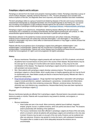
• The primary lesion of pemphigus vulgaris is a flaccid blister filled with clear fluid that arises on
healthy skin or on an erythematous base, as shown in the images below.
•
Early, small blister filled with clear fluid arises on healthy
skin.
•
Flaccid blister filled with clear fluid arises on healthy skin.
• The blisters are fragile; therefore, intact blisters may be sparse. The contents
soon become turbid, or the blisters rupture, producing painful erosions,
which is the most common skin presentation and is shown in the image
below. Erosions often are large because of their tendency to extend
peripherally with the shedding of the epithelium.
•
An erosion.
Vegetating pemphigus vulgaris: Ordinary pemphigus vulgaris erosions may develop
•
vegetation. Lesions in skin folds readily form vegetating granulations. In some
patients, erosions tend to develop excessive granulation tissue and crusting, and
these patients display more vegetating lesions. This type of lesion tends to occur
more frequently in intertriginous areas and on the scalp or face. The vegetating type
of response can be more resistant to therapy and can remain in one place for long
periods.
• Nails: Acute or chronic paronychia, subungual hematomas, and nail dystrophies affecting one or
several fingers or toes have been reported with pemphigus vulgaris.17,18 Patients with paronychial
pemphigus usually also have oral involvement.
3. • Pemphigus in pregnancy: Pemphigus vulgaris occurring in pregnancy is rare. When present, maternal
autoantibodies may cross the placenta, resulting in neonatal pemphigus. Neonatal pemphigus is
transient and improves with clearance of maternal autoantibodies.19 Treatment of pemphigus vulgaris
in pregnancy is with oral corticosteroids; however, prednisone and its metabolites cross the placenta
and have been associated with low birth weight, prematurity, infection, and adrenal insufficiency.
• Nikolsky sign: In patients with active blistering, firm sliding pressure with a finger separates normal-
appearing epidermis, producing an erosion. This sign is not specific for pemphigus vulgaris and is
found in other active blistering diseases.
• Asboe-Hansen sign: Lateral pressure on the edge of a blister may spread the blister into clinically
unaffected skin.
Age: Peak age of onset is from 50-60 years. Infants with neonatal pemphigus remit with clearance of
maternal autoantibodies. The disease may develop in children or in older persons.
• Disease association: Pemphigus occurs in patients with other autoimmune diseases,
particularly myasthenia gravis and thymoma.
Treatment: Prednisone
May decrease inflammation by reversing increased capillary permeability and suppressing PMN activity.
Immunosuppressive agents
Useful adjuvants in patients with pemphigus vulgaris with generalized disease unresponsive to steroids and/or
other anti-inflammatory agents or in patients unable to tolerate prednisone.
Azathioprine (Imuran)
Antagonizes purine metabolism and inhibits synthesis of DNA, RNA, and proteins.
4. 3. Erythema multiforme, Stevens Johnson
syndrome, Lyell’s syndrome
Erythema multiforme is a skin condition of unknown cause, possibly mediated by deposition of immune
complex (mostly IgM) in the superficial microvasculature of the skin and oral mucous membrane that usually
follows an infection or drug exposure. It is a common disorder, with peak incidence in the second and third
decades of life.
The condition varies from a mild, self-limited rash (E. multiforme minor)[1] to a severe, life-threatening form
known as erythema multiforme major (or erythema multiforme majus) that also involves mucous
membranes. This severe form may be related to Stevens-Johnson syndrome. The mild form is far more
common than the severe form. Diagnosis is confirmed by biopsy.
The mild form usually presents with mildly itchy, pink-red blotches, symmetrically arranged and starting on the
extremities. It often takes on the classical "target lesion" appearance,[2] with a pink-red ring around a pale
center. Resolution within 7–10 days is the norm.
Individuals with persistent (chronic) erythema multiforme will often have a sore form at an injury site, eg. a
minor scratch or abrasion, within a week. Irritation or even pressure from clothing will cause the erythema sore
to continue to expand along its margins for weeks or months, long after the original sore at the center heals.
The most common predisposing infection is Herpes simplex, but bacterial infections (commonly Mycoplasma)
and fungal diseases are also implicated. It has been shown that Herpes simplex virus suppression and even
prophylaxis (with acyclovir) can prevent recurrent erythema multiforme eruption.[3]
Other causes include drug reactions, most commonly to sulfa drugs, phenytoin, barbiturates, penicillin,
and allopurinol, or a host of internal ailments.
Stevens–Johnson syndrome (SJS) and toxic epidermal necrolysis (TEN)[1] are two forms of a life-
threatening condition affecting the skin in which cell death causes theepidermis to separate from the dermis.
The syndrome is thought to be a hypersensitivity complex affecting the skin and the mucous membranes.
Although the majority of cases are idiopathic, the main class of known causes is medications, followed by
infections and (rarely) cancers.
There is agreement in the medical literature that Stevens–Johnson syndrome (SJS) can be considered a milder
form of toxic epidermal necrolysis (TEN). These conditions were first recognised in 1922
Both diseases can be mistaken for erythema multiforme. Erythema multiforme is sometimes caused by a
reaction to a medication but is more often a type III hypersensitivity reaction to an infection (caused most often
by Herpes simplex) and is relatively benign. Although both SJS and TEN can also be caused by infections, they
are most often adverse effects of medications. Their consequences are potentially more dangerous than those
of erythema multiforme.
SJS usually begins with fever, sore throat, and fatigue, which is misdiagnosed and usually treated with
antibiotics. Ulcers and other lesions begin to appear in the mucous membranes, almost always in the mouth
and lips but also in the genital and anal regions. Those in the mouth are usually extremely painful and reduce
the patient's ability to eat or drink. Conjunctivitis of the eyes occurs in about 30% of children who develop SJS.
A rash of round lesions about an inch across arises on the face, trunk, arms and legs, and soles of the feet, but
usually not the scalp
SJS is thought to arise from a disorder of the immune system
It can be caused by infections (usually following infections such as herpes simplex virus, influenza, mumps, cat-
scratch fever, histoplasmosis, Epstein-Barr virus,mycoplasma pneumoniae or similar).
Although Stevens–Johnson Syndrome can be caused by viral infections, malignancies or severe allergic
reactions to medication, the leading cause appears to be the use of antibiotics and sulfa drugs.
Medications that have traditionally been known to lead to SJS, erythema multiforme and toxic epidermal
necrolysis
5. include sulfonamides (antibiotics), penicillins (antibiotics), barbiturates (sedatives), lamotrigine andphenytoin (e.
g. Dilantin) (anticonvulsants). Combining lamotrigine with sodium valproate increases the risk of SJS.
SJS constitutes a dermatological emergency. All medications should be discontinued, particularly those known
to cause SJS reactions.
Initially, treatment is similar to that for patients with thermal burns, and continued care can only be supportive
(e.g. intravenous fluids and nasogastric or parenteral feeding) and symptomatic (e.g. analgesic mouth rinse
for mouth ulcer). Dermatologists and surgeons tend to disagree about whether the skin should be debrided.[3]
Beyond this kind of supportive care, there is no accepted treatment for SJS. Treatment with corticosteroids is
controversial.
Toxic epidermal necrolysis (also known as "Lyell's syndrome"[1]) is a rare, life-
threatening dermatological condition that is usually induced by a reaction to medications.[2] It is characterized
by the detachment of the top layer of skin (the epidermis) from the lower layers of the skin (the dermis) all over
the body.
There is broad agreement in medical literature that TEN can be considered a more severe form of Stevens-
Johnson syndrome, and debate whether it falls on a spectrum of disease that includes erythema multiforme
TEN affects many parts of the body, but it most severely affects the mucous membranes, such as
the mouth, eyes, and vagina. The severe findings of TEN are often preceded by 1 to 2 weeks of fever. These
symptoms may mimic those of a common upper respiratory tract infection. When the rash appears it may be
over large and varied parts of the body, and it is usually warm and appears red. The dermal layer fills with fluid
being deposited there by the body's immune system, usually as a result of a negative reaction to an antibiotic.
The skin then begins to sag from the body and can be peeled off in great swaths. The mouth becomes blistered
and eroded, making eating difficult and sometimes necessitating feeding through a nasogastric tube through the
nose or a gastric tube directly into the stomach. The eyes are affected, becoming swollen, crusted, and
ulcerated and blindness may occur.
Toxic epidermal necrolysis is a rare and usually severe adverse reaction to certain drugs. History of medication
use exists in over 95% of patients with TEN.[2] The drugs most often implicated in TEN are antibiotics such as
sulfonamides, nonsteroidal anti-inflammatory drugs, allopurinol, antimetabolites (methotrexate), antiretroviral
drugs, corticosteroids, chlormezanone (anxiolytic) and anticonvulsants such
as phenobarbital,phenytoin, carbamazepine, and valproic acid.[2]
The condition might also result from infection with agents such as Mycoplasma pneumoniae or the herpes
virus; and transplants of bone marrow or organs
Microscopically, TEN causes cell death throughout the epidermis. Keratinocytes, which are the cells found
lower in the epidermis, specializing in holding the skin cells together, undergo necrosis (cell death).
Often, the diagnosis can be made clinically. Generally, if the clinical history is consistent with Stevens-Johnson
syndrome, and the skin lesion covers greater than 30% of the body surface area, the diagnosis of TEN is
appropriate. Sometimes, however, examination of affected tissue under the microscope may be needed to
distinguish it between other entities such as staphylococcal scalded skin syndrome. Typical histological criteria
of TEN include mild infiltrate of lymphocytes which may obscure the dermoepidermal junction and prominent
cell death with basal vacuolar change and individual cell necrosis.[5]
Nikolsky's sign is almost always present in toxic epidermal necrolysis
The first line of treatment is early withdrawal of culprit drugs, early referral and management in burn
units or intensive care units, supportive management, and nutritional support.
The second line is Intravenous immunoglobulin (IVIG)
6. The mortality for toxic epidermal necrolysis is 30-40 per cent.[2] Loss of the skin leaves patients vulnerable to
infections from fungi and bacteria, and can result in sepsis, the leading cause of death in the disease.[2]Death
is caused either by infection or by respiratory distress which is either due to pneumonia or damage to the
linings of the airway
7. 6. Erysipelas
Erysipelas is a superficial bacterial skin infection that characteristically extends into the cutaneous lymphatics.
Historically, erysipelas occurred on the face and was caused by Streptococcus pyogenes.However, a shift in the
distribution and etiology of erysipelas has occurred, with most erysipelas infections now occurring on the legs
and with non–group A streptococci sometimes being identified as the etiologic agents.
Bacterial inoculation into an area of skin trauma is the initial event in developing erysipelas. Thus, local factors,
such as venous insufficiency, stasis ulcerations, inflammatory dermatoses, dermatophyte infections, insect
bites, and surgical incisions, have been implicated as portals of entry. The source of the bacteria in facial
erysipelas is often the host's nasopharynx, and a history of recent streptococcal pharyngitis has been reported
in up to one third of cases. Other predisposing factors include diabetes, alcohol abuse,1 HIV infection,
nephrotic syndrome, other immunocompromising conditions, and vagrant lifestyle.
Preexisting lymphedema is a clear-cut risk factor for erysipelas.
Patients often cannot recall an inciting event, but a history of recent trauma or pharyngitis may be elicited.
Prodromal symptoms, such as malaise, chills, and high fever, often begin before the onset of the skin lesions
and usually are present within 48 hours of cutaneous involvement. Pruritus, burning, and tenderness are typical
complaints.
Physical
Erysipelas begins as a small erythematous patch that progresses to a fiery-red, indurated, tense, and shiny
plaque, as shown in the image below.
Facial erysipelas exhibiting classic fiery-red plaque with raised, well-demarcated
borders.
The lesion classically exhibits raised sharply demarcated advancing margins. Local signs of
inflammation, such as warmth, edema, and tenderness, are universal. Lymphatic involvement
often is manifested by overlying skin streaking and regional lymphadenopathy. More severe
infections may exhibit numerous vesicles and bullae along with petechiae and even frank
necrosis.
Streptococci cause most cases of erysipelas; thus, penicillin has remained first-line
therapy.8,9 Penicillin administered orally or intramuscularly is sufficient for most cases of
classic erysipelas and should be given for 10-20 days.
• A first-generation cephalosporin or macrolide, such as erythromycin or azithromycin, may be used if the
patient has an allergy to penicillin. Cephalosporins may cross-react with penicillin and should be used
with caution in patients with a history of severe penicillin allergy such as anaphylaxis.
8. 10) Types Of Contact Dermatitis:
Contact dermatitis is a term for a skin reaction (dermatitis) resulting from exposure
to allergens (allergic contact dermatitis) or irritants (irritant contact dermatitis).Phototoxic dermatitis
occurs when the allergen or irritant is activated by sunlight.
Contact dermatitis is a localized rash or irritation of the skin caused by contact with a foreign
substance. Only the superficial regions of the skin are affected in contact dermatitis. Inflammation of
the affected tissue is present in the epidermis (the outermost layer of skin) and the outer dermis (the
layer beneath the epidermis).[1] Unlikecontact urticaria, in which a rash appears within minutes of
exposure and fades away within minutes to hours, contact dermatitis takes days to fade away. Even
then, contact dermatitis fades only if the skin no longer comes in contact with the allergen or irritant.
[2] Contact dermatitis results in large, burning, and itchy rashes, and these can take anywhere from
several days to weeks to heal. Chronic contact dermatitis can develop when the removal of the
offending agent no longer provides expected relief.
There are three types of contact dermatitis: irritant contact dermatitis, allergic contact dermatitis, and
photocontact dermatitis. Photocontact dermatitis is divided into two categories that is, phototoxic and
photoallergic.
Irritant contact dermatitis
Irritant contact dermatitis can be divided into forms caused by chemical irritants and those caused by
physical irritants. Common chemical irritants implicated include solvents (alcohol, xylene, turpentine,
esters,acetone, ketones, and others); metalworking fluids (neat oils, water-based metalworking fluids
with surfactants); latex; kerosene; ethylene oxide; surfactants in topical medications and cosmetics
(sodium lauryl sulfate); alkalies (drain cleaners, strong soap with lye residues). Physical irritant contact
dermatitis may most commonly be caused by low humidity from air conditioning.[5] Also,
many plants are directly irritating the skin.
Allergic contact dermatitis
Although less common than ICD, ACD (Allergic Contact Dermatitis) is accepted to be the most
prevalent form of immunotoxicity found in humans.[6] By its allergic nature, this form of contact
dermatitis is a hypersensitive reaction that is atypical within the population. The mechanisms by which
these reactions occur are complex, with many levels of fine control. Their immunology centres around
the interaction of immunoregulatory cytokines and discrete subpopulations of T lymphocytes. Allergens
include nickel, gold
Photocontact dermatitis
ivided into two categories, phototoxic and photoallergic, PCD is the eczematous condition which is
triggered by an interaction between an otherwise unharmful or less harmful substance on the skin and
ultraviolet light (320-400 nm UVA) therefore manifesting itself only in regions where the sufferer has
been exposed to such rays. Without the presence of these rays, the photosensitiser is not harmful.
For this reason, this form of contact dermatitis is usually associated only with areas of skin which are
left uncovered by clothing.
9. Allergic dermatitis is usually confined to the area where the trigger actually touched the skin, whereas
irritant dermatitis may be more widespread on the skin. Symptoms of both forms include the following:
• Red rash. This is the usual reaction. The rash appears immediately in irritant contact dermatitis; in
allergic contact dermatitis, the rash sometimes does not appear until 24–72 hours after exposure to the
allergen.
• Blisters or wheals. Blisters, wheals (welts), and urticaria (hives) often form in a pattern where skin was
directly exposed to the allergen or irritant.
• Itchy, burning skin. Irritant contact dermatitis tends to be more painful than itchy, while allergic contact
dermatitis often itches.
While either form of contact dermatitis can affect any part of the body, irritant contact dermatitis often affects the
hands, which have been exposed by resting in or dipping into a container (sink, pail, tub, swimming pools with
high chlorine) containing the irritant.
Self-care at home
• Immediately after exposure to a known allergen or irritant, wash with soap and cool water to remove or
inactivate most of the offending substance.
• Weak acid solutions [lemon juice, vinegar] can be used to counteract the effects of dermatitis
contracted by exposure to basic irritants.
• If blistering develops, cold moist compresses applied for 30 minutes 3 times a day can offer relief.
• Calamine lotion and cool colloidal oatmeal baths may relieve itching.
• Oral antihistamines such as diphenhydramine (Benadryl, Ben-Allergin) can also relieve itching.
• For mild cases that cover a relatively small area, hydrocortisone cream in nonprescription strength may
be sufficient.
• Avoid scratching, as this can cause secondary infections.
• A barrier cream such as those containing zinc oxide (e.g. Desitin, etc.) may help to protect the skin and
retain moisture.
Medical care
If the rash does not improve or continues to spread after 2-3 of days of self-care, or if the itching and/or pain is
severe, the patient should contact a dermatologist or other physician or physician assistant. Medical treatment
usually consists of lotions, creams, or oral medications.
• Corticosteroids. A corticosteroid medication similar to hydrocortisone may be prescribed to combat
inflammation in a localized area. This medication may be applied to your skin as a cream or ointment. If
the reaction covers a relatively large portion of the skin or is severe, a corticosteroid in pill or injection
form may be prescribed.
• Antihistamines. Prescription antihistamines may be given if nonprescription strengths are inadequate.
Since contact dermatitis relies on an irritant or an allergen to initiate the reaction, it is important for the
patient to identify the responsible agent and avoid it. This can be accomplished by having patch tests,
a method commonly known as allergy testing. The patient must know where the irritant or allergen is
found to be able to avoid it.
10. 11) Atopic Dermatitis
Atopic dermatitis (AD; a type of eczema) is an inflammatory, chronically relapsing, non-contagious
and pruritic skin disorder.
The skin of a patient with atopic dermatitis reacts abnormally and easily to irritants, food, and
environmental allergens and becomes red, flaky and very itchy. It also becomes vulnerable to surface
infections caused by bacteria. The skin on the flexural surfaces of the joints (for example inner sides of
elbows and knees) are the most commonly affected regions in people.
Atopic dermatitis often occurs together with other atopic diseases like hay fever, asthma and allergic
conjunctivitis. It is a familial and chronic disease and its symptoms can increase or disappear over time. Atopic
dermatitis in older children and adults is often confused with psoriasis. Atopic dermatitis afflicts humans,
particularly young children; it is also a well-characterized disease in domestic dogs.
Although there is no cure for atopic eczema, and its cause is not well understood, it can be treated very
effectively in the short term through a combination of prevention (learning what triggers the allergic reactions)
and drug therapy.
Atopic dermatitis most often begins in childhood before age 5 and may persist into adulthood. For some, it
flares periodically and then subsides for a time, even up to several years
Although atopic dermatitis can theoretically affect any part of the body, it tends to be more frequent on
the hands and feet, on the ankles, wrists, face, neck and upper chest. Atopic dermatitis can also affect
the skin around the eyes, including the eyelids
n most patients, the usual symptoms that occur with this type of dermatitis are aggravated by
a Staphylococcus aureus infection, dry skin, stress,
low humidity and sweating, dust or sand or cigarette smoke. Also, the condition can be worsened by
having long and hot baths or showers, solvents, cleaners or detergents and wool fabrics or clothing.
Atopic dermatitis is also known as infantile eczema, when it occurs in infants. Infantile eczema may continue
into childhood and adolescence and it often involves an oozing, crusting rash mainly on the scalp and face,
although it can occur anywhere on the body
Symptoms may vary from person to person but they are usually present as a red, inflamed, and itchy
rash and can quickly develop into raised and painful bumps.[8] The first sign of atopic dermatitis is the
red to brownish-gray colored patches that are usually very itchy. Itching may become more intense
during the night. The skin may present small and raised bumps which may be crusting or oozing if
scratched, which will also worsen the itch. The skin tends to be more sensitive and may thicken, crack
or scale.
When appearing in the area next to the eyes, scratching can cause redness and swelling around them and
sometimes, rubbing or scratching in this area causes patchy loss of eyebrow hair and eyelashes.[3]
The symptoms of atopic dermatitis vary with the age of the patients. Usually, in infants, the condition causes
red, scaly, oozy and crusty cheeks and the symptoms may also appear on their legs, neck and arms.
Symptoms clear in about half of these children by the time they are 2 or 3 years old.[9] In older children, the
symptoms include dry and thick, scaly skin with a very persistent itch, which is more severe than in infants.
Since there is no cure for atopic eczema, treatment should mainly involve discovering the triggers of
allergic reactions and learning to avoid them.
11. The primary treatment involves prevention, includes avoiding or minimizing contact with (or intake of)
known allergens. Once that has been established, topical treatments can be used. Topical treatments
focus on reducing both the dryness and inflammation of the skin.
To combat the severe dryness associated with atopic dermatitis, a high-quality, dermatologist-approved
moisturizer should be used daily. Moisturizers should not have any ingredients that may further aggravate the
condition. Moisturizers are especially effective if applied 5–10 minutes after bathing. As a rule of thumb the
thicker the moisturizer the better it is at retaining moisture. Petroleum jelly is considered one of the most
effective moisturizers by reducing transepidermal water loss by up to 98%
If moisturizers on their own don't help and the eczema is severe, a doctor may prescribe
topical corticosteroid ointments, creams, or injections. Corticosteroids have traditionally been
considered the most effective method of treating severe eczema. Disadvantages of using steroid
creams include stretch marks and thinning of the skin.
Atopy is an allergic hypersensitivity[1] affecting parts of the body not in direct contact with
the allergen. A familial predisposition to a localized reaction.
Atopy is a disease characterized by a tendency to be “hyperallergic”. Atopy is a word taken from the
Greek meaning “special” or “unusual”. A patient with atopic allergies has atopic eczema or atopic
dermatitis since infancy. Atopic eczema is an extremely itchy skin condition with a hallmark rash that
appears most often over the flexural regions (e.g., back of knees, crook of elbows) but can involve
almost every region of the body. Crusty, scaly, flattened, erythematous lesions of atopic eczema can
appear almost everywhere, but are worse in certain areas or after exposure to certain irritants or
allergens (e.g., washing hands with a perfumed or otherwise allergenic soap, wearing a wool or
scratchy sweater or skirt, rolling across freshly cut lawns). The single most important feature
associated with atopic eczema lesions is that they are extremely itchy, and the itch can occur even
before the lesions erupt on the skin and are visible.
It is localized immediate hypersensitivity reaction to an allergen. It may involve eczema (atopic
dermatitis [AD]), allergic conjunctivitis, allergic rhinitis and asthma. There appears to be a
strong hereditary component. One study concludes that "the general risk of developing AD (3%) and
atopy (7%) increases by a factor of two with each first-degree family member already suffering from
atopy."[5] Environmental factors are also known to play a major role and the 'hygiene hypothesis' is
one of the best paradigms available to date to explain the steep rise observed in atopic diseases. This
supports that it is the excess 'cleanliness' of our environments that has led to the decline in the
number of infectious stimuli that are necessary for the proper development of our immune system.
Patients with atopic eczema usually develop what is referred to as the “allergic triad” of symptoms i.e.,
eczema, hayfever, and asthma. They also have a tendency to have food allergies, and other
symptoms characterized by their hyperallergic state. For example, eosinophilic esophagitis is found
associated with atopic allergies. Atopy and atopic eczema can be considered a genetic disease
because of its strong genetic component, but atopy does not segregate like an autosomal dominant
trait. There are certain environmental factors that contribute to its appearance in infants and children,
but the underlying cause is a genetic tendency to be hyperallergic. Atopic eczema cannot be
prevented in infants because of its genetic origins.
Atopic syndrome can be fatal for those who experience serious allergic reactions, such as anaphylaxis, brought
on by reactions to food or environment.
The individual components are all caused at least in part by allergy (type I hypersensitivity reactions). These
responses appear after the body is exposed to various allergens, for example specific kinds of
food,pollen, dander or insect venoms. Although atopy has various definitions, most consistently it is defined by
the presence of elevated levels of total and allergen-specific IgE in the serum of patient, leading to positive
skin-prick tests to common allergens.
12. 22) Skin in Systemic disease
Thyrotoxicosis may lead to multiple cutaneous manifestations, including hair loss, pretibial myxedema,
onycholysis and acropachy. In patients with hypothyroidism, there is hair loss, the skin is cold and pale, with
myxedematous changes, mainly in the hands and in the periorbital region. The striking features of Cushing
syndrome are centripetal obesity, moon facies, buffalo hump, supraclavicular fat pads, and abdominal striae. In
Addison disease, the skin is hyperpigmented, mostly on the face, neck and back of the hands. Virtually all
patients with acromegaly have acral and soft tissue overgrowth, with characteristic findings, like macrognathia
and enlarged hands and feet. The skin is thickened, and facial features are coarser. Conditions leading to
hyperandrogenism in females present as acne, hirsutism and signs of virilization (temporal balding,
clitoromegaly).A prominent feature of hypopituitarism is a pallor of the skin with a yellowish tinge. The skin is
also thinner, resulting in fine wrinkling around the eyes and mouth, making the patient look older. Primary
hyperparathyroidism is rarely associated with pruritus and chronic urticaria. In hypoparathyroidism, the skin is
dry, scaly and puffy. Nails become brittle and hair is coarse and sparse. Pseudohypoparathyroidism may have
a special somatic phenotype known as Albright osteodystrophy. This consists of short stature, short neck,
brachydactyly and subcutaneous calcifications. Some of the cutaneous manifestations of diabetes mellitus
include necrobiosis lipoidica diabeticorum, diabetic dermopathy, scleredema adultorum and acanthosis
nigricans.
Dermatologic Manifestations of Hematologic Disease
http://emedicine.medscape.com/article/1096183-overview
13. 29) Paraneoplastic Diseases
http://emedicine.medscape.com/article/1095113-overview
A wide range of cutaneous signs may be related to internal malignancy. Cancer may manifest in
the skin as metastasis (eg, leukemia cutis, cutaneous T-cell lymphoma, Paget disease of the
breast), nonspecific metabolic effects related to inanition (eg, wasting, alopecia, xerosis), infections
related to immunosuppression (eg, herpes zoster), signs resulting from compromise or dysfunction
of the affected organ (eg, jaundice), or diverse dermatologic entities called paraneoplastic
syndromes, which signal that a remote malignancy is present. Cutaneous manifestations may
develop before a diagnosis of malignancy is determined; thus, these findings may aid the physician
in the early identification of malignancy.
Acanthosis nigricans (AN) manifests as a hyperpigmented, velvety thickening of the skin that
usually occurs in the intertriginous zones, including the axillae, groin, neck, and inframammary
folds. AN may develop in areas subjected to trauma, such as the extensor surfaces of the knees
and elbows. As many as 30% of patients also have papillomatous thickening of the oral mucosa.
The subset of patients with AN associated with malignancies also has skin changes involving the
scalp, areolae, and eyelids.
Dermatomyositis
DM is an inflammatory proximal myopathy with characteristic skin changes; it is often associated with an
occult malignancy. The rash is characteristic and diagnostic and usually accompanies or precedes the onset of
the myopathy.
Skin manifestations include the following:
• Heliotrope rash (so named because of the similarity to the color of the blue-purple flower, the
heliotrope) on the upper eyelids
• Periorbital edema
• A macular red rash on the face and the V of the upper trunk that may become shiny and atrophic,
with variable pigmentation and telangiectasias (poikiloderma)
• Violaceous scaly papules over the interphalangeal and metacarpophalangeal joints (ie, Gottron
papules) that evolve into atrophic telangiectatic macules
Sweet Syndrome
In 1964, Robert Sweet, MD, described a syndrome involving an acute onset of febrile neutrophilic
dermatosis in 8 women he encountered over a 15-year period. At the time, the classic patient was a middle-
aged woman in good general health whose skin lesions developed within days of an upper respiratory tract
infection or minor illness. While this entity came to be known by the eponym Sweet syndrome, it has now
been expanded to include older patients with an underlying internal malignancy. Fever, neutrophilia, and
sterile erythematous plaques or nodules that respond to steroid therapy manifest it. Skin lesions most
commonly involve the upper extremities and face and begin as tender, erythematous plaques or nodules. The
lesions may evolve into vesicles, bullae, or pustules. Extracutaneous manifestations are not infrequent and
commonly involve the eyes, lungs, liver, kidneys, muscles, and bones. Laboratory features include
neutrophilia, anemia, and an elevated erythrocyte sedimentation rate.
Diagnosis is based on the clinical presentation and characteristic findings at skin biopsy. Histologic
evaluation reveals a neutrophilic infiltrate in the dermis, without evidence of infection, vasculitis, or
malignant cells. Patients frequently have a positive perinuclear antineutrophil cytoplasmic antibody titer.
The clinical syndrome can mimic several other entities; the differential diagnoses include erythema
multiforme, cellulitis, and leukemia cutis. Sweet syndrome can usually be distinguished from these other
lesions on the basis of the biopsy results; however, patients are often initially treated for an infectious process
before the proper diagnosis is realized.
Paraneoplastic pemphigus a member of a heterogeneous group of autoimmune paraneoplastic
syndromes. It is characterized by painful, intractable erosive ulcerative stomatitis and a
polymorphic cutaneous eruption consisting of erythema, papules, iris lesions, bullae, and erosions.