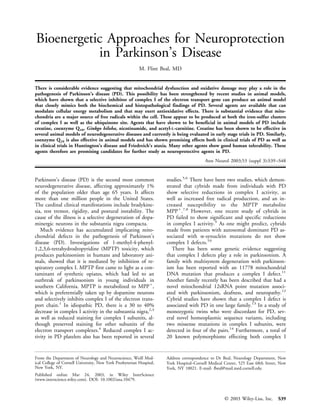
2. and transfer RNA mutations were found. Mitochon- Table. Bioenergetic Agents Effective in Parkinson’s Disease
drial DNA sequences, however, tended to be identical, Models
and the disease did not affect siblings of each pair. The
Agent Proposed Mechanism of Action
pathogenic relevance of several of these mutations
therefore is questionable. In addition, an out-of-frame Coenzyme Q10 Cofactor of complex I, II, III and anti-
cytochrome b gene deletion has been detected in a pa- oxidant
tient with parkinsonism that was associated with im- Creatine Increases PCr, inhibits the MPT
paired complex III assembly and an increase in free Ginkgo biloba Antioxidant and preserves mitochon-
drial function
radical production.15 Carnitine Facilitates fatty acid transport, increases
In a direct sequencing study of complex I in transfer repiration
RNA mutations, we recently observed no homoplasmic Nicotinamide Precursor of NADH, inhibitor of poly-
mutations, suggesting either that the observed complex ADP-ribose polymerase
I defects are caused by heteroplasmic mutations or that Lipoic acid Coenzyme for ␣-ketoglutarate dehydro-
genase, antioxidant
they may involve interactions between the nuclear ge-
nome and the environment.16 We also recently directly PCr ϭ creatine/phosphocreatine; MPT ϭ mitochondrial permeabil-
sequenced mitochondrial DNA from postmortem ity transition pore; NADH ϭ nicotinamide adenine dinucleotide.
brain tissue of neuropathologically confirmed PD pa-
tients.17 Once again, we did not detect any homoplas- with a loss of immunoreactivity for tyrosine hydroxy-
mic mitochondrial DNA mutations associated with lase, dopamine transporter, and vesicular monoamine
PD. This suggests that if mitochondrial DNA muta- transporter. Furthermore, the nigral neurons showed
tions play a role in PD, the pathogenetic effects may be cytoplasmic inclusions that were highly suggestive of
very complicated. It recently has been demonstrated Lewy bodies in that they stained with antibodies to
that nuclear background determines the biochemical ubiquitin and ␣-synuclein, and electron microscopy
phenotype of the deafness-associated mitochondrial 12s showed a dense core surrounded by fibrillar elements
RNA mutation.18 A nuclear mitochondrial DNA mu- similar to Lewy bodies. The rats showed bradykinesia,
tation affecting hearing impairment also has been dem- postural instability, unsteady gait, and some evidence
onstrated in mice.19 Furthermore, mitochondrial DNA of tremor that improved after treatment with the do-
variant susceptibility to dilated cardiomyopathy is dif- pamine agonist, apomorphine. These findings suggest
ferent in two different human populations.20 These that rotenone can produce a selective degeneration of
findings suggest that there are complex interactions be- nigrostriatal neurons consistent with the neuropatho-
tween the nuclear and mitochondrial DNA, and that logical and clinical manifestations of PD. They are re-
expression of a mitochondrial disease may occur only markable because they show that an inhibitor of com-
in selective nuclear DNA backgrounds. This may make plex I of the electron transport chain, which acts
the study of mitochondrial DNA defects in parkinson- uniformly throughout the brain, produces a selective
ism extremely complex. degeneration of nigrostriatal neurons. They therefore
A major finding suggesting that a complex I defect indicate the substantia nigra neurons are particularly
may play a critical role in the pathogenesis of PD susceptible to complex I inhibitors. This is consistent
comes from recent studies with the environmental with the findings of decreased complex I activity in PD
toxin rotenone. The possibility that pesticides and postmortem tissue and platelets. It has been suggested
other environmental toxins are involved in the patho- that the selective effects of rotenone may be mediated
genesis of PD is suggested by several epidemiological by oxidative damage. This is also consistent with prior
studies.21,22 Patients with certain glutathione trans- studies showing extensive oxidative damage in the sub-
ferase polymorphisms and exposure to pesticides seem stantia nigra of PD patients.
to have an increased incidence of PD.23 Furthermore, If mitochondrial defects and oxidative damage play a
an atypical PD syndrome has been described in associ- role in the pathogenesis of PD, then one would suspect
ation with the consumption of fruits and herbal tea that agents that may improve mitochondrial function or
containing insecticides in the French West Indies.24 exert antioxidative effects could be neuroprotective.
Rotenone is a natural occurring compound derived There are several agents that currently are under inves-
from the roots of certain plant species, which has been tigation for their potential neuroprotective effects based
used as an insecticide for vegetables and to kill fish on their capacity to modify mitochondrial dysfunction.
populations in lakes or reservoirs. Rotenone is known These include creatine, coenzyme Q10 (CoQ10), Ginkgo
to be a high-affinity–specific inhibitor of complex I of biloba, nicotinamide, riboflavin, acetyl-carnitine, and li-
the electron transport chain. poic acid (Table). Of these creatine, CoQ10, G. biloba
A recent study examined the effects of rotenone and nicotinamide have all been assessed in the MPTP
when infused intravenously into rats.25 The rats devel- model of PD. As noted above, MPTP toxicity in pri-
oped progressive degeneration of nigrostriatal neurons mates replicates all the clinical signs of PD, including
S40 Annals of Neurology Vol 53 (suppl 3) 2003
3. tremor, rigidity, akinesia, and postural instability (re- creatine requires the amino acids arginine and glycine
viewed in Beal26). as well as methionine. L-Arginine:glycine amidinotrans-
ferase results in the production of guanidinoacetate,
Mitochondria and Reactive Oxygen Species which, in turn, is methylated by S-adenosyl-
In addition to their critical role in ATP synthesis, mi- methionine to produce creatine.32 Creatine is taken up
tochondria are also the major source of reactive oxygen into brain and cardiac and skeletal muscle by a
species (ROS) in most cell types. ROS include super- sodium-dependent transporter that has been cloned
oxide, hydrogen peroxide (H2O2), and hydroxyl free and sequenced.33 The creatine/phosphocreatine (PCr)
radical (•OH). It has been suggested that as much as system functions as a spatial energy buffer between the
2% of the oxygen consumed by mitochondria is con- cytosol and mitochondria, using a unique mitochon-
verted to superoxide, which then is converted by man- drial creatine kinase (CK) isoform.34 The mitochon-
ganese superoxide dismutase into H2O2. Recently, drial CK isoform exists in the intermembrane space of
CuZn superoxide dismutase has been localized in the the mitochondria35 where it can convert from an oc-
intermembrane space of mitochondria.27 This enzyme tameric to a dimeric form. The octameric form facili-
may be important in preventing the exit of mitochon- tates the functional coupling between the porin mole-
drially derived superoxide into the cytoplasm where it cule on the outer mitochondrial membrane and the
could damage critical cellular components. Approxi- adenine nucleotide translocase in the inner mitochon-
mately 50% of superoxide derived from the electron drial membrane. Together, they form components of
transport chain is directed toward the intermembrane the mitochondrial permeability transition pore, whose
space.28 opening (which promotes apoptosis) is inhibited when
The principal sites of production of ROS are mitochondrial CK is in the octameric form.36 It has
thought to be ubiquinone and an as yet undetermined been demonstrated that the octameric form is con-
site in complex I. A recent study of rat brain mito- verted into the dimeric form in the presence of free
chondria showed that the highest rate of mitochondrial radicals such as peroxynitrite thereby promoting open-
ROS generation was observed in mitochondria respir- ing of the pore and apoptosis.37 Creatine administra-
ing on the complex II substrate succinate.29 This pro- tion can protect mitochondrial CK from being con-
duction of ROS appeared to be dependent on reverse verted into the dimeric form. Both creatine and PCr
electron transport through complex I, because it was can attenuate peroxynitrite-mediated mitochondrial
inhibited by rotenone. It was also very sensitive to CK inactivation with consequent dimerization and
changes in mitochondrial membrane potential, being opening of the PTP.38 Another potential neuroprotec-
inhibited by reductions in membrane potential such as tive effect of creatine administration is increasing glu-
those associated with ATP generation. Mitochondria tamate uptake into synaptic vesicles, which has been
respiring on the complex I substrates glutamate and shown to be energy dependent and which can be fu-
malate produce very little ROS unless complex I is in- eled by PCr.39
hibited by rotenone. It is noteworthy that although The potential of creatine to be protective can be il-
ubiquinone produces ROS with both substrates, they lustrated in numerous models of neurodegeneration.
represent a relatively minor component of the overall Creatine administration protects against glutamate and
ROS generation. -amyloid toxicity in rat hippocampal neurons.40 Cre-
Another recent study of isolated rat brain mitochon- atine is also beneficial in animal models of traumatic
dria also showed that most of ROS generation pro- brain injury and cerebral ischemia.41,42 In addition,
duced by succinate occurs at complex I through reverse preincubation of anoxic rat hippocampal slices with
electron transfer rather than at the ubiquinone site.30 creatine attenuated the decrease in PCr and ATP con-
Similarly, complex I substrates produced very little tent.43
ROS unless rotenone or antimycin A were present. In We initially studied the effects of oral creatine sup-
these studies, the authors used the flavoprotein inhibi- plementation on striatal lesions produced by malonate
tor diphenyliodonium, which has been shown to block and 3-nitropropionic acid, which are reversible and ir-
succinate-induced H202 production, consistent with reversible inhibitors of complex II, respectively, and
flavin mononucleotide being the source of mitochon- which model Huntington’s disease (HD).44 After ad-
drial ROS rather than complex I iron-sulfur clusters. ministration of 3-nitropropionic acid there was attenu-
Other data, however, favor some of the distal complex ation of ATP and phosphocreatine depletion, reduced
I iron-sulfur clusters in generation of ROS. lactate accumulation, and reduced oxidative stress. We
also examined the effects of creatine supplementation
Bioenergetics on MPTP-induced parkinsonism.45 We found that cre-
Creatine is a guanidine compound found in meat- atine produced dose-dependent protection against do-
containing products and produced endogenously by pamine loss, as well as an attenuation of neuron loss in
the liver, kidneys, and pancreas.31 The production of the substantia nigra of mice treated with MPTP. Sub-
Beal: Bioenergetics in Parkinson’s S41
4. sequent work has shown that creatine significantly im- sevenfold increase in mitochondrial ␣-tocopherol con-
proves survival and neuronal survival in transgenic tent, whereas CoQ10 administration increased both to-
mouse models of both amyotrophic lateral sclerosis tal CoQ content and ␣-tocopherol by approximately
(ALS) and HD.46 – 48 In the transgenic mouse model of fivefold. In these mice, the rate of superoxide radical
ALS, there is also a delayed onset loss of neurons in the generation from submitochondrial particles was in-
substantia nigra of approximately 20 to 25%. This loss versely related to ␣-tocopherol content, but unrelated
of neurons is of particular interest because it is late in to CoQ content. This study therefore provides in vivo
onset and slowly progressive, similar to the cell loss evidence that at least part of the antioxidant effects of
that occurs in human PD. This cell loss was completely CoQ are mediated by its ability to reduce the
prevented by 1% creatine administration in mice stud- ␣-tocopheroxyl radical.
ied at 110 days of age. A potentially very interesting effect of CoQ is its in-
Another potential bioenergetic treatment for PD is teraction with mitochondrial uncoupling proteins.
CoQ10, which recently has been studied in a small pi- CoQ has been shown to be an obligatory cofactor for
lot clinical trial. CoQ10 is an important cofactor of the uncoupling protein function.61,62 This has been dem-
electron transport chain where it accepts electrons from onstrated for uncoupling proteins 1, 2, and 3. The ef-
complexes I and II.49,50 It consists of a quinone head fect originally was examined in liposomes; it subse-
attached to a chain of isoprene units numbering 9 to quently was demonstrated that CoQ increased proton
10 in various mammalian species. The quinone head conductance in rat kidney mitochondria that are oxi-
can alternately assume three different redox states, dizing succinate.62 This increase required fatty acids
namely, ubiquinone (Q) the fully oxidized form; the and was prevented by guanosine diphosphate. CoQ ac-
free radical ubisemiquinone (•QH), which is the par- tivated proton conductance in these studies only when
tially reduced form; and ubiquinol (QH2), the fully re- it was likely to be reduced to CoQH2. Activation was
duced form. Ubiquinone initially is reduced to the abolished by superoxide dismutase, indicating that
semiquinone radical and then transfers electrons one at CoQ might mediate uncoupling through the produc-
a time to complex III of the electron transport chain. tion of superoxide. This subsequently was shown to be
CoQ10, which is also known as ubiquinone, serves as the case when CoQ was replaced by an exogenous sys-
an important antioxidant in both mitochondrial and tem that generates superoxide using xanthine plus xan-
lipid membranes.51,52 It is a particularly important an- thine oxidase.
tioxidant in the inner mitochondrial membrane where This effect is important because uncoupling proteins
it can directly scavenge free radicals.53 Ubiquinol has may reduce the generation of free radicals,63 important
also recently been documented to directly interact with mediators of oxidative damage. Through an interaction
nitric oxide.54 There is also substantial evidence that with CoQ, uncoupling proteins (UCPs) may adjust
ubiquinol also may act as an antioxidant in concert electron transfer by regulating the quinone pool ac-
with ␣-tocopherol,55 because it reduces ␣-tocopheroxyl cording to cellular context and needs.62 This may be
radical back to ␣-tocopherol.53,56,57 In rat liver subject an adjustment in response to the formation of ROS
to oxidant stress, mitochondrial CoQ9 levels are oxi- and biological parameters such as the need for ATP
dized before the onset of massive lipid peroxidation production.64
and the subsequent depletion of ␣-tocopherol.58 In rat CoQ10 has been shown to exert neuroprotective ef-
mitochondria, supplementation with succinate results fects in the central nervous system in several in vivo
in a reduction of CoQ to ubiquinol, thereby preserving models. It produces significant protection against ex-
␣-tocopherol concentrations during oxidation.51 This perimental ischemia,65 attenuating ATP and glutathi-
suggests that ␣-tocopherol is the direct radical scaven- one depletion as well as neuronal injury in the hip-
ger, and ubiquinol primarily acts to regenerate pocampus. We found that oral administration of
␣-tocopherol. Another interaction occurs between di- CoQ10 significantly attenuated ATP depletion and
hydrolipoic acid and CoQ.59 Dihydrolipoic acid re- produced dose-dependent neuroprotective effects
duces ubiquinone to ubiquinol by the transfer of a pair against striatal lesions produced by the mitochondrial
of electrons, thereby increasing the antioxidant capacity toxin malonate.66 CoQ10 administration also signifi-
of ubiquinol in biomembranes. Lipoic acid has been cantly attenuated striatal lesions produced by aminoxy-
shown to maintain a normal ratio of reduced to oxi- acetic acid.67 The role of CoQ10 has also been studied
dized ubiquinone after MPTP administration in in MPTP toxicity. We demonstrated significant protec-
vivo.60 tion against dopamine depletion and loss of tyrosine
The effects of oral supplementation with CoQ or hydroxylase immunostained neurons in 24-month-old
␣-tocopherol on the rate of mitochondrial superoxide mice treated with MPTP.68 We also found that CoQ10
radical generation have been examined in skeletal mus- produces marked neuroprotective effects against the
cle, liver, and kidney of 24-month-old mice.51 In this systemic administration of the mitochondrial toxin
study, the administration of ␣-tocopherol produced a 3-nitroproprionic acid.69 This is an irreversible inhibi-
S42 Annals of Neurology Vol 53 (suppl 3) 2003
5. tor of succinate dehydrogenase that produces selective 44% as assessed by the UPDRS. A larger phase III
striatal lesions in both rats and primates, closely resem- study is required to determine whether these results
bling those found in HD. Administration of CoQ10 can be replicated. Interestingly, there was a dose-
for 1 week before coadministration of 3-nitropropionic dependent increase in plasma CoQ10 levels, with the
acid resulted in a 90% neuroprotection against the stri- largest increase occurring between the 600 and
atal lesions and significantly attenuated the reductions 1,200mg doses, consistent with the magnitude of
in reduced CoQ9 and reduced CoQ10. More recently, changes in clinical efficacy. These findings indicate that
we have demonstrated that CoQ10 produces neuropro- CoQ10 is an extremely promising agent for study as a
tective effects in transgenic mouse models of both ALS neuroprotectant for PD.
and HD.69,70 CoQ10 and its analog, idebenone, also have been
On the basis of these results, we, and others, have studied in patients with Friedreich’s ataxia where it has
examined the effects of CoQ10 in patients with neuro- been reported to significantly reduce cardiac mass76,77
degenerative diseases. We initially tested the oral ad- and to significantly improve cardiac and skeletal mus-
ministration of 360mg daily of CoQ10 on elevated oc- cle bioenergetics.78 The latter study examined the ef-
cipital cortex lactate concentrations in patients with fects of 6 months of treatment with 400mg daily of
HD.71 In this study, we obtained lactate concentra- CoQ10 and vitamin E 2,100IU/day in 10 Friedreich’s
tions before, during, and after the discontinuation of ataxia patients using in vivo phosphorous magnetic res-
CoQ therapy. CoQ10 treatment produced a 37% re- onance spectroscopy. After 3 months of treatment, the
duction in occipital cortex lactate concentrations, cardiac PCr to ATP ratio showed a mean increase of
which was reversed after discontinuation of therapy. 178%, and the maximum rate of skeletal muscle mito-
Recently, a clinical trial was performed by the Hun- chondrial ATP production was increased by 139% in
tington’s Study Group, which examined the effects of comparison with their respective baseline values. These
CoQ10 with or without the N-methyl-D-aspartate re- improvements were sustained after 6 months of ther-
ceptor antagonist remacemide.72 The trial encompassed apy. There were, however, no significant improvements
340 patients who were treated for 30 months. Patients
on neurological or echocardiographic evaluation. These
were randomized to CoQ10 600mg daily, remacemide,
findings also warrant a larger trial of Friedreich’s ataxia
or a combination of the two in a 2 ϫ 2 factorial de-
patients who can be studied over a longer time frame.
sign. In this study, remacemide demonstrated no effi-
Several other agents that modulate cerebral energy
cacy. Administration of CoQ10 resulted in a 14% slow-
metabolism or that exert antioxidant effects are also
ing of disease progression as assessed by a total
potential neuroprotective treatments for PD. G. biloba
functional capacity rating scale, but the effect did not
reach significance because the study was not powered is a plant extract composed of a complex chemical mix-
to detect an effect of this magnitude. Nevertheless, ture that exerts neuroprotective effect against models of
there was significant improvement on several secondary mitochondrial damage and oxidative stress. It has been
outcome measurements. shown to significantly reduce the generation of lipid
Studies of PD patients have shown that the ratio of peroxides in brain homogenates and in rat brain syn-
reduced to oxidized CoQ10 is significantly reduced in aptosomes,79 and to protect primary cultures of cere-
platelets,73 although in another study serum levels were bellar neurons against oxidative damage80 and hip-
unaltered.74 We measured CoQ10 levels in mitochon- pocampal neurons from toxicity produced by either
dria isolated from platelets of PD patients and found hydrogen peroxide or nitric oxide.81 G. biloba has been
significant reductions that directly correlated with de- reported to protect dopamine neurons from MPTP-
creases in complex I activity.75 Oral administration of induced neurotoxicity82 and to be effective in models
CoQ10 to PD patients was well tolerated and resulted of focal and global ischemia. Finally, we found that G.
in significant, dose-dependent increases in plasma biloba extract has beneficial effects on survival in trans-
CoQ10 levels. genic mice that model ALS.83
We recently completed a phase II clinical study of Nicotinamide is a precursor of nicotinamide adenine
CoQ10 in de novo PD patients (Parkinson Study dinucleotide (NADH), which is a substrate for com-
Group, unpublished findings). Patients were treated plex I of the electron transport chain. It is also an in-
with placebo or 300, 600, or 1,200mg of CoQ10 for hibitor of polyADP-ribose polymerase, an enzyme that
10 months. The primary outcome measure was the is activated by DNA damage and that, in turn, depletes
change in the Unified Parkinson’s Disease Rating Scale both NADH and ATP. Several studies have shown
(UPDRS) between baseline and final visits. Secondary that nicotinamide, like other polyADP-ribose polymer-
outcome measures were changes in complex I activity ase inhibitors, protects against MPTP neurotoxicity.84
of the mitochondrial electron transport chain in plate- Similar results have been observed in mice with a
lets and serum CoQ10 levels. This study demonstrated knockout of polyADP-ribose polymerase.85 Our stud-
a dose-dependent reduction in disease progression of ies further demonstrate that nicotinamide attenuates
Beal: Bioenergetics in Parkinson’s S43
6. neuronal injury and ATP depletion produced by focal Conclusions
ischemia, malonate, and MPTP.66,86,87 There is substantial evidence based on postmortem
Carnitine and acetyl-L-carnitine are agents that facil- studies of PD tissue as well as experimental animal
itate the entry and exit of fatty acids from mitochon- models indicating that mitochondrial dysfunction and
dria. Carnitine facilitates the entry of long chain fatty oxidative damage play a role in the pathogenesis of PD.
acids into mitochondria for subsequent -oxidation In the laboratory, experimental animal models of PD
and the removal of short chain and medium chain fatty have been produced with both MPTP and rotenone,
acids that accumulate during normal and abnormal which are known to inhibit complex I of the electron
metabolism. Short and medium chain fatty acids are transport chain and to increase oxidative damage. Sev-
eral agents are now available that can modulate cellular
esterified to carnitine by the action of carnitine acetyl-
energy metabolism and that thereby may exert antioxi-
transferase. The acetylcarnitine esters are then trans-
dative and protective effects. Several of these agents
ported out of mitochondria by carnitine acetylcarnitine have been shown to produce significant neuroprotec-
translocase. Acetyl- L-carnitine may have better brain tive effects in the MPTP model of PD, including cre-
penetration and may be useful as an agent for elevating atine, CoQ10, G. biloba, nicotinamide, and acetyl-L-
brain carnitine levels. carnitine. Creatine has been shown to produce
Carnitine delays mitochondrial depolarization in re- significant neuroprotective effects in several animal
sponse to a variety of stressors including oxidative models of neurodegenerative diseases and is well toler-
damage.88 Acetyl- L-carnitine increases cellular respira- ated in man. Similarly, CoQ10 is effective in several
tion, mitochondrial membrane potential, and cardio- animal models of neurodegenerative diseases and re-
lipin levels in hepatocytes of 24-month-old rats.89 cently has shown very promising results in a phase II
These biochemical effects are paralleled by increases in study in PD patients. Many of the other agents de-
ambulatory activity of aged rats. Carnitine and acetyl- scribed above also show good human tolerability.
L-carnitine attenuate neuronal damage produced by These observations raise the possibility that these
3-nitroproprionic acid, rotenone, and MPTP in agents, either alone or in combination, are worthy of
vitro.90,91 After ischemia reperfusion in rats, acetyl-L- further study as possible neuroprotective agents in PD.
carnitine resulted in a more rapid recovery of ATP and
PCr and lactate levels.92
Lipoic acid is a disulfide compound that is found This work was supported by grants from National Institute of Neu-
naturally in mitochondria as a coenzyme for pyruvate rological Disorders and Stroke, the Department of Defense, and the
Parkinson’s Disease Foundation.
dehydrogenase and ␣-ketoglutarate dehydrogenase and
also has antioxidant effects. It has been shown to pro- The secretarial assistance of S. Melanson is gratefully acknowledged.
tect against peroxynitrite-induced nitration and
␣-antiproteinase inactivation and is neuroprotective in
rodent models of both focal and global cerebral isch- References
emia.93–96 We found that ␣-lipoic acid exerts signifi- 1. Nicklas WJ, Vyas I, Heikkila RE. Inhibition of NADH-linked
oxidation in brain mitochondria by 1-methyl-4-phenyl-
cant neuroprotective effects in a transgenic mouse pyridine, a metabolite of the neurotoxin, 1-methyl-4-phenyl-
model of HD.97 In humans, a dose of 600mg/day de- 1,2,3,6-tetrahydropyridine. Life Sci 1985;36:2503–2508.
creased plasma indices of oxidative stress, low-density 2. Bindoff LA, Birch-Martin M, Cartlidge NEF, et al. Mitochon-
lipoprotein oxidation, and urinary isoprostanes.98 drial function in Parkinson’s disease. Lancet 1989;1:49.
3. Schapira AHV, Cooper JM, Dexter D, et al. Mitochondrial
Supplementation with ␣-lipoic acid in old rats im- complex I deficiency in Parkinson’s disease. J Neurochem
proved ambulatory activity, decreased oxidative damage, 1990;54:823– 827.
and improved mitochondrial function.99,100 Recent 4. Hattori N, Tanaka M, Ozawa T, Mizuno Y. Immunohisto-
studies of lipoic acid in combination with acetyl-L- chemical studies on complexes I, II, III and IV of mitochon-
dria in Parkinson’s disease. Ann Neurol 1991;30:563–571.
carnitine have demonstrated significant improvements in 5. Haas RH, Nasirian F, Nakano K, et al. Low platelet mito-
mitochondrial function in old rats.101 This was shown chondrial complex I and complex II/III activity in early un-
to occur in the absence of any increase in oxidative dam- treated Parkinson’s disease. Ann Neurol 1995;37:714 –722.
age, which is observed when acetyl-L-carnitine is admin- 6. Parker WD Jr, Boyson SJ, Parks JK. Abnormalities of the elec-
tron transport chain in idiopathic Parkinson’s disease. Ann
istered alone. Furthermore, examination of aged rats Neurol 1989;26:719 –723.
treated with acetyl-L-carnitine and lipoic acid showed 7. Gu M, Cooper JM, Taanman JW, Schapira AHV. Mitochon-
significant improvements on cognitive tasks,100 includ- drial DNA transmission of the mitochondrial defect in Par-
kinson’s disease. Ann Neurol 1998;44:177–186.
ing the Morris water maze test. These findings suggest
8. Swerdlow RH, Parks JK, Miller SW, et al. Origin and func-
that this combination of agents could be beneficial for tional consequences of the complex I defect in Parkinson’s dis-
treating age-related cognitive deficits. ease. Ann Neurol 1996;40:663– 671.
S44 Annals of Neurology Vol 53 (suppl 3) 2003
7. 9. Aomi Y, Chen CS, Nakada K, et al. Cytoplasmic transfer of 28. Han D, Antunes F, Daneri F, Cadenas E. Mitochondrial su-
platelet mtDNA from elderly patients with Parkinson’s disease peroxide anion production and release into intermembrane
to mtDNA-less HeLa cells restores complete mitochondrial re- space. Methods Enzymol 2002;349:271–280.
spiratory function. Biochem Biophys Res Commun 2001;280: 29. Votyakova TV, Reynolds IJ. ␦m-Dependent and -independent
265–273. production of reactive oxygen species by rat brain mitochondria.
10. Swerdlow RH, Parks JK, Cassarino DS, et al. Biochemical J Neurochem 2001;79:266 –277.
analysis of cybrids expressing mitochondrial DNA from Con- 30. Liu Y, Fiskum G, Schubert D. Generation of reactive oxygen
tursi kindred Parkinson’s subjects. Exp Neurol 2001;169: species by the mitochondrial electron transport chain. J Neu-
479 – 485. rochem 2002;80:780 –787.
11. Simon DK, Pulst SM, Sutton JP, et al. Familial multisystem 31. Tarnopolsky MA, Beal MF. Potential for creatine and other
degeneration with parkinsonism associated with the 11778 mi- therapies targeting cellular energy dysfunction in neurological
tochondrial DNA mutation. Neurology 1999;53:1787–1793. disorders. Ann Neurol 2001;49:561–574.
12. Thyagarajan D, Bressman S, Bruno C, et al. A novel mitho- 32. Guthmiller P, Van Pilsum JF, Boen JR, McGuire DM. Clon-
chondrial 12SrRNA point mutation in parkinsonism, deaf- ing and sequencing of rat kidney L-arginine:glycine amidino-
ness, and neuropathy. Ann Neurol 2000;48:730 –736. transferase. Studies on the mechanism of regulation by growth
13. Swerdlow RH, Parks JK, Cassarino DS, et al. Mitochondria in hormone and creatine. J Biol Chem 1994;269:17556 –17560.
sporadic amyotrophic lateral sclerosis. Exp Neurol 1998;153: 33. Sora I, Richman J, Santoro G, et al. The cloning and expres-
135–142. sion of a human creatine transporter. Biochem Biophys Res
14. Kosel S, Grasbon-Frodl EM, Hagenash JM, et al. Parkinson Commun 1994;204:419 – 427.
disease: analysis of mitochondrial DNA in monozygotic twins. 34. Brdiczka D, Wallimann T. The importance of the outer mi-
Neurogenetics 2000;2:227–230. tochondrial compartment in regulation of energy metabolism.
15. Rana M, de Coo I, Diaz F, et al. An out-of-frame cytochrome Mol Cell Biochem 1994;133–134:69 – 83.
b gene deletion from a patient with parkinsonism is associated 35. Rojo M, Hovius R, Demel RA, et al. Mitochondrial creatine
with impaired complex III assembly and an increase in free kinase mediates contact formation between mitochondrial
radical production. Ann Neurol 2000;48:774 –781. membranes. J Biol Chem 1991;266:20290 –20295.
16. Simon DK, Mayeux R, Marder K, et al. Mitochondrial DNA 36. Brdiczka D, Beutner G, Ruck A, et al. The molecular struc-
mutations in complex I and tRNA genes in Parkinson’s dis- ture of mitochondrial contact sites. Their role in regulation of
ease. Neurology 2000;54:703–709. energy metabolism and permeability transition. Biofactors
17. Vives-Bauza C, Andreu AL, Manfredi G, et al. Sequence anal- 1998;8:235–242.
37. Stachowiak O, Dolder M, Wallimann T, Richter C. Mito-
ysis of the entire mitochondrial genome in Parkinson’s disease.
chondrial creatine kinase is a prime target of peroxynitrite-
Biochem Biophys Res Commun 2002;290:1593–1601.
induced modification and inactivation. J Biol Chem 1998;
18. Guan MX, Fischel-Ghodsian N, Attardi G. Nuclear back-
273:16694 –16699.
ground determines biochemical phenotype in the deafness-
38. O’Gorman E, Beutner G, Dolder M, et al. The role of crea-
associated mitochondrial 12S rRNA mutation. Hum Mol
tine kinase inhibition of mitochondrial permeability transition.
Genet 2001;10:573–580.
FEBS Lett 1997;414:253–257.
19. Johnson KR, Zheng QY, Bykhovskaya Y, et al. A nuclear-
39. Xu CJ, Klunk WE, Kanfer JN, et al. Phosphocreatine-
mitochondrial DNA interaction affecting hearing impairment
dependent glutamate uptake by synaptic vesicles. J Biol Chem
in mice. Nat Genet 2001;27:191–194.
1996;271:13435–13440.
20. Khogali SS, Mayosi BM, Beattie JM, et al. A common mito- 40. Brewer GJ, Wallimann TW. Protective effect of the energy
chondrial DNA variant associated with susceptibility to dilated precursor creatine against toxicity of glutamate and -amyloid
cardiomyopathy in two different populations. Lancet 2001; in rat hippocampal neurons. J Neurochem 2000;74:
357:1265–1267. 1968 –1978.
21. Gorell JM, Johnson CC, Rybicki BA, et al. The risk of Par- 41. Sullivan PG, Geiger JD, Mattson MP, Scheff SW. Dietary
kinson’s disease with exposure to pesticides, farming, well wa- supplement creatine protects against traumatic brain injury.
ter, and rural living. Neurology 1998;50:1346 –1350. Ann Neurol 2000;48:723–729.
22. Seidler A, Hellenbrand W, Robra B-P, et al. Possible environ- 42. Wilken B, Ramirez JM, Probst I, et al. Anoxic ATP depletion
mental, occupational, and other etiologic factors for Parkin- in neonatal mice brainstem is prevented by creatine supple-
son’s disease: a case-control study in Germany. Neurology mentation. Arch Dis Child Fetal Neonatal Ed 2000;82:
1996;46:1275–1284. F224 –F227.
23. Menegon A, Board PG, Blackburn AC, et al. Parkinson’s dis- 43. Carter AJ, Muller RE, Pschorn U, Stransky W. Preincubation
ease, pesticides, and glutathione transferase polymorphisms. with creatine enhances levels of creatine phosphate and pre-
Lancet 1998;352:1344 –1346. vents anoxic damage in rat hippocampal slices. J Neurochem
24. Caparros-Lefebvre D, Elbaz A. Possible relation of atypical 1995;64:2691–2699.
parkinsonism in the French West Indies with consumption of 44. Matthews RT, Yang L, Jenkins BG, et al. Neuroprotective ef-
tropical plants: a case-control study. Caribbean Parkinsonism fects of creatine and cyclocreatine in animal models of Hun-
Study Group. Lancet 1999;354:281–286. tington’s disease. J Neurosci 1998;18:156 –163.
25. Betarbet R, Sherer TB, MacKenzie G, et al. Chronic systemic 45. Matthews RT, Ferrante RJ, Klivenyi P, et al. Creatine and
pesticide exposure reproduces features of Parkinson’s disease. cyclocreatine attenuate MPTP neurotoxicity. Exp Neurol
Nat Neurosci 2000;3:1301–1306. 1999;157:142–149.
26. Beal MF. Experimental models of Parkinson’s disease. Nat 46. Andreassen OA, Dedeoglu A, Ferrante RJ, et al. Creatine in-
Rev Neurosci 2001;2:325–334. crease survival and delays motor symptoms in a transgenic an-
27. Sturtz LA, Diekert K, Jensen LT, et al. A fraction of yeast imal model of Huntington’s disease. Neurobiol Dis 2001;8:
Cu,Zn-superoxide dismutase and its metallochaperone, CCS, 479 – 491.
localize to the intermembrane space of mitochondria. A phys- 47. Ferrante RJ, Andreassen OA, Jenkins BG, et al. Neuroprotec-
iological role for SOD1 in guarding against mitochondrial ox- tive effects of creatine in a transgenic mouse model of Hun-
idative damage. J Biol Chem 2001;276:38084 –38089. tington’s disease. J Neurosci 2000;20:4389 – 4397.
Beal: Bioenergetics in Parkinson’s S45
8. 48. Klivenyi P, Ferrante RJ, Matthews RT, et al. Neuroprotective 67. Brouillet E, Henshaw DR, Schulz JB, Beal MF. Aminooxyace-
effects of creatine in a transgenic animal model of amyotro- tic acid striatal lesions attenuated by 1,3-butanediol and coen-
phic lateral sclerosis. Nat Med 1999;5:347–350. zyme Q10. Neurosci Lett 1994;177:58 – 62.
49. Beyer RE. An analysis of the role of coenzyme Q in free rad- 68. Beal MF, Matthews RT, Tieleman A, Shults CW. Coenzyme
ical generation and as an antioxidant. Biochem Cell Biol Q10 attenuates the 1-methyl-4-phenyl-1,2,3,tetrahydropyridine
1992;70:390 – 403. (MPTP) induced loss of striatal dopamine and dopaminergic
50. Dallner G, Sindelar PJ. Regulation of ubiquinone metabolism. axons in aged mice. Brain Res 1998;783:109 –114.
Free Radic Biol Med 2000;29:285–294. 69. Matthews RT, Yang S, Browne S, et al. Coenzyme Q10 ad-
51. Lass A, Sohal RS. Electron transport-linked ubiquinone- ministration increases brain mitochondrial concentrations and
dependent recycling of ␣-tocopherol inhibits autooxidation of exerts neuroprotective effects. Proc Natl Acad Sci USA 1998;
mitochondrial membranes. Arch Biochem Biophys 1998;352: 95:8892– 8897.
229 –236. 70. Ferrante RJ, Andreassen OA, Dedeoglu A, et al. Therapeutic
52. Noack H, Kube U, Augustin W. Relations between tocoph- effects of coenzyme Q10 and remacemide in transgenic mouse
erol depletion and coenzyme Q during lipid peroxidation in models of Huntington’s disease. J Neurosci 2002;22:
rat liver mitochondria. Free Radic Res 1994;20:375–386. 1592–1599.
53. Kagan V, Serbinova E, Packer L. Antioxidant effects of 71. Koroshetz WJ, Jenkins BG, Rosen BR, Beal MF. Energy me-
ubiquinones in microsomes and mitochondria are mediated by tabolism defects in Huntington’s disease and effects of coen-
tocopherol recycling. Biochem Biophys Res Commun 1990; zyme Q10. Ann Neurol 1997;41:160 –165.
169:851– 857. 72. Huntington’s Study Group. A randomized, placebo-controlled
54. Poderoso JJ, Carreras MC, Schopfer F, et al. The reaction of trial of coenzyme Q10 and remacemide in Huntington’s dis-
nitric oxide with ubiquinol: kinetic properties and biological ease. Neurology 2001;57:397– 404.
significance. Free Radic Biol Med 1999;26:925–935. 73. Gotz ME, Gerstner A, Harth R, et al. Altered redox state of
55. Kagan VE, Serbinova EA, Koynova GM, et al. Antioxidant platelet coenzyme Q10 in Parkinson’s disease. J Neural
action of ubiquinol homologues with different isoprenoid Transm 2000;107:41– 48.
chain length in biomembranes. Free Radic Biol Med 1990;9: 74. Jimenez-Jimenez FJ, Molina JA, de Bustos F, et al. Serum lev-
117–126. els of coenzyme Q10 in patients with Parkinson’s disease.
56. Maguire JJ, Kagan V, Ackrell BA, et al. Succinate-ubiquinone J Neural Transm 2000;107:177–181.
reductase linked recycling of alpha-tocopherol in reconstituted 75. Shults CW, Haas RH, Passov D, Beal MF. Coenzyme Q10
systems and mitochondria: requirement for reduced ubiqui- levels correlate with the activities of complexes I and II/III in
none. Arch Biochem Biophys 1992;292:47–53. mitochondria from parkinsonian and nonparkinsonian sub-
57. Mukai K, Morimoto H, Kikuchi S, Nagaoka S. Kinetic study jects. Ann Neurol 1997b;42:261–264.
of free-radical-scavenging action of biological hydroquinones 76. Hausse AO, Aggoun Y, Bonnet D, et al. Idebenone and re-
(reduced forms of ubiquinone, vitamin K and tocopherol qui- duced cardiac hypertrophy in Friedreich’s ataxia. Heart 2002;
none) in solution. Biochim Biophys Acta 1993;1157:313–317. 87:346 –349.
58. Noack H, Kube U, Augustin W. Relations between tocoph- 77. Rustin P, von Kleist-Retzow JC, Chantrel-Groussard K, et al.
erol depletion and coenzyme Q during lipid peroxidation in Effect of idebenone on cardiomyopathy in Friedreich’s ataxia:
rat liver mitochondria. Free Radic Res 1994;20:375–386. a preliminary study. Lancet 1999;354:477– 479.
59. Kozlov AV, Gille L, Staniek K, Nohl H. Dihydrolipoic acid 78. Lodi R, Rajagopalan B, Blamire AM, et al. Cardiac energetics
maintains ubiquinone in the antioxidant active form by two- are abnormal in Friedreich ataxia patients in the absence of
electron reduction of ubiquinone and one-electron reduction cardiac dysfunction and hypertrophy: an in vivo 31P magnetic
of ubisemiquinone. Arch Biochem Biophys 1999;363: resonance spectroscopy study. Cardiovasc Res 2001;52:
148 –154. 111–119.
60. Gotz ME, Dirr A, Burger R, et al. Effect of lipoic acid on 79. Sram RJ, Binkova B, Stejskalova J, Topinka J. Effect of EGb
redox state of coenzyme Q in mice treated with 1-methyl-4- 761 on lipid peroxidation, DNA repair and antioxienzyme ac-
phenyl-1,2,3,6-tetrahydropyridine and diethyldithiocarbamate. tivity. In: Ferradini C, Droy-Lefaix MT, Christen Y, eds. Ad-
Eur J Pharmacol 1994;266:291–300. vances in Ginkgo biloba extract research. Ginkgo biloba extract
61. Echtay KS, Winkler E, Frischmuth K, Klingenberg M. Un- (EGb 761) as a free-radical scavenger. Vol 2. Paris: Elsevier,
coupling proteins 2 and 3 are highly active HϮ transporters 1993:27–38.
and highly nucleotide sensitive when activated by coenzyme Q 80. Kobuchi H, Droy-Lefaix MT, Christen Y, Packer L. Ginkgo
(ubiquinone). Proc Natl Acad Sci USA 2001;98:1416 –1421. biloba extract (EGb 761): inhibitory effect on nitric oxide pro-
62. Echtay KS, Roussel D, St-Pierre J, et al. Superoxide activates duction in the macrophage cell line RAW 264.7. Biochem
mitochondrial uncoupling proteins. Nature 2002;415:96 –99. Pharmacol 1997;53:897–903.
63. Casteilla L, Rigoulet M, Penicaud L. Mitochondrial ROS 81. Oyama Y, Chikahisa L, Ueha T, et al. Ginkgo biloba extract
metabolism: modulation by uncoupling proteins. IUBMB Life protects brain neurons against oxidative stress induced by hy-
2001;52:181–188. drogen peroxide. Brain Res 1996;712:349 –352.
64. Krauss S, Zhang CY, Lowell BB. A significant portion of mi- 82. Ramassamy C, Clostre F, Christen Y, Costentin J. In vivo
tochondrial proton leak in intact thymocytes depends on ex- Ginkgo biloba extract (EGb 761) protects against neurotoxic
pression of UCP2. Proc Natl Acad Sci USA 2002;99: effects induced by MPTP: investigations into its mechanism(s)
118 –122. of action. In: Christen Y, Costentin J, Lacour M, eds. Effects
65. Ostrowski RP. Effect of coenzyme Q10 on biochemical and of Ginkgo biloba extract (EGb 761) on the central nervous
morphological changes in experimental ischemia in the rat system. Paris: Elsevier; 1992:27–36.
brain. Brain Res Bull 2000;53:399 – 407. 83. Ferrante RJ, Klein AM, Dedeoglu A, Beal MF. Therapeutic
66. Beal MF, Henshaw R, Jenkins BG, et al. Coenzyme Q10 and efficacy of EGb761 (Ginkgo biloba extract) in a transgenic
nicotinamide block striatal lesions produced by the mitochon- mouse model of amyotrophic lateral sclerosis. J Mol Neurosci
drial toxin malonate. Ann Neurol 1994;36:882– 888. 2001;17:89 –96.
S46 Annals of Neurology Vol 53 (suppl 3) 2003
9. 84. Cosi C, Marien M. Decreases in mouse brain NADϩ and ATP 101. Liu J, Killilea DW, Ames BN. Age-associated mitochondrial
induced by 1-methyl-4-phenyl-1, 2,3,6-tetrahydropyridine oxidative decay: improvement of carnitine acetyltransferase
(MPTP): prevention by the poly(ADP-ribose) polymerase in- substrate-binding affinity and activity in brain by feeding old
hibitor, benzamide. Brain Res 1998;809:58 – 67. rats acetyl-L-carnitine and/or R-alpha-lipoic acid. Proc Natl
85. Mandir AS, Przedborski S, Jackson-Lewis V, et al. Poly(ADP- Acad Sci USA 2002;99:1876 –1881.
ribose) polymerase activation mediates 1-methyl-4-phenyl-1,
2,3,6-tetrahydropyridine (MPTP)-induced parkinsonism. Proc
Natl Acad Sci USA 1999;96:5774 –5779. Discussion
86. Ayoub IA, Lee EJ, Ogilvy CS, et al. Nicotinamide reduces Rascol: Do you have any experiments combining mul-
infarction up to two hours after the onset of permanent focal tiple possible neuroprotective agents that are thought
cerebral ischemia in Wistar rats. Neurosci Lett 1999;259: to act via different mechanisms? Do they act in an ad-
21–24.
87. Schulz JB, Henshaw DR, Matthews RT, Beal MF. Coenzyme
ditive or synergistic way?
Q10 and nicotinamide and a free radical spin trap protect Beal: Yes. In the Huntington’s mice we have been
against MPTP neurotoxicity. Exp Neurol 1995;132:279 –283. able to show that there are additive effects of remace-
88. Di Lisa F, Bobyleva-Guarriero V, Jocelyn P, et al. Stabilising mide and CoQ. You can go from a 15 to 20% effect
action of carnitine on energy linked processes in rat liver mi- on survival to a 33% effect. You also can demonstrate
tochondria. Biochem Biophys Res Commun 1985;131: additive effects for behavior and weight loss. We also
968 –973. have tested a combination of four different agents in
89. Hagen TM, Ingersoll RT, Wehr CM, et al. Acetyl-L-carnitine
fed to old rats partially restores mitochondrial function and
this model: a transglutamenase inhibitor, a nitric oxide
ambulatory activity. Proc Natl Acad Sci USA 1998;95: synthase inhibitor, remacemide, and CoQ . When we
9562–9566. use the four agents, we can get even better protective
90. Snyder JW, Kyle ME, Ferraro TN. L-carnitine delays the kill- effects with increases in survival up to 46% in the
ing of cultured hepatocytes by 1-methyl-4-phenyl-1,2,3,6- Huntington’s transgenics. Therefore, it appears that we
tetrahydropyridine. Arch Biochem Biophys 1990;276: can get increased benefits with multiple agents just as
132–138. they have found with cancer chemotherapy.
91. Virmani MA, Biselli R, Spadoni A, et al. Protective actions of
L-carnitine and acetyl-L-carnitine on the neurotoxicity evoked
Marek: In the study that was performed in Hunting-
by mitochondrial uncoupling or inhibitors. Pharmacol Res ton patients, remacemide and CoQ were ineffective.
1995;32:383–389. So, how reliable are these models in predicting the re-
92. Aureli T, Miccheli A, Di Cocco ME, et al. Effect of acetyl-L- sponse in humans?
carnitine on recovery of brain phosphorus metabolites and lac- Beal: The problem may have been dosing. We chose
tic acid level during reperfusion after cerebral ischemia in the a dose in the mice that was based on what we previ-
rat—study by 13P- and 1H-NMR spectroscopy. Brain Res ously had found to be protective against acute excito-
1994;643:92–99.
93. Muller U, Krieglstein J. Prolonged pretreatment with alpha-
toxic lesions. In the humans, the dose was limited by
lipoic acid protects cultured neurons against hypoxic, tolerability. Patients became drowsy and developed hal-
glutamate-, or iron-induced injury. J Cereb Blood Flow Metab lucinations as has been found with other N-methyl-D-
1995;15:624 – 630. aspartate receptor antagonists. The problem therefore
94. Panigrahi M, Sadguna Y, Shivakumar BR, et al. ␣-Lipoic acid may be that in humans you cannot get up to those
protects against reperfusion injury following cerebral ischemia dose levels that are neuroprotective in rodents.
in rats. Brain Res 1996;717:184 –188.
Olanow: Have you tried any specific N-methyl-D-
95. Whiteman M, Tritschler H, Halliwell B. Protection against
peroxynitrite-dependent tyrosine nitration and ␣1-antiproteinase
aspartate receptor subunit blockers that might avoid
inactivation by oxidized and reduced lipoic acid. FEBS Lett the side effects that occur when the entire receptor is
1996;379:74 –76. blocked?
96. Wolz P, Krieglstein J. Neuroprotective effects of alpha-lipoic Beal: I think that is a promising strategy that might
acid and its enantiomers demonstrated in rodent models of focal work. Some have been tested in animals and they do
cerebral ischemia. Neuropharmacology 1996;35:369 –375. have neuroprotective effects, but none have yet been
97. Andreassen OA, Ferrante RJ, Dedeoglu A, Beal MF. Lipoic
tested in humans.
acid improves survival in transgenic mouse models of Hun-
tington’s disease. Neuroreport 2001;12:3371–3373.
Kordower: In the Huntington’s mouse model that
98. Marangon K, Devaraj S, Tirosh O, et al. Comparison of the you use, I was very impressed by the loss of cells and
effect of alpha-lipoic acid and alpha-tocopherol supplementa- the loss of striatal volume, and yet my understanding is
tion on measures of oxidative stress. Free Radic Biol Med that there is very little striatal degeneration in the R6/2
1999;27:1114 –1121. mice. Could you expand upon that?
99. Kriegstein AR. Cortical neurogenesis and its disorders. Curr Beal: Well, they do in fact have profound striatal
Opin Neurol 1996;9:113–117.
atrophy, but initial reports did suggest there was no
100. Liu J, Head E, Gharib AM, et al. Memory loss in old rats is
associated with brain mitochondrial decay and RNA/DNA
cell loss. Now, the reason you have striatal atrophy is
oxidation: partial reversal by feeding acetyl-L-carnitine and/or twofold. One is the overall cell bodies shrink and the
R-alpha -lipoic acid. Proc Natl Acad Sci USA 2002;99: other is cell loss. There is good evidence for cell shrink-
2356 –2361. age in this model, and this is probably the major factor
Beal: Bioenergetics in Parkinson’s S47
10. leading to striatal atrophy. However, electronmicros- bral cortex and specific increases in the motor cortex in
copy studies also indicate that there is some degree of ALS patients. As to the mechanism responsible for
cell loss, but you cannot pick it up by routine light neuroprotection? One possibility is that it simply in-
microscopy. creases levels of phosphocreatine. It is also possible that
Kordower: In your neuroprotection studies in Hun- it could have direct effects on the mitochondrial tran-
tington’s disease, do you use cell size as the primary sition pore working through the mitochondrial CK.
outcome measure? However, we have mice now that have a knockout of
Beal: We have shown that we can protect against the mitochondrial CK in whom we still see protection with
loss of cell size in these studies with some neuroprotec- creatine. This would argue that creatine is not acting
tive agents. by way of a direct effect on mitochondria.
Kordower: Does creatine treatment lead to hypertro- Tatton: We examined the capacity of creatine to
phy in addition to preventing cell shrinkage? block apoptosis in four different types of cells in tissue
Beal: We protected against shrinkage, and the cells culture. We found that maximal protection was ob-
were not larger than normal. tained with concentrations of approximately 10Ϫ6 mo-
Isacson: In the MPTP-treated mice that you study, lar. However, antiapoptotic effects were largely blocked
you presented data indicating that many different by protein synthesis inhibitors, suggesting that the drug
agents can block degeneration. However, if you do acts through a transcriptional mechanism. We did find
nothing, most of the dopaminergic neurons will re- that creatine upregulated CK, but I believe that the
cover. So, it is an appealing model because you can effects are caused by a transcriptional action of creatine
demonstrate that some agents have powerful neuropro- and not by an energetic action.
tective effects, but as Ken Marek pointed out, I am not
Beal: That is very possible, I agree.
sure that the same conditions apply in PD patients or
Olanow: Have you tested creatine as well as Co-
that you can assume that you will obtain comparable
enzyme Q in clinical trials of PD?
results.
Beal: Schults and colleagues have tested CoQ in a
Stocchi: Does oral creatine gain access to the central
prospective double-blind clinical trial in PD. The study
nervous system and what do you think is the mecha-
is now completed but not yet published. Creatine, I
nism of action for neuroprotection? There was one
study in Italy of athletes that failed to demonstrate any am told, is in a trial for PD in Munich but I have no
increase in power, although they felt less fatigue. data to provide. There are also two trials of creatine in
Beal: I think the data are relatively solid that the ALS and a pilot trial in Huntington’s disease that is
drug has no effect on long-term athletic performance. combined with imaging. None of these results are cur-
On the other hand, with very high output short-term rently available.
athletic performance there are data indicating improved Olanow: In the CoQ study, which type of patient
performance and an enhanced rate of regeneration of was studied and what was the primary endpoint?
phosphocreatine as demonstrated by phosphorus nu- Beal: We studied patients with early PD who were
clear magnetic resonance studies. As a result, most high untreated and remained untreated throughout the
output athletes in the United States, such as sprinters study. Change from baseline in UPDRS is the primary
and baseball, football, and hockey players, are taking it. end point. Secondary end point is complex I activity in
As to whether it get into the central nervous system? platelets.
We have demonstrated that it does based on direct bio- Olanow: Are there renal complications with high-
chemical measurements and phosphorus nuclear mag- dose creatine and are there any problems with CoQ?
netic resonance. We have performed these studies in Beal: There have been reports of renal problems at
both the mouse and patients and have shown that we doses of 20gm/day, probably because such a large load
get an approximately 10 to 15% increase in creatine was being placed on the kidneys. In the trial, we used
and phophocreatine levels in the brain. a dose of 5gm/day, which is very well tolerated. CoQ
Schapira: In what brain area does this occur? can be administered in doses of up to 1,200mg/day
Beal: We have primarily found increases in the cere- without tolerability problems.
S48 Annals of Neurology Vol 53 (suppl 3) 2003