
Recommandé
Contenu connexe
Tendances
Tendances (20)
En vedette
Similaire à section 1, chapter 9: muscular system
Similaire à section 1, chapter 9: muscular system (20)
Plus de Michael Walls
Plus de Michael Walls (20)
Dernier
Dernier (20)
section 1, chapter 9: muscular system
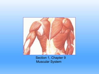
- 1. Section 1, Chapter 9 Muscular System
- 2. Muscle is derived from Musculus, for “Mouse” Imagine a mouse running beneath the skin. Functions of Muscles: 1. Body movement 2. Maintain posture 3. Produces heat 4. Propel substances through body 5. Heartbeat Types of muscles include: 1. Smooth muscle 2. Cardiac muscle 3. Skeletal muscle
- 3. Smooth Muscle Characteristics of smooth muscles • Involuntary control • Tapered cells with a single, central nucleus • Lack striations
- 4. Smooth Muscle There are two types of smooth muscles • Multi-unit Smooth Muscle • unorganized cells that contract as individual cells •Located within the iris of eye and the walls of blood vessels • Visceral (single-unit) Smooth Muscle • Form sheets of muscle • Cells are connected by gap junctions • Muscle fibers contract as a group • Rhythmic contractions • Within walls of most hollow organs (viscera)
- 5. Cardiac Muscle •Located only in the heart •Striated cells •Intercalated discs • Muscle fibers branch •Muscle fibers contract as a unit • Self-exciting and rhythmic
- 6. Skeletal Muscle • Usually attached to bone • Voluntary control • Striated (light & dark bands) • Muscle fibers form bundles • Several peripheral nuclei
- 7. Coverings of Skeletal Muscle Fascia • Dense connective tissue surrounding skeletal muscles Tendons • Dense connective tissue that attaches muscle to bones • Continuation of muscle fascia and bone periosteum Aponeurosis • Broad sheet of connective tissue attaching muscles to bone, or to other muscles.
- 9. Coverings of Skeletal Muscle Epimysium • Connective tissue that covers the entire muscle • Lies deep to fascia Perimysium • Surrounds organized bundles of muscle fibers, called fascicles Endomysium • Connective tissue that covers individual muscle fibers (cells)
- 10. Figure 9.3 Scanning electron micrograph of a fascicle surrounded by its perimysium. Muscle fibers within the fascicle are surrounded by endomysium.
- 11. Organization of Skeletal Muscle Fascicle Organized bundle of muscle fibers Muscle Fiber Single muscle cell Collection of myofibrils Myofibrils Collection of myofilaments Myofilaments Actin filament Myosin filament Figure 9.2 Skeletal muscle organization
- 13. Skeletal Muscle Fibers Sarcolemma • Cell membrane of muscle fibers Sarcoplasm • Cytoplasm of muscle fibers Sarcoplasmic Reticulum • Modified Endoplasmic Reticulum • Stores large deposits of Calcium sarcolemma
- 14. Skeletal Muscle Fibers (Transverse)T-tubules: • invaginations of sarcolemma, extending into the sarcoplasm. Cisternae: • enlarged region of sarcoplasmic reticulum, adjacent to the t-tubules Triad • T-tubule + adjacent cisternae Openings into t-tubules
- 15. Myofibrils Myofibrils are bundles of actin and myosin filaments. • Actin – thin filament • Myosin – thick filament Striations appear from the organization of actin and myosin filaments Figure 9.4 Organization of actin and myosin filaments
- 16. Sarcomere A sarcomere is the functional unit of skeletal muscle • A sarcomere is the area between adjacent Z-lines. •During a muscle contraction the z-lines move together and the sarcomere shortens.
- 17. Sarcomere
- 18. Striations appear from alternate light and dark banding patterns. Z Line is the attachment site of actin filaments (center of I bands) I Bands (light band): consists of only actin filaments A Bands (dark band) : consists of myosin filaments and the overlapping portion of actin filaments Figure 9.5 thin and thick filaments in a sarcomere.
- 19. filaments Thin filaments composed of actin proteins Thin filaments are associated with troponin and tropomyosin proteins Thick filaments composed of myosin proteins During muscle contraction the heads on myosin filaments bind to actin filaments forming a Cross-bridge
- 20. Cross-Bridges When a muscle is at rest, myosin heads are extended in the “cocked” position. During a contraction, myosin heads bind to actin, forming a cross-bridge and the myosin head pivot forward (Power Stroke) and back (Recovery stroke)
- 21. Troponin-Tropomyosin Complex The troponin-tropomyosin complex prevents crossbridge formation when the muscle is at rest. Tropomyosin Blocks binding sites on actin when a muscle is at rest Troponin Ca2+ binds to troponin during a muscle contraction. Troponin moves repositions the tropomyosin filaments, so the myosin and actin filaments can interact.