4.16.24 21st Century Movements for Black Lives.pptx
What makes me nervous?
1. Trick or Treat at Nerveville
What makes you nervous?
Four groups consisting of five to ten people will rotate between four different haunted
houses each having a unique theme related to the Nervous System located in
Nerveville.This activity will educate the students on what exactly makes us nervous and
what our Nervous System consists of.
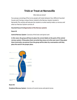
Haunted House #1 (Organization of the Nervous system)
Room #1
Central Nervous System- Consists of the brain and spinal cord
In this room, the group will have to place the correct labels on the parts of the central
nervous system. If they place them correctly they move on to the next room if they place
them incorrectly, one person from the group will be taken by a werewolve until they
place the word in the proper place.
(3)
Room #2
Peripheral Nervous System- Consists of cranial nerves and spinal nerves
2. The group must place the parts of the PNS on the correct locations of the body. They must do
this under 20 minutes or someone from their group will get fake blood poured all over them.
(4)
Room #3
The group will be divided in half, to create an afferent group and an efferent group. The
Afferent group will begin a relay race by running to the “autonomic integration center”,
where the Efferent group will be exiting pathways to finish the race as a “flight response”.
Afferent: Consists of all incoming sensory or afferent pathways. Pathways belong to the
visceral sensory division which carries feedback information to the autonomic
integration centers in the central nervous system.
Efferent: Consists of all outgoing motor or efferent pathways. Pathway of the autonomic
nervous system can be divided into sympathetic and parasympathetic divisions.
Pathways exit the middle portions of the spinal cord which produces the “fight-or-flight”
response. 5 (page 344)
Room #4
Next, the afferent group must carry the sensory information they just received to generate a
response signal. There will be a life-sized model gland that consists of smooth, cardiac
3. muscles which they will have to carry to the autonomic system to see how voluntary control
works.
Somatic Nervous System- carries information to the somatic effectors which are the
skeletal muscles. This system includes afferent pathways that receive sensory
information which then generate the efferent response signal. 5 (page 344)
Autonomic Nervous System- Carry information to the autonomic or visceral effectors
which consists of smooth and cardiac muscles and glands. Consists of voluntary control
meaning it functions without our knowledge. Also consists of efferent pathways. 5(page
344)
Haunted House #2 (Nerve Impulses)
Room #1
The Students must match each definition with the correct term, if they match it correctly they
move on through an obstacle course which will lead them to their next definition, if they were to
fail, a zombie will throw fake eyeballs at them.
Nerve Impulses: self-propagating wave of electrical depolarization carries information
along nerves; also called action potential. (5)
Membrane Potential: difference in electrical charge between inside and outside of the
plasma membrane. (5)
Resting Membrane Potential: electric charge difference inside a cell membrane,
measured relative to just outside the cell membrane. (6)
Local Potential: slight shift from resting membrane potential in a specific region of the
plasma membrane. (5)
Action Potential: change in membrane potential in an excitable tissue that acts as an
electric signal and is propagated in an all-or-none fashion. (6)
Room #2
The group will draw an illustration of the action potential.
4. Mechanism that produces the action potential: The action potential consists of rapid
dramatic changes in membrane potential that occur due to opening and closing of
voltage-gated ion channels. It begins with a steady depolarization called the generator
potential. If the generator potential reaches a critical voltage called the threshold, the
membrane will continue to depolarize, followed by a period of repolarization and then a
short period of hyperpolarization.(6)
(7)
Haunted House #3 (Cells of the nervous System)
Room #1
The group will be quizzed on the five types of Glia after reading about them, and will
receive prizes for correct answers.
5 types of Glia and where to find them:
Astrocytes they form feet that cover the surfaces of neurons and blood vessels they
provide support and help form the blood brain barrier.
Oligodendrocytes are responsible for providing the myelin sheath around nerve
fibers in the CNS.
Microglia are found within the central nervous system ,microglia phagocytize dead
nervous tissue, microorganisms, and other foreign matter.
Ependymal cells are on the surface of the choroid plexus secrete cerebrospinal fluid.
Schwann cells forms a myelin sheath called the neurilemma around each axon
within the peripheral nervous system.
5. Satellite cells little is known about their function, it is located in the nervous
system.
(6)
Room #2 (6)
The group will color a picture of a neuron.
Room #3
The group will make models out of toothpicks and marshmallows to demonstrate
the difference between the types of neurons. The group with the best model wins
a free pass to skip one of the rooms.
Types of Neurons (6)
1. Bipolar neurons - The cell body of a bipolar neuron has only two nerve fibers, one
arising from each end. Although these fibers are structurally similar, one is an axon and
the other is a dendrite. Neurons within specialized parts of the eyes, nose, and ears are
bipolar.
2. Unipolar neurons - A unipolar neuron has a single nerve fiber that extends from the
cell body and then divides into two branches, one connected to a peripheral body part
and functions as a dendrite, and the other entering the brain or spinal cord and
functions as an axon. The cell bodies of some unipolar neurons aggregate in
6. specialized masses of nervous tissue called ganglia (singular, ganglion), located outside
the brain and spinal cord.
3. Multipolar neurons - Multipolar neurons have many nerve fibers arising from their cell
bodies. Only one fiber of each neuron is an axon; the rest are dendrites. Most neurons
whose cell bodies lie within the brain or spinal cord are multipolar.
Room #4
The group will be divided into Group A and B, they will have to act out different
functions of the neurons, the group with the highest score wins a prize.
Functions or Neurons (6)
On the basis of functional differences, neurons are grouped as follows:
A. Sensory neurons (afferent neurons) - These carry nerve impulses from peripheral
body parts into the brain or spinal cord. These neurons either have specialized receptor
ends at the tips of their dendrites, or they have dendrites that are closely associated
with receptor cells in the skin or in sensory organs.
1. Changes that occur inside or outside the body stimulate receptor ends or receptor
cells, triggering sensory nerve impulses. 2. The impulses travel along sensory neuron
fibers, which lead to the brain or spinal cord, where other neurons continue to process
the impulses. Most sensory neurons are unipolar, although some are bipolar.
B. Interneurons (also called internuncial or association neurons) - These neurons lie
within the brain or spinal cord. They are multipolar and link other neurons. 3.
Interneurons transmit impulses from one part of the brain or spinal cord to another. That
is, they may direct incoming sensory impulses to appropriate parts for processing and
interpreting. Other incoming impulses are transferred to motor neurons.
C. Motor neurons (efferent neurons) - 4. Motor neurons are multipolar and carry nerve
impulses out of the brain or spinal cord to effectors. 5. Motor impulses stimulate
muscles to contract and glands to release secretions.
Haunted House #4 (Synapse)
Room #1
The group will have a distance jumping competition to simulate information
crossing a synapse. The group with the longest distance will get to eliminate one
of the other team’s members.
7. Information from one neuron flows to another neuron across a synapse. The synapse contains a small
gap separating neurons.
Synapse consists of:
Pre-synaptic ending that contains neurotransmitters, mitochondria, and other cell organelles.
Post-synaptic ending that contains receptor sites for neurotransmitters.
Synaptic cleft (or space) between pre and post-synaptic endings.
Room #2
The group will play a ball game to simulate neurotransmission. The team with the
highest score will get to proceed to the next round, the losing team will have to
throw a rotten egg towards a target and hit it in order to move on.
Neurotransmitters
Communication of info between neurons is accomplished by movement of chemicals across a small gap
(synapse).
Chemicals, called neurotransmitters, are released from one neuron at the pre-synaptic nerve terminal.
They then cross the synapse where they may be accepted by the next neuron at a specialized site called
the receptor.
Room #3
The group will have to crawl through tunnels one at a time to simulate Spatial
Summation, and then try crawling through at the same time to simulate temporal
summation. The group that does it the fastest will obtain a bag of candy corn.
Summation
Spatial Summation
Several impulses arrive at one neuron via several synapses
Cause sufficient depolarization/ open sufficient sodium ion channels
Temporal Summation
Several impulses arrive at the same neuron via same synapse
What Makes Me Nervous in Nerveville Lab
8. After all the groups of kids have finished going through all four houses they must then go to
an open field in which they will do an experiment to put all of what they’ve learned into use.
They will experiment on one of their team members and record their partners’ reaction to
sound stimulus and to contact stimulus, by studying their reflex. They must record their
answers on bar graphs and answer questions. The team that finishes the fastest and does it
correctly will get a free pass to Six Flags Freight Fest.
20
15
Kick 1
10 Kick 5
5 Kick 3 Kick 2
0 Kick 3
Kick 1
Kick 4
Kick 5
Average
30 Reflex 1
20 Reflex 2
10 Reflex 5 Reflex 3
Reflex 3
0 Reflex 4
Reflex 1
Reflex 5
Average
9. 3
2
1 Max (mV)
0 Min (mV)
Max (mV)
1
2
∆mV
3
4
5
Average
6
4
2 Max (mV)
0 Min (mV)
Max (mV) ∆mV
1
2
3
4
5
Average
10. 1. Compare the reaction times for voluntary vs. involuntary activation of the
quadriceps muscle. What might account for the observed differences in reaction
times?
Voluntary activation of the quadriceps muscle were a lot slower than the
involuntary activation of the quadriceps muscle.The voluntary was the
reaction time after hearing a sound and involuntary was the reaction time
after being hit with a tool.
2. Using data from Table 2, calculate speed at which a stimulus traveled from the
patellar tendon to the spinal cord and back to the quadriceps muscle (a complete
reflex arc). To do this, you must estimate the distance traveled. Using a cloth
tape measure, measure the distance in cm from the mark on the patellar tendon
to the spinal cord at waist level (straight across from the anterior superior iliac
spine–see Figure 9). Multiply the distance by two to obtain the total distance
traveled in the reflex arc. Once this value has been obtained, divide by the
average Δt from Table 2 and divide by 100 to obtain the speed, in m/s, at which
the stimulus traveled.
Total distance traveled in the reflex arc: 100m
Speed: 29.41m/s
3. Nerve impulses have been found to travel as fast as 100 m/s. What could
account for the
difference between your answer to Question 2 and this value obtained by
researchers?
The difference between our answer and the researchers depends on
how tall the person they experimented on was and the reaction spam
that the person’s body has when it its hit with a tool.
4. Assume the speed of a nerve impulse is 100 m/s. How does this compare to the
speed of electricity in a copper wire (approx. 3.00 108 m/s)?
The speed of electricity is slower than the speed at which we react to
the nerve impulses in our bodies.
5. Compare the data you obtained in this experiment with other members of your
group/class. Can individual differences be attributed to any physical differences
(body shape/size, muscle mass, physical fitness level)?
Individual differences can be attributed to any physical difference like
physical fitness level due to the fact that athletes tend to have a faster
reaction spam than that of a non-athlete.