A pictorial review of “signs in thoracic imaging 01”
•Télécharger en tant que PPTX, PDF•
12 j'aime•2,230 vues

Air bronchogram sign Air crescent sign

Recommandé
Recommandé
Contenu connexe
Tendances
Tendances (20)
En vedette
En vedette (16)
Similaire à A pictorial review of “signs in thoracic imaging 01”
Similaire à A pictorial review of “signs in thoracic imaging 01” (20)
Dernier
Dernier (20)
A pictorial review of “signs in thoracic imaging 01”
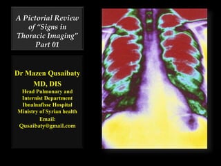
- 1. A Pictorial Review of “Signs in Thoracic Imaging” Part 01 Dr Mazen Qusaibaty MD, DIS Head Pulmonary and Internist Department Ibnalnafisse Hospital Ministry of Syrian health Email: Qusaibaty@gmail.com
- 2. Topic Outline 1. Air bronchogram sign 2. Air crescent sign 2
- 3. Air bronchogram sign الهوائي القصبي االرتسام عالمة 3
- 4. Air bronchogram sign الهوائي القصبي االرتسام عالمة •مشاهدة على القدرة هي ضمن القصبية الحدود المصاب الرئوي المتن تقيحية مرضية بحديثة (قيح)وذمية أو(ماء) •الرئة ذات. 4
- 5. Air bronchogram sign RUL Consolidation Sterp Pneumonia 5
- 6. Air bronchogram sign Pneumococcal pneumonia • Consolidation with multiple air bronchograms ( black branching structures) in the: A. RUL B. RML C. RLL 6
- 7. Air bronchogram sign Pneumococcal pneumonia • Consolidation with multiple air bronchograms in the: A. Segment anterior of RUL B. RML C. RLL 7
- 8. Air bronchogram sign Diffuse Alveolar Hemorrhage 8
- 9. 9 Air bronchogram sign Acute eosinophilic pneumonia
- 10. Air Bronchogram Sign DD: Pneumonia Lymphoma Bronchoalveolar cell carcinoma.
- 11. Conclusion: Air bronchogram Indicates a parenchymal process Including non- obstructive atelectasis Distinguished from pleural or mediastinal processes 11
- 12. 12 Air crescent sign الغازي الهالل عالمة
- 13. Crescent-shaped area of air within: Nodule Mass Consolidation Crescent-shaped area of air surrounding round or oval opacity within cavity seen in CT and radiograph 13
- 14. • Pulmonary cavity • Ectatic bronchus • Angioinvasive aspergillosis in patients with severe neutropenia Air crescent sign results most commonly from mycetoma (fungus ball) within: 14
- 15. Air crescent sign الغازي الهالل عالمة The air surrounding a radio opaque material 15
- 16. Air crescent sign الغازي الهالل عالمة In a crescentric manner along both its inner and outer margins indicating cavitary disease 16
- 17. Air crescent sign الغازي الهالل عالمة Aspergilloma in an old TB cavity 17
- 18. o The arrows denote an ill-defined nodular opacity in medial aspect of right upper lobe with ill-defined rim of lucency surrounding it. 18
- 19. Invasive aspergillosis in a 6-year-old girl with neutropenia and acute lymphocytic leukemia 19
- 20. Frontal chest radiograph shows two air crescent signs (arrowheads) 20
- 21. Chest roentgenogram PA view Right pneumothorax Mycetoma with air-crescent sign The tip of intercostal drain is also visible near the base. 21
- 22. Computed tomogram of the thorax Various views of the mycetoma Rupture into the pleural cavity leading to pneumothorax22
- 23. Coronal reformation image Intracavitary aspergilloma (*) in the left upper lobe 23
- 24. Air crescent –shaped surrounding oval opacity within cavity 24 • Subpleural honeycombing in the lower lobes consistent with idiopathic pulmonary fibrosis.
- 25. What is your diagnosis? The patient was a 58-year-old man with previous tuberculosis and intracavitary aspergilloma 25
- 26. Sagittal reformat from a CT scan of the chest 26 • A 14 year-old female with acute myeloblastic leukaemia
- 27. Where is located this lesion? 27 A. Left Lung B. Right Lung
- 28. Where is located this lesion? 28 A. Left Lung B. Apical segment of Right Upper Lobe in lateral position
- 29. Correct description A Mycetoma with air-crescent sign located in apical segment of right upper lobe in lateral position 29
- 30. Air crescent sign in anterior segment of right upper lobe Aspergilloma 30
- 31. PA Projection radiograph 31 • Patchy airspace disease with areas of lucency suggestive of cavitation in the right upper lobe.
- 32. PA Projection radiograph 32 • Emphysematous changes are seen bilaterally.
- 33. Chest CT scan Air crescent sign within the consolidated right upper lobe. 33
- 34. Chest CT scan Paraseptal emphysema 34
- 35. Chest CT scan A cavity with an intracavitary mass within the consolidated right upper lobe 35
- 36. Conclusion: Air crescent sign • indicates a lung cavity, often due to fungal infection 36
Notes de l'éditeur
- A Pictorial Review of “Signs in Thoracic Imaging” Karuppasamy, K.1, Abhyankar-Gupta, M.1, Fewins, H.1, Curtis, J.2 1The Cardiothoracic Centre - Liverpool NHS Trust, 2Aintree University Hospitals NHS Foundation Trust, Liverpool, United Kingdom
- علامة الارتسام القصبي الهوائي: هو القدرة على مشاهدة الحدود القصبية ضمن المتن الرئوي المصاب بحديثة مرضية تقيحية (قيح) أو وذمية ( ماء)
- Pneumococcal pneumonia produces consolidation in the right upper lobe with multiple air bronchograms (black branching structures) present since the spaces surrounding the air-filled bronchi normally contain air but now are filled with inflammatory exudate. There is no cavitation, the disease is in the lower lobe and it contains air bronchograms, all characteristics of pneumonia caused by Streptococcus Pneumoniae (formerly known as Diplococcus Pneumoniae)
- Pneumococcal pneumonia produces consolidation in the right upper lobe with multiple air bronchograms (black branching structures) present since the spaces surrounding the air-filled bronchi normally contain air but now are filled with inflammatory exudate. There is no cavitation, the disease is in the lower lobe and it contains air bronchograms, all characteristics of pneumonia caused by Streptococcus Pneumoniae (formerly known as Diplococcus Pneumoniae)
- Pneumococcal pneumonia produces consolidation in the right upper lobe with multiple air bronchograms (black branching structures) present since the spaces surrounding the air-filled bronchi normally contain air but now are filled with inflammatory exudate. There is no cavitation, the disease is in the lower lobe and it contains air bronchograms, all characteristics of pneumonia caused by Streptococcus Pneumoniae (formerly known as Diplococcus Pneumoniae)
- Branching, linear, tubular lucency representing a bronchus or bronchiole passing through airless lung parenchyma . This sign indicates that the underlying opacity must be parenchymal rather than pleural or mediastinal in location.
- air bronchogram - indicates a parenchymal process, including non-obstructive atelectasis, as distinguished from pleural or mediastinal processes
- mycetoma :ورم فطري
- Refers to the air surrounding a radio opaque material in a crescentric manner along both its inner and outer margins indicating cavitary disease Aspergilloma in an old TB cavity
- Refers to the air surrounding a radio opaque material in a crescentric manner along both its inner and outer margins indicating cavitary disease Aspergilloma in an old TB cavity
- Refers to the air surrounding a radio opaque material in a crescentric manner along both its inner and outer margins indicating cavitary disease Aspergilloma in an old TB cavity
- علامة هلال الهوا In radiology, the air crescent sign is a finding on chest radiograph and computed tomography that is crescenteric and radiolucent, due to a lung cavity that is filled with air and has a round radioopaque mass.[1] Classically, it is due to an aspergilloma, a form of aspergillosis, that occurs when the fungus Aspergillus grows in a cavity in the lung.[2]
- علامة هلال الهوا In radiology, the air crescent sign is a finding on chest radiograph and computed tomography that is crescenteric and radiolucent, due to a lung cavity that is filled with air and has a round radioopaque mass.[1] Classically, it is due to an aspergilloma, a form of aspergillosis, that occurs when the fungus Aspergillus grows in a cavity in the lung.[2]
- علامة هلال الهوا In radiology, the air crescent sign is a finding on chest radiograph and computed tomography that is crescenteric and radiolucent, due to a lung cavity that is filled with air and has a round radioopaque mass.[1] Classically, it is due to an aspergilloma, a form of aspergillosis, that occurs when the fungus Aspergillus grows in a cavity in the lung.[2]
- Right pneumothorax Mycetoma with air-crescent sign The tip of intercostal drain is also visible near the base.
- Computed tomogram of the thorax showing the various views of the mycetoma with rupture into the pleural cavity leading to pneumothorax over adjacent CT sections.
- Aspergilloma in tuberculous cavity. Coronal reformation image better shows the intracavitary aspergilloma (asterisk) in the left upper lobe. Also noted is subpleural honeycombing in the lower lobes consistent with idiopathic pulmonary fibrosis. The patient was a 58-year-old man with previous tuberculosis and intracavitary aspergilloma.
- Aspergilloma in tuberculous cavity. Coronal reformation image better shows the intracavitary aspergilloma (asterisk) in the left upper lobe. Also noted is subpleural honeycombing in the lower lobes consistent with idiopathic pulmonary fibrosis. The patient was a 58-year-old man with previous tuberculosis and intracavitary aspergilloma.
- Aspergilloma in tuberculous cavity. Coronal reformation image better shows the intracavitary aspergilloma (asterisk) in the left upper lobe. Also noted is subpleural honeycombing in the lower lobes consistent with idiopathic pulmonary fibrosis. The patient was a 58-year-old man with previous tuberculosis and intracavitary aspergilloma.
- “Sagittal reformat from a CT scan of the chest, performed on a 14 year-old female with acute myeloblastic leukaemia. The image shows a rounded cavi...”
- “Sagittal reformat from a CT scan of the chest, performed on a 14 year-old female with acute myeloblastic leukaemia. The image shows a rounded cavi...”
- “Sagittal reformat from a CT scan of the chest, performed on a 14 year-old female with acute myeloblastic leukaemia. The image shows a rounded cavi...”
- “Sagittal reformat from a CT scan of the chest, performed on a 14 year-old female with acute myeloblastic leukaemia. The image shows a rounded cavi...”
- Aspergilloma in tuberculous cavity. Coronal reformation image better shows the intracavitary aspergilloma (asterisk) in the left upper lobe. Also noted is subpleural honeycombing in the lower lobes consistent with idiopathic pulmonary fibrosis. The patient was a 58-year-old man with previous tuberculosis and intracavitary aspergilloma.
- showing patchy airspace disease with areas of lucency suggestive of cavitation in the right upper lobe. Emphysematous changes are seen bilaterally.
- showing patchy airspace disease with areas of lucency suggestive of cavitation in the right upper lobe. Emphysematous changes are seen bilaterally.
- Aspergilloma in tuberculous cavity. Coronal reformation image better shows the intracavitary aspergilloma (asterisk) in the left upper lobe. Also noted is subpleural honeycombing in the lower lobes consistent with idiopathic pulmonary fibrosis. The patient was a 58-year-old man with previous tuberculosis and intracavitary aspergilloma.
- Aspergilloma in tuberculous cavity. Coronal reformation image better shows the intracavitary aspergilloma (asterisk) in the left upper lobe. Also noted is subpleural honeycombing in the lower lobes consistent with idiopathic pulmonary fibrosis. The patient was a 58-year-old man with previous tuberculosis and intracavitary aspergilloma.
- Aspergilloma in tuberculous cavity. Coronal reformation image better shows the intracavitary aspergilloma (asterisk) in the left upper lobe. Also noted is subpleural honeycombing in the lower lobes consistent with idiopathic pulmonary fibrosis. The patient was a 58-year-old man with previous tuberculosis and intracavitary aspergilloma.