SlideShare une entreprise Scribd logo
1  sur  93
Dr Mazen Qusaibaty
MD, DIS
Head Pulmonary and Internist
Department
Ibnalnafisse Hospital
Ministry of Syrian health
Email: Qusaibaty@gmail.com
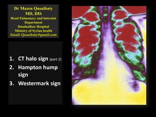
1. CT halo sign (part 2)
2. Hampton hump
sign
3. Westermark sign
Topic Outline
1. CT halo sign (part 2)
2. Hampton hump sign
3. Westermark sign
2
Useful links
3
http://bjr.birjournals.org
http://radiographics.rsna.org
http://radiology.rsna.org
CT halo sign (part 2)
Bronchiolitis Obliterans with Organizing
Pneumonia
• An idiopathic disease
that produces
polypoid granulation
tissue in:
 Bronchioles
 alveolar ducts
5
ColbyTV. Pathologic aspects of bronchiolitis obliterans organizing
pneumonia. Chest 1992; 102(1 suppl): 38S–43S.
Bronchiolitis Obliterans with Organizing
Pneumonia
• Variable degrees of
interstitial and
airspace infiltration
by mononuclear cells
and foamy
macrophages
6
ColbyTV. Pathologic aspects of bronchiolitis obliterans organizing
pneumonia. Chest 1992; 102(1 suppl): 38S–43S.
The most common CT finding in patients with
BOOP
• Asymmetric bilateral
ground-glass opacity
• with a
predominantly
peripheral
distribution (arrows)
7 Courtesy of Paul Stark, MD.
The most common CT finding in patients with
BOOP
Consolidation
8
The most common CT finding in patients with
BOOP
Consolidation
Peribronchovascular
distribution
(peripheral)
9
The most common CT finding in patients with
BOOP
Consolidation
Peribronchovascular
distribution
(peripheral)
Lower zones
localization
10
The most common CT finding in patients with
BOOP
Consolidation
Peribronchovascular
distribution
(peripheral)
Lower zones
localization
Air bronchogram
11
The most common CT finding in patients with
BOOP
Consolidation
Peribronchovascular
distribution
(peripheral)
Lower zones
localization
Air bronchogram
One or more
nodules
12
The most common CT finding in patients with
BOOP
Consolidation
Peribronchovascular
distribution
(peripheral)
Lower zones
localization
Air bronchogram
One or more
nodules
One or more masses
13
BOOP
• Kim et al reported that
CT images of 06 of 31
patients with BOOP
showed nodular
ground-glass opacity
14
KimSJ, Lee KS, Ryu YH, et al. Reversed halo sign on high-resolution CT of cryptogenic organizing
pneumonia: diagnostic implications. AJR Am J Roentgenol 2003; 180: 1251–1254.
BOOP
A 46-year-old woman
• Axial CT image at the
level of the aortic arch
15
KimSJ, Lee KS, Ryu YH, et al. Reversed halo sign on high-resolution CT of cryptogenic organizing
pneumonia: diagnostic implications. AJR Am J Roentgenol 2003; 180: 1251–1254.
BOOP
A 46-year-old woman
• Shows multiple bilateral
areas of ill-defined
nodular ground-glass
opacity, some of which
contain solid
components (arrows).
16
KimSJ, Lee KS, Ryu YH, et al. Reversed halo sign on high-resolution CT of cryptogenic organizing
pneumonia: diagnostic implications. AJR Am J Roentgenol 2003; 180: 1251–1254.
Case
• 48-year-old woman with an eight-week
history of cough, dyspnea with exertion,
fatigue, and slight weight loss.
17
Case/ High-resolution CT scan of this patient
• RLL reticular and
hazy opacities that
are subpleural
18 Courtesy of Talmadge E King Jr, MD
Quiz conti…………..
• This picture is look like:
A. Idiopathic pulmonary
fibrosis.
B. Sarcoidosis stage IV
C. TB
D. BOOP
E. All above
19
Quiz conti…………..
• This picture is look like:
A. Idiopathic pulmonary
fibrosis.
B. Sarcoidosis stage IV
C. TB
D. BOOP
E. All above
20
Reverse halo sign: BOOP
CT of localized organizing pneumonia
manifesting as a solitary nodule of the
left lower lobe
21
Reverse halo sign: BOOP
This pattern may be diagnosed as
primary or metastatic lung tumor
22
Thoracic Endometriosis
Catamenial syndrome
23
Monthly recurrence
Hemoptysis, during
the menstrual
period
24
AlifanoM, Trisolini R, Cancellieri A, Regnard JF. Thoracic endometriosis: current knowledge. Ann Thorac
Surg 2006; 81: 761
Monthly recurrence
Hemoptysis, during
the menstrual
period
History
25
AlifanoM, Trisolini R, Cancellieri A, Regnard JF. Thoracic endometriosis: current knowledge. Ann Thorac
Surg 2006; 81: 761
Monthly recurrence
Hemoptysis, during
the menstrual
period
History
Pregnancy
26
AlifanoM, Trisolini R, Cancellieri A, Regnard JF. Thoracic endometriosis: current knowledge. Ann Thorac
Surg 2006; 81: 761
Monthly recurrence
Hemoptysis, during
the menstrual
period
History
Pregnancy
Obstetric-
gynecologic surgery
27
AlifanoM, Trisolini R, Cancellieri A, Regnard JF. Thoracic endometriosis: current knowledge. Ann Thorac
Surg 2006; 81: 761
Catamenial syndrome
• This disease group includes four well-
recognized clinical entities
1. Catamenial pneumothorax
2. Catamenial hemothorax
3. Catamenial hemoptysis
4. Lung nodules
28
AlifanoM, Trisolini R, Cancellieri A, Regnard JF. Thoracic endometriosis: current knowledge. Ann
Thorac Surg 2006; 81: 761
CPT
• Catamenial
pneumothorax is a
rare condition
characterized by a
reoccurrence of air
in the pleural space
coinciding with the
onset of menses.
29
http://www.catamenial-pneumothorax.com/id15.htm
CPT
• CPT was first
described in
literature in 1958
30
http://www.catamenial-pneumothorax.com/id2.htm
CPT
• It is almost always
right-sided
31
http://www.catamenial-pneumothorax.com/id2.htm
CPT
• Generally affects
women in their
thirties and forties
32
http://www.catamenial-pneumothorax.com/id2.htm
CPT
• Although the exact etiology of the condition is
unknown
• Most physicians agree that endometriosis is
involved.
33
http://www.catamenial-pneumothorax.com/id2.htm
CPT
• Documented case
studies have described
endometrial implants
on the:
 Lung
 Pleura
 Diaphragmatic
fenestrations (holes in the
diaphragm)
34
http://www.catamenial-pneumothorax.com/id2.htm
CPT
• Case studies report
women with CPT
experiencing monthly
 Chest pain
 Shortness of breath
 Dizziness and fatigue
35
http://www.catamenial-pneumothorax.com/id2.htm
CPT
• Case studies report women with CPT
experiencing monthly
Some women have experienced multiple lung
collapses over a period of several years.
Many of these women have also been diagnosed
with pelvic endometriosis.
36
http://www.catamenial-pneumothorax.com/id2.htm
Case 1
• In a 42-year-old woman who presented with:
 Three episodes of spontaneous pneumothorax,
each associated with the onset of menses.
37
http://radiographics.rsna.org/content/21/1/193/F43.expansion.html
Case 1
 She had
undergone a prior
hysterectomy for
endometriosis
38
http://radiographics.rsna.org/content/21/1/193/F43.expansion.html
Case 1
Had a 2-3-year history of:
• Episodic cough
• Hemoptysis
• Pleuritic chest pain
39
http://radiographics.rsna.org/content/21/1/193/F43.expansion.html
Posteroanterior chest radiograph
• Shows a right-sided
pneumothorax
• A nodular opacity in
the right lung base
(arrow)
40
http://radiographics.rsna.org/content/21/1/193/F43.expansion.html
Case 2
Chest CT scan (lung windows)
• Shows a 2.5-cm solitary
pulmonary nodule in
the right lower lobe
(arrow).
41
http://radiographics.rsna.org/content/21/1/193/F43.expansion.html
Parenchymal endometrioma in a 74-year-old
woman
• The lesion was found
on a routine
preoperative chest
radiograph obtained
for cataract surgery.
42
http://radiographics.rsna.org/content/21/1/193/F43.expansion.html
Parenchymal endometrioma in a 74-year-old
woman
• She had been receiving
estrogen replacement
therapy since
undergoing
hysterectomy
• She had no known
history of
endometriosis
43
http://radiographics.rsna.org/content/21/1/193/F43.expansion.html
Case 3
Catamenial hemoptysis syndrome in a 24-
year-old woman with recurrent monthly
hemoptysis during menstruation
44
http://radiographics.rsna.org/content/27/2/391/F29.expansion.html
Axial CT image at the level of the
diaphragmatic dome
Multiple areas of ill-defined nodular
ground-glass opacity (arrows)
45 http://radiographics.rsna.org/content/27/2/391/F29.expansion.html
The patient underwent a
bronchoscopic examination, and
endometrial tissue was found at
bronchial lavage
46 http://radiographics.rsna.org/content/27/2/391/F29.expansion.html
Focal Traumatic Lung Injury
47
Focal Traumatic Lung Injury
• Traumatic lung injury may be manifested as
nodular ground-glass opacity at CT during
subsequent disease progression
48
Focal Traumatic Lung Injury
• A transthoracic lung
biopsy
• A transbronchial
biopsy
49
KazerooniEA, Cascade PN, Gross BH. Transplanted lungs: nodules following transbronchial
biopsy. Radiology 1995; 194: 209–212.
Pseudonodule in a 56-year-old woman who
underwent a previous percutaneous lung biopsy
• Thin-section CT
image obtained at
the level of the
aortic arch
50
Pseudonodule in a 56-year-old woman who
underwent a previous percutaneous lung biopsy
• Shows a 9-mm well-
defined nodular
ground-glass opacity
(arrow) in the right
upper lobe
51
Pseudonodule in a 56-year-old woman who
underwent a previous percutaneous lung biopsy
• Axial image obtained
during CT-guided
percutaneous
transthoracic biopsy
with the patient in the
supine position shows
the biopsy needle
(arrow), which has
been inserted near the
nodule
52
Pseudonodule in a 56-year-old woman who
underwent a previous percutaneous lung biopsy
• Axial CT image,
obtained after the
biopsy, shows a poorly
defined pseudonodule
represented by ground-
glass opacity (arrow)
along the biopsy tract
53
Pseudonodule in a 56-year-old woman who
underwent a previous percutaneous lung biopsy
• The pathologic
diagnosis, obtained
after a wedge
resection, was focal
interstitial fibrosis
54
Pulmonary Embolism Signs
55
Radiographic signs with a relatively high
specificity but low sensitivity for PTE
1. Pleura-based areas of increased opacity
(Hampton sign)
2. Decreased vascularity in the peripheral lung
(Westermarck sign)
3. Enlargement of the central pulmonary artery
(Fleischner sign)
4. Hemidiaphragm elevation
56
Worsley DF, Alavi A, Aronchick JM, Chen JT, Greenspan RH, Ravin CE.Chest radiographic findings in patients
with acute pulmonary embolism: observations from the PIOPED Study. Radiology 1993; 189:
Helical CT Findings in Acute PTE
57
Helical CT Findings in Acute PTE
58
Helical CT Findings in Acute PTE
59
Helical CT Findings in Acute PTE
60
Helical CT Findings in Acute PTE
61
Helical CT Findings in Acute PTE
62
Helical CT Findings in Acute PTE
63
Acute PE
Helical CT Findings in Chronic PTE
65
Pulmonary Embolism
Acute PE
Central intraluminal filling defects
Parenchyma: rounded pleural-based densities, pleural
effusion
Subacute PE
Convex mural filling defects
Parenchyma: oval pleural-based densities, pleural effusion
Chronic PE
Concave mural filling defects, intraluminal “rope ladder”
Irregular wall thickening, abnormal vascular tapering,
variation in size of segmental vessels
Parenchyma: translobular lines, mosaic perfusion, pleural
effusion
BC: Wacker_Gustav_19330609_BC_zentral/Akute LE: Sauer_Erna_DOB19211226_LE
CTEPH: Prell/Sarkom: Jpn. J. Clin. Oncol. Uchida et al. 35 (7): 417
Chronic PE
Concave mural filling defects
67
Chronic PE
Intraluminal “rope ladder”
68
Hampton hump sign
69
Hampton hump sign
A homogeneous
wedge-shaped
consolidation in the
lung periphery
Hampton hump sign
A base contiguous to a
visceral pleural
Hampton hump sign
A rounded convex apex
directed toward the
hilum
Hampton hump sign
Associated with
pulmonary infarct
Hampton hump sign
• Left intraluminal filling defects in left pulmonary artery
• Pulmonary infarction secondary to pulmonary embolism
• Bilateral pleural effusion
74
Computed tomography angiogram in a 53-year-
old man with acute pulmonary embolism
• This image shows an
intraluminal filling
defect that occludes :
The anterior basal
segmental artery of
the right lower lobe
Acute Pulmonary Embolism (Helical CT): Imaging Contributor Information and Disclosures
Updated: May Author: Kavita Garg, MD, Professor, Department of Radiology, University of Colorado Health
Sciences Center- 14, 2008
Computed tomography angiogram in a 53-year-
old man with acute pulmonary embolism
• An infraction of the
corresponding lung,
which is indicated by:
o A triangular, pleura-
based consolidation
(Hampton hump)
Acute Pulmonary Embolism (Helical CT): Imaging Contributor Information and Disclosures
Updated: May Author: Kavita Garg, MD, Professor, Department of Radiology, University of Colorado Health
Sciences Center- 14, 2008
Case 3
• A young man who
experienced:
o Acute chest pain
o Shortness of breath
after a
transcontinental
flight
77
Acute Pulmonary Embolism (Helical CT): Imaging Contributor Information and Disclosures
Updated: May Author: Kavita Garg, MD, Professor, Department of Radiology, University of Colorado Health
Sciences Center- 14, 2008
Case 3
Computed tomography angiography
• This image
demonstrates a clot in :
o The anterior
segmental artery in
the left upper lung
o A clot in the anterior
segmental artery in
the right upper lung
78
Acute Pulmonary Embolism (Helical CT): Imaging Contributor Information and Disclosures
Updated: May Author: Kavita Garg, MD, Professor, Department of Radiology, University of Colorado Health
Sciences Center- 14, 2008
Westermark sign
79
Westermark sign
Refers to an area of
oligemia with minimal
change in lung volume
distal to a large PE
Westermark sign
This regional oligemia
is caused either by:
•Mechanical
obstruction to blood
flow by the clot
•Reflex vasoconstriction
Case 4
• A 41-year-old woman presented to the
emergency department complaining of a
three-day history of left-sided chest pain.
82
Case 4
• The pain was described as pressure-like,
pleuritic, made worse with ambulation and
when supine.
83
Case 4
• The pain was constant, increasing in intensity
and not associated with any alleviating
factors, hence her request for evaluation
84
Case 4
• Six weeks prior to the onset of her chest pain
she had complained of:
Right hip pain, radiating down the leg.
This was diagnosed as radicular pain secondary to
sciatica for which she had been under bed rest
and analgesics
85
Case 4
• Her past medical history was remarkable for:
Depression
Occasional migraine headaches
Two cesarean sections
Oral contraceptive
86
Case
• Physical examination was normal
87
Axial CT image of the chest
A mass extending outside the vessel
wall + a large filling defect involving
the left main pulmonary artery and
extending into all sub-branches.
88
Coronal reformatted CT image of the chest
Demonstrating a mass extending
outside the vessel wall
89
Sagittal reformatted CT image of the chest
Demonstrating a mass extending
outside the vessel wall
90
What diagnosis that do you expect?
A. Left large cell
carcinoma
B. Bronchioloalveolar
carcinoma
C. Left small cell
carcinoma
D. Chronic left chronic
pulmonary embolism
E. Pulmonary
angiosarcoma
91
What diagnosis that do you expect?
A. Left large cell
carcinoma
B. Bronchioloalveolar
carcinoma
C. Left small cell
carcinoma
D. Chronic left chronic
pulmonary embolism
E. Pulmonary
angiosarcoma
92
‫خلقنا‬ ‫داق‬ ‫بقا‬ ‫حاجه‬

Contenu connexe

Tendances

Collapse- RADIOLOGY
Collapse- RADIOLOGYCollapse- RADIOLOGY
Collapse- RADIOLOGYNavdeep Shah
 
IMAGING IN ABDOMINAL TUBERCULOSIS
IMAGING IN ABDOMINAL TUBERCULOSISIMAGING IN ABDOMINAL TUBERCULOSIS
IMAGING IN ABDOMINAL TUBERCULOSISNavni Garg
 
Mediastinum-RADIOLOGY
Mediastinum-RADIOLOGYMediastinum-RADIOLOGY
Mediastinum-RADIOLOGYNavdeep Shah
 
Presentation1.pptx, radiological imaging of pulmonary infection.
Presentation1.pptx, radiological imaging of pulmonary infection.Presentation1.pptx, radiological imaging of pulmonary infection.
Presentation1.pptx, radiological imaging of pulmonary infection.Abdellah Nazeer
 
Normal chest x ray- Radiology Basics
Normal chest x  ray- Radiology BasicsNormal chest x  ray- Radiology Basics
Normal chest x ray- Radiology BasicsSandeep Awal
 
Presentation1.pptx radiological imaging of mediastinal masses .
Presentation1.pptx radiological imaging of mediastinal masses .Presentation1.pptx radiological imaging of mediastinal masses .
Presentation1.pptx radiological imaging of mediastinal masses .Abdellah Nazeer
 
4 diffuse reticular or reticulonodular pattern
4 diffuse reticular or reticulonodular pattern4 diffuse reticular or reticulonodular pattern
4 diffuse reticular or reticulonodular patternDr. Muhammad Bin Zulfiqar
 
Presentation1.pptx, radiological imaging of bronchiectasis.
Presentation1.pptx, radiological imaging of bronchiectasis.Presentation1.pptx, radiological imaging of bronchiectasis.
Presentation1.pptx, radiological imaging of bronchiectasis.Abdellah Nazeer
 
Hrct anatomy
Hrct anatomyHrct anatomy
Hrct anatomyRMLIMS
 
Ct chest developmental anomalies , airways
Ct chest developmental anomalies , airwaysCt chest developmental anomalies , airways
Ct chest developmental anomalies , airwaysRikin Hasnani
 
Chest XRay and other imaging investigations of chest, CT chest, HRCT Chest
Chest XRay and other imaging investigations of chest, CT chest, HRCT ChestChest XRay and other imaging investigations of chest, CT chest, HRCT Chest
Chest XRay and other imaging investigations of chest, CT chest, HRCT ChestBishnu Khatiwada
 
Chest radiology part 2
Chest radiology part 2Chest radiology part 2
Chest radiology part 2Gamal Agmy
 
Radiographic manifestations of pulmonary tuberculosis
Radiographic manifestations of pulmonary tuberculosisRadiographic manifestations of pulmonary tuberculosis
Radiographic manifestations of pulmonary tuberculosisDev Lakhera
 

Tendances (20)

Normal chest ct
Normal chest ctNormal chest ct
Normal chest ct
 
Lung cancer radiology
Lung cancer radiologyLung cancer radiology
Lung cancer radiology
 
CT CHEST ANATOMY
CT CHEST ANATOMYCT CHEST ANATOMY
CT CHEST ANATOMY
 
Collapse- RADIOLOGY
Collapse- RADIOLOGYCollapse- RADIOLOGY
Collapse- RADIOLOGY
 
IMAGING IN ABDOMINAL TUBERCULOSIS
IMAGING IN ABDOMINAL TUBERCULOSISIMAGING IN ABDOMINAL TUBERCULOSIS
IMAGING IN ABDOMINAL TUBERCULOSIS
 
Abnormal Chest xray
Abnormal Chest xray Abnormal Chest xray
Abnormal Chest xray
 
HRCT Chest
HRCT ChestHRCT Chest
HRCT Chest
 
Basics of CT chest
Basics of CT chestBasics of CT chest
Basics of CT chest
 
Mediastinum-RADIOLOGY
Mediastinum-RADIOLOGYMediastinum-RADIOLOGY
Mediastinum-RADIOLOGY
 
Presentation1.pptx, radiological imaging of pulmonary infection.
Presentation1.pptx, radiological imaging of pulmonary infection.Presentation1.pptx, radiological imaging of pulmonary infection.
Presentation1.pptx, radiological imaging of pulmonary infection.
 
Normal chest x ray- Radiology Basics
Normal chest x  ray- Radiology BasicsNormal chest x  ray- Radiology Basics
Normal chest x ray- Radiology Basics
 
Presentation1.pptx radiological imaging of mediastinal masses .
Presentation1.pptx radiological imaging of mediastinal masses .Presentation1.pptx radiological imaging of mediastinal masses .
Presentation1.pptx radiological imaging of mediastinal masses .
 
4 diffuse reticular or reticulonodular pattern
4 diffuse reticular or reticulonodular pattern4 diffuse reticular or reticulonodular pattern
4 diffuse reticular or reticulonodular pattern
 
Presentation1.pptx, radiological imaging of bronchiectasis.
Presentation1.pptx, radiological imaging of bronchiectasis.Presentation1.pptx, radiological imaging of bronchiectasis.
Presentation1.pptx, radiological imaging of bronchiectasis.
 
Hrct anatomy
Hrct anatomyHrct anatomy
Hrct anatomy
 
Ct chest developmental anomalies , airways
Ct chest developmental anomalies , airwaysCt chest developmental anomalies , airways
Ct chest developmental anomalies , airways
 
Chest XRay and other imaging investigations of chest, CT chest, HRCT Chest
Chest XRay and other imaging investigations of chest, CT chest, HRCT ChestChest XRay and other imaging investigations of chest, CT chest, HRCT Chest
Chest XRay and other imaging investigations of chest, CT chest, HRCT Chest
 
Chest radiology part 2
Chest radiology part 2Chest radiology part 2
Chest radiology part 2
 
Chest xray
Chest xray  Chest xray
Chest xray
 
Radiographic manifestations of pulmonary tuberculosis
Radiographic manifestations of pulmonary tuberculosisRadiographic manifestations of pulmonary tuberculosis
Radiographic manifestations of pulmonary tuberculosis
 

En vedette

Pulmonary Embolism- Diagnosis by Dr.Tinku Joseph
Pulmonary Embolism- Diagnosis  by Dr.Tinku JosephPulmonary Embolism- Diagnosis  by Dr.Tinku Joseph
Pulmonary Embolism- Diagnosis by Dr.Tinku JosephDr.Tinku Joseph
 
Pulmonary embolism 2
Pulmonary embolism 2Pulmonary embolism 2
Pulmonary embolism 2samirelansary
 
Emergency cadiopulmonary imaging
Emergency cadiopulmonary imagingEmergency cadiopulmonary imaging
Emergency cadiopulmonary imagingAmir Mahmoud
 
How read chest ct 1
How  read  chest ct  1How  read  chest ct  1
How read chest ct 1ANAS ALSOHLE
 
Imaging of Pulmonary Embolism
Imaging of Pulmonary Embolism  Imaging of Pulmonary Embolism
Imaging of Pulmonary Embolism Gamal Agmy
 
57 filling defect in pulmonary artery on computed
57 filling defect in pulmonary artery on computed57 filling defect in pulmonary artery on computed
57 filling defect in pulmonary artery on computedDr. Muhammad Bin Zulfiqar
 
How read chest xr 8
How  read  chest xr  8How  read  chest xr  8
How read chest xr 8ANAS ALSOHLE
 
How read chest xr 1
How read chest xr 1How read chest xr 1
How read chest xr 1ANAS ALSOHLE
 
Xaheer shah...pulmonary embolism
Xaheer shah...pulmonary embolismXaheer shah...pulmonary embolism
Xaheer shah...pulmonary embolismzaheer shah
 
Pulmonary Embolism Power Point
Pulmonary Embolism Power PointPulmonary Embolism Power Point
Pulmonary Embolism Power PointTodd Peterson
 
Investigation of suspected pulmonary embolism in pregnancy
Investigation of suspected pulmonary embolism in pregnancyInvestigation of suspected pulmonary embolism in pregnancy
Investigation of suspected pulmonary embolism in pregnancySCGH ED CME
 
Pulmonary thromboembolism
Pulmonary thromboembolismPulmonary thromboembolism
Pulmonary thromboembolismcamiij1
 
Chest and cvs radiology mocks fcps
Chest  and cvs radiology mocks fcpsChest  and cvs radiology mocks fcps
Chest and cvs radiology mocks fcpsdrneelammalik
 
granulomatosis with polyangiitis (Wegener’s granulomatosis)
granulomatosis with polyangiitis (Wegener’s granulomatosis) granulomatosis with polyangiitis (Wegener’s granulomatosis)
granulomatosis with polyangiitis (Wegener’s granulomatosis) Ameen Rageh
 
How read chest ct 2
How  read  chest ct  2How  read  chest ct  2
How read chest ct 2ANAS ALSOHLE
 

En vedette (20)

Pulmonary Embolism- Diagnosis by Dr.Tinku Joseph
Pulmonary Embolism- Diagnosis  by Dr.Tinku JosephPulmonary Embolism- Diagnosis  by Dr.Tinku Joseph
Pulmonary Embolism- Diagnosis by Dr.Tinku Joseph
 
Pulmonary embolism 2
Pulmonary embolism 2Pulmonary embolism 2
Pulmonary embolism 2
 
Emergency cadiopulmonary imaging
Emergency cadiopulmonary imagingEmergency cadiopulmonary imaging
Emergency cadiopulmonary imaging
 
Figure 3 sign fleischner sign-fissure sign-headcheese sign-hilar overlay sign...
Figure 3 sign fleischner sign-fissure sign-headcheese sign-hilar overlay sign...Figure 3 sign fleischner sign-fissure sign-headcheese sign-hilar overlay sign...
Figure 3 sign fleischner sign-fissure sign-headcheese sign-hilar overlay sign...
 
How read chest ct 1
How  read  chest ct  1How  read  chest ct  1
How read chest ct 1
 
Imaging of Pulmonary Embolism
Imaging of Pulmonary Embolism  Imaging of Pulmonary Embolism
Imaging of Pulmonary Embolism
 
57 filling defect in pulmonary artery on computed
57 filling defect in pulmonary artery on computed57 filling defect in pulmonary artery on computed
57 filling defect in pulmonary artery on computed
 
Pulmonary embolism
Pulmonary embolismPulmonary embolism
Pulmonary embolism
 
How read chest xr 8
How  read  chest xr  8How  read  chest xr  8
How read chest xr 8
 
How read chest xr 1
How read chest xr 1How read chest xr 1
How read chest xr 1
 
Ct halo sign (part 1)
Ct halo sign (part 1)Ct halo sign (part 1)
Ct halo sign (part 1)
 
Rib notching
Rib notching Rib notching
Rib notching
 
Xaheer shah...pulmonary embolism
Xaheer shah...pulmonary embolismXaheer shah...pulmonary embolism
Xaheer shah...pulmonary embolism
 
Pulmonary Embolism Power Point
Pulmonary Embolism Power PointPulmonary Embolism Power Point
Pulmonary Embolism Power Point
 
Pulmonary Embolism - (PE)
Pulmonary Embolism - (PE)Pulmonary Embolism - (PE)
Pulmonary Embolism - (PE)
 
Investigation of suspected pulmonary embolism in pregnancy
Investigation of suspected pulmonary embolism in pregnancyInvestigation of suspected pulmonary embolism in pregnancy
Investigation of suspected pulmonary embolism in pregnancy
 
Pulmonary thromboembolism
Pulmonary thromboembolismPulmonary thromboembolism
Pulmonary thromboembolism
 
Chest and cvs radiology mocks fcps
Chest  and cvs radiology mocks fcpsChest  and cvs radiology mocks fcps
Chest and cvs radiology mocks fcps
 
granulomatosis with polyangiitis (Wegener’s granulomatosis)
granulomatosis with polyangiitis (Wegener’s granulomatosis) granulomatosis with polyangiitis (Wegener’s granulomatosis)
granulomatosis with polyangiitis (Wegener’s granulomatosis)
 
How read chest ct 2
How  read  chest ct  2How  read  chest ct  2
How read chest ct 2
 

Similaire à Ct halo sign (part 2) hampton hump sign-westermark sign

Chest case conference March 1, 2017
Chest case conference March 1, 2017 Chest case conference March 1, 2017
Chest case conference March 1, 2017 Jayanth Hiremagalur
 
The teaching files chest 1
The teaching files chest 1 The teaching files chest 1
The teaching files chest 1 Double M
 
Tracheomalacia
TracheomalaciaTracheomalacia
Tracheomalaciagagsol
 
Pneumothorax case based
Pneumothorax case basedPneumothorax case based
Pneumothorax case basedSiruhan Ali
 
23205036
2320503623205036
23205036radgirl
 
IMAGING IN LUNG CANCER
IMAGING IN LUNG CANCERIMAGING IN LUNG CANCER
IMAGING IN LUNG CANCERKanhu Charan
 
Drs. Milam and Thomas's CMC X-Ray Mastery Project: June Cases
Drs. Milam and Thomas's CMC X-Ray Mastery Project: June CasesDrs. Milam and Thomas's CMC X-Ray Mastery Project: June Cases
Drs. Milam and Thomas's CMC X-Ray Mastery Project: June CasesSean M. Fox
 
23204992
2320499223204992
23204992radgirl
 
Pediatric chest infection imaging considerations
Pediatric chest infection imaging considerationsPediatric chest infection imaging considerations
Pediatric chest infection imaging considerationsAhmed Bahnassy
 
Pulmonary nocardiosis
Pulmonary nocardiosisPulmonary nocardiosis
Pulmonary nocardiosiscarlosalejo20
 
Bronchoscopy in kyphoscoliosis.pptx
Bronchoscopy in kyphoscoliosis.pptxBronchoscopy in kyphoscoliosis.pptx
Bronchoscopy in kyphoscoliosis.pptxVijishVenugopal
 
BRONCHOPLEURAL fistula pptx.............
BRONCHOPLEURAL fistula pptx.............BRONCHOPLEURAL fistula pptx.............
BRONCHOPLEURAL fistula pptx.............venkateshchowdary71
 
Diagnosis of ruptured pulmonary hydatid cyst by means of flexible fiberoptic ...
Diagnosis of ruptured pulmonary hydatid cyst by means of flexible fiberoptic ...Diagnosis of ruptured pulmonary hydatid cyst by means of flexible fiberoptic ...
Diagnosis of ruptured pulmonary hydatid cyst by means of flexible fiberoptic ...Abdulsalam Taha
 
A female patient with intra operative pulmonary edema and bilateral spontaneo...
A female patient with intra operative pulmonary edema and bilateral spontaneo...A female patient with intra operative pulmonary edema and bilateral spontaneo...
A female patient with intra operative pulmonary edema and bilateral spontaneo...Jack Michel MD
 
Bronchopleural fistula anesthetic concerns
Bronchopleural fistula  anesthetic concernsBronchopleural fistula  anesthetic concerns
Bronchopleural fistula anesthetic concernspriyanka andal
 
Differential diagnosis of pulmonary cyst: 1. Bullous Emphysema. 2. Cystic Bro...
Differential diagnosis of pulmonary cyst: 1. Bullous Emphysema. 2. Cystic Bro...Differential diagnosis of pulmonary cyst: 1. Bullous Emphysema. 2. Cystic Bro...
Differential diagnosis of pulmonary cyst: 1. Bullous Emphysema. 2. Cystic Bro...Dr.Bijay Yadav
 

Similaire à Ct halo sign (part 2) hampton hump sign-westermark sign (20)

A pictorial review of “signs in thoracic imaging 01”
A pictorial review of “signs in thoracic imaging 01”A pictorial review of “signs in thoracic imaging 01”
A pictorial review of “signs in thoracic imaging 01”
 
Fibrosis Quistica
Fibrosis QuisticaFibrosis Quistica
Fibrosis Quistica
 
Chest case conference March 1, 2017
Chest case conference March 1, 2017 Chest case conference March 1, 2017
Chest case conference March 1, 2017
 
The teaching files chest 1
The teaching files chest 1 The teaching files chest 1
The teaching files chest 1
 
Tracheomalacia
TracheomalaciaTracheomalacia
Tracheomalacia
 
Pneumothorax case based
Pneumothorax case basedPneumothorax case based
Pneumothorax case based
 
23205036
2320503623205036
23205036
 
Secondary pulmonary lobule crazy paving sign
Secondary pulmonary lobule crazy paving signSecondary pulmonary lobule crazy paving sign
Secondary pulmonary lobule crazy paving sign
 
IMAGING IN LUNG CANCER
IMAGING IN LUNG CANCERIMAGING IN LUNG CANCER
IMAGING IN LUNG CANCER
 
Drs. Milam and Thomas's CMC X-Ray Mastery Project: June Cases
Drs. Milam and Thomas's CMC X-Ray Mastery Project: June CasesDrs. Milam and Thomas's CMC X-Ray Mastery Project: June Cases
Drs. Milam and Thomas's CMC X-Ray Mastery Project: June Cases
 
23204992
2320499223204992
23204992
 
Pediatric chest infection imaging considerations
Pediatric chest infection imaging considerationsPediatric chest infection imaging considerations
Pediatric chest infection imaging considerations
 
Pulmonary nocardiosis
Pulmonary nocardiosisPulmonary nocardiosis
Pulmonary nocardiosis
 
Bronchoscopy in kyphoscoliosis.pptx
Bronchoscopy in kyphoscoliosis.pptxBronchoscopy in kyphoscoliosis.pptx
Bronchoscopy in kyphoscoliosis.pptx
 
BRONCHOPLEURAL fistula pptx.............
BRONCHOPLEURAL fistula pptx.............BRONCHOPLEURAL fistula pptx.............
BRONCHOPLEURAL fistula pptx.............
 
Diagnosis of ruptured pulmonary hydatid cyst by means of flexible fiberoptic ...
Diagnosis of ruptured pulmonary hydatid cyst by means of flexible fiberoptic ...Diagnosis of ruptured pulmonary hydatid cyst by means of flexible fiberoptic ...
Diagnosis of ruptured pulmonary hydatid cyst by means of flexible fiberoptic ...
 
Chest CT.pptx
Chest CT.pptxChest CT.pptx
Chest CT.pptx
 
A female patient with intra operative pulmonary edema and bilateral spontaneo...
A female patient with intra operative pulmonary edema and bilateral spontaneo...A female patient with intra operative pulmonary edema and bilateral spontaneo...
A female patient with intra operative pulmonary edema and bilateral spontaneo...
 
Bronchopleural fistula anesthetic concerns
Bronchopleural fistula  anesthetic concernsBronchopleural fistula  anesthetic concerns
Bronchopleural fistula anesthetic concerns
 
Differential diagnosis of pulmonary cyst: 1. Bullous Emphysema. 2. Cystic Bro...
Differential diagnosis of pulmonary cyst: 1. Bullous Emphysema. 2. Cystic Bro...Differential diagnosis of pulmonary cyst: 1. Bullous Emphysema. 2. Cystic Bro...
Differential diagnosis of pulmonary cyst: 1. Bullous Emphysema. 2. Cystic Bro...
 

Plus de Minstry of health ,Ibn alnafis hoapital, Damascus (8)

The adequate standard of care in tract respiratory infection in daily practice
The adequate standard of care in tract respiratory infection in daily practiceThe adequate standard of care in tract respiratory infection in daily practice
The adequate standard of care in tract respiratory infection in daily practice
 
Types of atelectasis occupational lung disease-comet tail sign
Types of atelectasis occupational lung disease-comet  tail signTypes of atelectasis occupational lung disease-comet  tail sign
Types of atelectasis occupational lung disease-comet tail sign
 
Tree in-bud sign golden s sign
Tree in-bud sign golden s signTree in-bud sign golden s sign
Tree in-bud sign golden s sign
 
Deep sulcus sign fallen lung sign-ct angiogram sign-flat-waist sign
Deep sulcus sign fallen lung sign-ct angiogram sign-flat-waist signDeep sulcus sign fallen lung sign-ct angiogram sign-flat-waist sign
Deep sulcus sign fallen lung sign-ct angiogram sign-flat-waist sign
 
Gloved finger sign and cervicothoracic sign
Gloved finger sign and cervicothoracic signGloved finger sign and cervicothoracic sign
Gloved finger sign and cervicothoracic sign
 
A pictorial review of “signs in thoracic imaging 02”
A pictorial review of “signs in thoracic imaging 02”A pictorial review of “signs in thoracic imaging 02”
A pictorial review of “signs in thoracic imaging 02”
 
Lines & mediastinal stripes 02
Lines & mediastinal stripes 02Lines & mediastinal stripes 02
Lines & mediastinal stripes 02
 
Lines & mediastinal stripes 01
Lines & mediastinal stripes 01Lines & mediastinal stripes 01
Lines & mediastinal stripes 01
 

Dernier

Top Rated Hyderabad Call Girls Erragadda ⟟ 9332606886 ⟟ Call Me For Genuine ...
Top Rated  Hyderabad Call Girls Erragadda ⟟ 9332606886 ⟟ Call Me For Genuine ...Top Rated  Hyderabad Call Girls Erragadda ⟟ 9332606886 ⟟ Call Me For Genuine ...
Top Rated Hyderabad Call Girls Erragadda ⟟ 9332606886 ⟟ Call Me For Genuine ...chandars293
 
Russian Call Girls Lucknow Just Call 👉👉7877925207 Top Class Call Girl Service...
Russian Call Girls Lucknow Just Call 👉👉7877925207 Top Class Call Girl Service...Russian Call Girls Lucknow Just Call 👉👉7877925207 Top Class Call Girl Service...
Russian Call Girls Lucknow Just Call 👉👉7877925207 Top Class Call Girl Service...adilkhan87451
 
Pondicherry Call Girls Book Now 9630942363 Top Class Pondicherry Escort Servi...
Pondicherry Call Girls Book Now 9630942363 Top Class Pondicherry Escort Servi...Pondicherry Call Girls Book Now 9630942363 Top Class Pondicherry Escort Servi...
Pondicherry Call Girls Book Now 9630942363 Top Class Pondicherry Escort Servi...GENUINE ESCORT AGENCY
 
Top Rated Bangalore Call Girls Ramamurthy Nagar ⟟ 9332606886 ⟟ Call Me For G...
Top Rated Bangalore Call Girls Ramamurthy Nagar ⟟  9332606886 ⟟ Call Me For G...Top Rated Bangalore Call Girls Ramamurthy Nagar ⟟  9332606886 ⟟ Call Me For G...
Top Rated Bangalore Call Girls Ramamurthy Nagar ⟟ 9332606886 ⟟ Call Me For G...narwatsonia7
 
Call Girls Rishikesh Just Call 8250077686 Top Class Call Girl Service Available
Call Girls Rishikesh Just Call 8250077686 Top Class Call Girl Service AvailableCall Girls Rishikesh Just Call 8250077686 Top Class Call Girl Service Available
Call Girls Rishikesh Just Call 8250077686 Top Class Call Girl Service AvailableDipal Arora
 
The Most Attractive Hyderabad Call Girls Kothapet 𖠋 9332606886 𖠋 Will You Mis...
The Most Attractive Hyderabad Call Girls Kothapet 𖠋 9332606886 𖠋 Will You Mis...The Most Attractive Hyderabad Call Girls Kothapet 𖠋 9332606886 𖠋 Will You Mis...
The Most Attractive Hyderabad Call Girls Kothapet 𖠋 9332606886 𖠋 Will You Mis...chandars293
 
Night 7k to 12k Chennai City Center Call Girls 👉👉 7427069034⭐⭐ 100% Genuine E...
Night 7k to 12k Chennai City Center Call Girls 👉👉 7427069034⭐⭐ 100% Genuine E...Night 7k to 12k Chennai City Center Call Girls 👉👉 7427069034⭐⭐ 100% Genuine E...
Night 7k to 12k Chennai City Center Call Girls 👉👉 7427069034⭐⭐ 100% Genuine E...hotbabesbook
 
Call Girls Service Jaipur {9521753030} ❤️VVIP RIDDHI Call Girl in Jaipur Raja...
Call Girls Service Jaipur {9521753030} ❤️VVIP RIDDHI Call Girl in Jaipur Raja...Call Girls Service Jaipur {9521753030} ❤️VVIP RIDDHI Call Girl in Jaipur Raja...
Call Girls Service Jaipur {9521753030} ❤️VVIP RIDDHI Call Girl in Jaipur Raja...Sheetaleventcompany
 
8980367676 Call Girls In Ahmedabad Escort Service Available 24×7 In Ahmedabad
8980367676 Call Girls In Ahmedabad Escort Service Available 24×7 In Ahmedabad8980367676 Call Girls In Ahmedabad Escort Service Available 24×7 In Ahmedabad
8980367676 Call Girls In Ahmedabad Escort Service Available 24×7 In AhmedabadGENUINE ESCORT AGENCY
 
Most Beautiful Call Girl in Bangalore Contact on Whatsapp
Most Beautiful Call Girl in Bangalore Contact on WhatsappMost Beautiful Call Girl in Bangalore Contact on Whatsapp
Most Beautiful Call Girl in Bangalore Contact on WhatsappInaaya Sharma
 
Call Girls Hosur Just Call 9630942363 Top Class Call Girl Service Available
Call Girls Hosur Just Call 9630942363 Top Class Call Girl Service AvailableCall Girls Hosur Just Call 9630942363 Top Class Call Girl Service Available
Call Girls Hosur Just Call 9630942363 Top Class Call Girl Service AvailableGENUINE ESCORT AGENCY
 
Top Rated Bangalore Call Girls Mg Road ⟟ 9332606886 ⟟ Call Me For Genuine S...
Top Rated Bangalore Call Girls Mg Road ⟟   9332606886 ⟟ Call Me For Genuine S...Top Rated Bangalore Call Girls Mg Road ⟟   9332606886 ⟟ Call Me For Genuine S...
Top Rated Bangalore Call Girls Mg Road ⟟ 9332606886 ⟟ Call Me For Genuine S...narwatsonia7
 
Top Quality Call Girl Service Kalyanpur 6378878445 Available Call Girls Any Time
Top Quality Call Girl Service Kalyanpur 6378878445 Available Call Girls Any TimeTop Quality Call Girl Service Kalyanpur 6378878445 Available Call Girls Any Time
Top Quality Call Girl Service Kalyanpur 6378878445 Available Call Girls Any TimeCall Girls Delhi
 
Call Girls in Delhi Triveni Complex Escort Service(🔝))/WhatsApp 97111⇛47426
Call Girls in Delhi Triveni Complex Escort Service(🔝))/WhatsApp 97111⇛47426Call Girls in Delhi Triveni Complex Escort Service(🔝))/WhatsApp 97111⇛47426
Call Girls in Delhi Triveni Complex Escort Service(🔝))/WhatsApp 97111⇛47426jennyeacort
 
Call Girls Ahmedabad Just Call 9630942363 Top Class Call Girl Service Available
Call Girls Ahmedabad Just Call 9630942363 Top Class Call Girl Service AvailableCall Girls Ahmedabad Just Call 9630942363 Top Class Call Girl Service Available
Call Girls Ahmedabad Just Call 9630942363 Top Class Call Girl Service AvailableGENUINE ESCORT AGENCY
 
Premium Call Girls In Jaipur {8445551418} ❤️VVIP SEEMA Call Girl in Jaipur Ra...
Premium Call Girls In Jaipur {8445551418} ❤️VVIP SEEMA Call Girl in Jaipur Ra...Premium Call Girls In Jaipur {8445551418} ❤️VVIP SEEMA Call Girl in Jaipur Ra...
Premium Call Girls In Jaipur {8445551418} ❤️VVIP SEEMA Call Girl in Jaipur Ra...parulsinha
 
All Time Service Available Call Girls Marine Drive 📳 9820252231 For 18+ VIP C...
All Time Service Available Call Girls Marine Drive 📳 9820252231 For 18+ VIP C...All Time Service Available Call Girls Marine Drive 📳 9820252231 For 18+ VIP C...
All Time Service Available Call Girls Marine Drive 📳 9820252231 For 18+ VIP C...Arohi Goyal
 
Best Rate (Guwahati ) Call Girls Guwahati ⟟ 8617370543 ⟟ High Class Call Girl...
Best Rate (Guwahati ) Call Girls Guwahati ⟟ 8617370543 ⟟ High Class Call Girl...Best Rate (Guwahati ) Call Girls Guwahati ⟟ 8617370543 ⟟ High Class Call Girl...
Best Rate (Guwahati ) Call Girls Guwahati ⟟ 8617370543 ⟟ High Class Call Girl...Dipal Arora
 
Model Call Girls In Chennai WhatsApp Booking 7427069034 call girl service 24 ...
Model Call Girls In Chennai WhatsApp Booking 7427069034 call girl service 24 ...Model Call Girls In Chennai WhatsApp Booking 7427069034 call girl service 24 ...
Model Call Girls In Chennai WhatsApp Booking 7427069034 call girl service 24 ...hotbabesbook
 
Call Girls Kakinada Just Call 9907093804 Top Class Call Girl Service Available
Call Girls Kakinada Just Call 9907093804 Top Class Call Girl Service AvailableCall Girls Kakinada Just Call 9907093804 Top Class Call Girl Service Available
Call Girls Kakinada Just Call 9907093804 Top Class Call Girl Service AvailableDipal Arora
 

Dernier (20)

Top Rated Hyderabad Call Girls Erragadda ⟟ 9332606886 ⟟ Call Me For Genuine ...
Top Rated  Hyderabad Call Girls Erragadda ⟟ 9332606886 ⟟ Call Me For Genuine ...Top Rated  Hyderabad Call Girls Erragadda ⟟ 9332606886 ⟟ Call Me For Genuine ...
Top Rated Hyderabad Call Girls Erragadda ⟟ 9332606886 ⟟ Call Me For Genuine ...
 
Russian Call Girls Lucknow Just Call 👉👉7877925207 Top Class Call Girl Service...
Russian Call Girls Lucknow Just Call 👉👉7877925207 Top Class Call Girl Service...Russian Call Girls Lucknow Just Call 👉👉7877925207 Top Class Call Girl Service...
Russian Call Girls Lucknow Just Call 👉👉7877925207 Top Class Call Girl Service...
 
Pondicherry Call Girls Book Now 9630942363 Top Class Pondicherry Escort Servi...
Pondicherry Call Girls Book Now 9630942363 Top Class Pondicherry Escort Servi...Pondicherry Call Girls Book Now 9630942363 Top Class Pondicherry Escort Servi...
Pondicherry Call Girls Book Now 9630942363 Top Class Pondicherry Escort Servi...
 
Top Rated Bangalore Call Girls Ramamurthy Nagar ⟟ 9332606886 ⟟ Call Me For G...
Top Rated Bangalore Call Girls Ramamurthy Nagar ⟟  9332606886 ⟟ Call Me For G...Top Rated Bangalore Call Girls Ramamurthy Nagar ⟟  9332606886 ⟟ Call Me For G...
Top Rated Bangalore Call Girls Ramamurthy Nagar ⟟ 9332606886 ⟟ Call Me For G...
 
Call Girls Rishikesh Just Call 8250077686 Top Class Call Girl Service Available
Call Girls Rishikesh Just Call 8250077686 Top Class Call Girl Service AvailableCall Girls Rishikesh Just Call 8250077686 Top Class Call Girl Service Available
Call Girls Rishikesh Just Call 8250077686 Top Class Call Girl Service Available
 
The Most Attractive Hyderabad Call Girls Kothapet 𖠋 9332606886 𖠋 Will You Mis...
The Most Attractive Hyderabad Call Girls Kothapet 𖠋 9332606886 𖠋 Will You Mis...The Most Attractive Hyderabad Call Girls Kothapet 𖠋 9332606886 𖠋 Will You Mis...
The Most Attractive Hyderabad Call Girls Kothapet 𖠋 9332606886 𖠋 Will You Mis...
 
Night 7k to 12k Chennai City Center Call Girls 👉👉 7427069034⭐⭐ 100% Genuine E...
Night 7k to 12k Chennai City Center Call Girls 👉👉 7427069034⭐⭐ 100% Genuine E...Night 7k to 12k Chennai City Center Call Girls 👉👉 7427069034⭐⭐ 100% Genuine E...
Night 7k to 12k Chennai City Center Call Girls 👉👉 7427069034⭐⭐ 100% Genuine E...
 
Call Girls Service Jaipur {9521753030} ❤️VVIP RIDDHI Call Girl in Jaipur Raja...
Call Girls Service Jaipur {9521753030} ❤️VVIP RIDDHI Call Girl in Jaipur Raja...Call Girls Service Jaipur {9521753030} ❤️VVIP RIDDHI Call Girl in Jaipur Raja...
Call Girls Service Jaipur {9521753030} ❤️VVIP RIDDHI Call Girl in Jaipur Raja...
 
8980367676 Call Girls In Ahmedabad Escort Service Available 24×7 In Ahmedabad
8980367676 Call Girls In Ahmedabad Escort Service Available 24×7 In Ahmedabad8980367676 Call Girls In Ahmedabad Escort Service Available 24×7 In Ahmedabad
8980367676 Call Girls In Ahmedabad Escort Service Available 24×7 In Ahmedabad
 
Most Beautiful Call Girl in Bangalore Contact on Whatsapp
Most Beautiful Call Girl in Bangalore Contact on WhatsappMost Beautiful Call Girl in Bangalore Contact on Whatsapp
Most Beautiful Call Girl in Bangalore Contact on Whatsapp
 
Call Girls Hosur Just Call 9630942363 Top Class Call Girl Service Available
Call Girls Hosur Just Call 9630942363 Top Class Call Girl Service AvailableCall Girls Hosur Just Call 9630942363 Top Class Call Girl Service Available
Call Girls Hosur Just Call 9630942363 Top Class Call Girl Service Available
 
Top Rated Bangalore Call Girls Mg Road ⟟ 9332606886 ⟟ Call Me For Genuine S...
Top Rated Bangalore Call Girls Mg Road ⟟   9332606886 ⟟ Call Me For Genuine S...Top Rated Bangalore Call Girls Mg Road ⟟   9332606886 ⟟ Call Me For Genuine S...
Top Rated Bangalore Call Girls Mg Road ⟟ 9332606886 ⟟ Call Me For Genuine S...
 
Top Quality Call Girl Service Kalyanpur 6378878445 Available Call Girls Any Time
Top Quality Call Girl Service Kalyanpur 6378878445 Available Call Girls Any TimeTop Quality Call Girl Service Kalyanpur 6378878445 Available Call Girls Any Time
Top Quality Call Girl Service Kalyanpur 6378878445 Available Call Girls Any Time
 
Call Girls in Delhi Triveni Complex Escort Service(🔝))/WhatsApp 97111⇛47426
Call Girls in Delhi Triveni Complex Escort Service(🔝))/WhatsApp 97111⇛47426Call Girls in Delhi Triveni Complex Escort Service(🔝))/WhatsApp 97111⇛47426
Call Girls in Delhi Triveni Complex Escort Service(🔝))/WhatsApp 97111⇛47426
 
Call Girls Ahmedabad Just Call 9630942363 Top Class Call Girl Service Available
Call Girls Ahmedabad Just Call 9630942363 Top Class Call Girl Service AvailableCall Girls Ahmedabad Just Call 9630942363 Top Class Call Girl Service Available
Call Girls Ahmedabad Just Call 9630942363 Top Class Call Girl Service Available
 
Premium Call Girls In Jaipur {8445551418} ❤️VVIP SEEMA Call Girl in Jaipur Ra...
Premium Call Girls In Jaipur {8445551418} ❤️VVIP SEEMA Call Girl in Jaipur Ra...Premium Call Girls In Jaipur {8445551418} ❤️VVIP SEEMA Call Girl in Jaipur Ra...
Premium Call Girls In Jaipur {8445551418} ❤️VVIP SEEMA Call Girl in Jaipur Ra...
 
All Time Service Available Call Girls Marine Drive 📳 9820252231 For 18+ VIP C...
All Time Service Available Call Girls Marine Drive 📳 9820252231 For 18+ VIP C...All Time Service Available Call Girls Marine Drive 📳 9820252231 For 18+ VIP C...
All Time Service Available Call Girls Marine Drive 📳 9820252231 For 18+ VIP C...
 
Best Rate (Guwahati ) Call Girls Guwahati ⟟ 8617370543 ⟟ High Class Call Girl...
Best Rate (Guwahati ) Call Girls Guwahati ⟟ 8617370543 ⟟ High Class Call Girl...Best Rate (Guwahati ) Call Girls Guwahati ⟟ 8617370543 ⟟ High Class Call Girl...
Best Rate (Guwahati ) Call Girls Guwahati ⟟ 8617370543 ⟟ High Class Call Girl...
 
Model Call Girls In Chennai WhatsApp Booking 7427069034 call girl service 24 ...
Model Call Girls In Chennai WhatsApp Booking 7427069034 call girl service 24 ...Model Call Girls In Chennai WhatsApp Booking 7427069034 call girl service 24 ...
Model Call Girls In Chennai WhatsApp Booking 7427069034 call girl service 24 ...
 
Call Girls Kakinada Just Call 9907093804 Top Class Call Girl Service Available
Call Girls Kakinada Just Call 9907093804 Top Class Call Girl Service AvailableCall Girls Kakinada Just Call 9907093804 Top Class Call Girl Service Available
Call Girls Kakinada Just Call 9907093804 Top Class Call Girl Service Available
 

Ct halo sign (part 2) hampton hump sign-westermark sign

  • 1. Dr Mazen Qusaibaty MD, DIS Head Pulmonary and Internist Department Ibnalnafisse Hospital Ministry of Syrian health Email: Qusaibaty@gmail.com 1. CT halo sign (part 2) 2. Hampton hump sign 3. Westermark sign
  • 2. Topic Outline 1. CT halo sign (part 2) 2. Hampton hump sign 3. Westermark sign 2
  • 4. CT halo sign (part 2)
  • 5. Bronchiolitis Obliterans with Organizing Pneumonia • An idiopathic disease that produces polypoid granulation tissue in:  Bronchioles  alveolar ducts 5 ColbyTV. Pathologic aspects of bronchiolitis obliterans organizing pneumonia. Chest 1992; 102(1 suppl): 38S–43S.
  • 6. Bronchiolitis Obliterans with Organizing Pneumonia • Variable degrees of interstitial and airspace infiltration by mononuclear cells and foamy macrophages 6 ColbyTV. Pathologic aspects of bronchiolitis obliterans organizing pneumonia. Chest 1992; 102(1 suppl): 38S–43S.
  • 7. The most common CT finding in patients with BOOP • Asymmetric bilateral ground-glass opacity • with a predominantly peripheral distribution (arrows) 7 Courtesy of Paul Stark, MD.
  • 8. The most common CT finding in patients with BOOP Consolidation 8
  • 9. The most common CT finding in patients with BOOP Consolidation Peribronchovascular distribution (peripheral) 9
  • 10. The most common CT finding in patients with BOOP Consolidation Peribronchovascular distribution (peripheral) Lower zones localization 10
  • 11. The most common CT finding in patients with BOOP Consolidation Peribronchovascular distribution (peripheral) Lower zones localization Air bronchogram 11
  • 12. The most common CT finding in patients with BOOP Consolidation Peribronchovascular distribution (peripheral) Lower zones localization Air bronchogram One or more nodules 12
  • 13. The most common CT finding in patients with BOOP Consolidation Peribronchovascular distribution (peripheral) Lower zones localization Air bronchogram One or more nodules One or more masses 13
  • 14. BOOP • Kim et al reported that CT images of 06 of 31 patients with BOOP showed nodular ground-glass opacity 14 KimSJ, Lee KS, Ryu YH, et al. Reversed halo sign on high-resolution CT of cryptogenic organizing pneumonia: diagnostic implications. AJR Am J Roentgenol 2003; 180: 1251–1254.
  • 15. BOOP A 46-year-old woman • Axial CT image at the level of the aortic arch 15 KimSJ, Lee KS, Ryu YH, et al. Reversed halo sign on high-resolution CT of cryptogenic organizing pneumonia: diagnostic implications. AJR Am J Roentgenol 2003; 180: 1251–1254.
  • 16. BOOP A 46-year-old woman • Shows multiple bilateral areas of ill-defined nodular ground-glass opacity, some of which contain solid components (arrows). 16 KimSJ, Lee KS, Ryu YH, et al. Reversed halo sign on high-resolution CT of cryptogenic organizing pneumonia: diagnostic implications. AJR Am J Roentgenol 2003; 180: 1251–1254.
  • 17. Case • 48-year-old woman with an eight-week history of cough, dyspnea with exertion, fatigue, and slight weight loss. 17
  • 18. Case/ High-resolution CT scan of this patient • RLL reticular and hazy opacities that are subpleural 18 Courtesy of Talmadge E King Jr, MD
  • 19. Quiz conti………….. • This picture is look like: A. Idiopathic pulmonary fibrosis. B. Sarcoidosis stage IV C. TB D. BOOP E. All above 19
  • 20. Quiz conti………….. • This picture is look like: A. Idiopathic pulmonary fibrosis. B. Sarcoidosis stage IV C. TB D. BOOP E. All above 20
  • 21. Reverse halo sign: BOOP CT of localized organizing pneumonia manifesting as a solitary nodule of the left lower lobe 21
  • 22. Reverse halo sign: BOOP This pattern may be diagnosed as primary or metastatic lung tumor 22
  • 24. Monthly recurrence Hemoptysis, during the menstrual period 24 AlifanoM, Trisolini R, Cancellieri A, Regnard JF. Thoracic endometriosis: current knowledge. Ann Thorac Surg 2006; 81: 761
  • 25. Monthly recurrence Hemoptysis, during the menstrual period History 25 AlifanoM, Trisolini R, Cancellieri A, Regnard JF. Thoracic endometriosis: current knowledge. Ann Thorac Surg 2006; 81: 761
  • 26. Monthly recurrence Hemoptysis, during the menstrual period History Pregnancy 26 AlifanoM, Trisolini R, Cancellieri A, Regnard JF. Thoracic endometriosis: current knowledge. Ann Thorac Surg 2006; 81: 761
  • 27. Monthly recurrence Hemoptysis, during the menstrual period History Pregnancy Obstetric- gynecologic surgery 27 AlifanoM, Trisolini R, Cancellieri A, Regnard JF. Thoracic endometriosis: current knowledge. Ann Thorac Surg 2006; 81: 761
  • 28. Catamenial syndrome • This disease group includes four well- recognized clinical entities 1. Catamenial pneumothorax 2. Catamenial hemothorax 3. Catamenial hemoptysis 4. Lung nodules 28 AlifanoM, Trisolini R, Cancellieri A, Regnard JF. Thoracic endometriosis: current knowledge. Ann Thorac Surg 2006; 81: 761
  • 29. CPT • Catamenial pneumothorax is a rare condition characterized by a reoccurrence of air in the pleural space coinciding with the onset of menses. 29 http://www.catamenial-pneumothorax.com/id15.htm
  • 30. CPT • CPT was first described in literature in 1958 30 http://www.catamenial-pneumothorax.com/id2.htm
  • 31. CPT • It is almost always right-sided 31 http://www.catamenial-pneumothorax.com/id2.htm
  • 32. CPT • Generally affects women in their thirties and forties 32 http://www.catamenial-pneumothorax.com/id2.htm
  • 33. CPT • Although the exact etiology of the condition is unknown • Most physicians agree that endometriosis is involved. 33 http://www.catamenial-pneumothorax.com/id2.htm
  • 34. CPT • Documented case studies have described endometrial implants on the:  Lung  Pleura  Diaphragmatic fenestrations (holes in the diaphragm) 34 http://www.catamenial-pneumothorax.com/id2.htm
  • 35. CPT • Case studies report women with CPT experiencing monthly  Chest pain  Shortness of breath  Dizziness and fatigue 35 http://www.catamenial-pneumothorax.com/id2.htm
  • 36. CPT • Case studies report women with CPT experiencing monthly Some women have experienced multiple lung collapses over a period of several years. Many of these women have also been diagnosed with pelvic endometriosis. 36 http://www.catamenial-pneumothorax.com/id2.htm
  • 37. Case 1 • In a 42-year-old woman who presented with:  Three episodes of spontaneous pneumothorax, each associated with the onset of menses. 37 http://radiographics.rsna.org/content/21/1/193/F43.expansion.html
  • 38. Case 1  She had undergone a prior hysterectomy for endometriosis 38 http://radiographics.rsna.org/content/21/1/193/F43.expansion.html
  • 39. Case 1 Had a 2-3-year history of: • Episodic cough • Hemoptysis • Pleuritic chest pain 39 http://radiographics.rsna.org/content/21/1/193/F43.expansion.html
  • 40. Posteroanterior chest radiograph • Shows a right-sided pneumothorax • A nodular opacity in the right lung base (arrow) 40 http://radiographics.rsna.org/content/21/1/193/F43.expansion.html
  • 41. Case 2 Chest CT scan (lung windows) • Shows a 2.5-cm solitary pulmonary nodule in the right lower lobe (arrow). 41 http://radiographics.rsna.org/content/21/1/193/F43.expansion.html
  • 42. Parenchymal endometrioma in a 74-year-old woman • The lesion was found on a routine preoperative chest radiograph obtained for cataract surgery. 42 http://radiographics.rsna.org/content/21/1/193/F43.expansion.html
  • 43. Parenchymal endometrioma in a 74-year-old woman • She had been receiving estrogen replacement therapy since undergoing hysterectomy • She had no known history of endometriosis 43 http://radiographics.rsna.org/content/21/1/193/F43.expansion.html
  • 44. Case 3 Catamenial hemoptysis syndrome in a 24- year-old woman with recurrent monthly hemoptysis during menstruation 44 http://radiographics.rsna.org/content/27/2/391/F29.expansion.html
  • 45. Axial CT image at the level of the diaphragmatic dome Multiple areas of ill-defined nodular ground-glass opacity (arrows) 45 http://radiographics.rsna.org/content/27/2/391/F29.expansion.html
  • 46. The patient underwent a bronchoscopic examination, and endometrial tissue was found at bronchial lavage 46 http://radiographics.rsna.org/content/27/2/391/F29.expansion.html
  • 47. Focal Traumatic Lung Injury 47
  • 48. Focal Traumatic Lung Injury • Traumatic lung injury may be manifested as nodular ground-glass opacity at CT during subsequent disease progression 48
  • 49. Focal Traumatic Lung Injury • A transthoracic lung biopsy • A transbronchial biopsy 49 KazerooniEA, Cascade PN, Gross BH. Transplanted lungs: nodules following transbronchial biopsy. Radiology 1995; 194: 209–212.
  • 50. Pseudonodule in a 56-year-old woman who underwent a previous percutaneous lung biopsy • Thin-section CT image obtained at the level of the aortic arch 50
  • 51. Pseudonodule in a 56-year-old woman who underwent a previous percutaneous lung biopsy • Shows a 9-mm well- defined nodular ground-glass opacity (arrow) in the right upper lobe 51
  • 52. Pseudonodule in a 56-year-old woman who underwent a previous percutaneous lung biopsy • Axial image obtained during CT-guided percutaneous transthoracic biopsy with the patient in the supine position shows the biopsy needle (arrow), which has been inserted near the nodule 52
  • 53. Pseudonodule in a 56-year-old woman who underwent a previous percutaneous lung biopsy • Axial CT image, obtained after the biopsy, shows a poorly defined pseudonodule represented by ground- glass opacity (arrow) along the biopsy tract 53
  • 54. Pseudonodule in a 56-year-old woman who underwent a previous percutaneous lung biopsy • The pathologic diagnosis, obtained after a wedge resection, was focal interstitial fibrosis 54
  • 56. Radiographic signs with a relatively high specificity but low sensitivity for PTE 1. Pleura-based areas of increased opacity (Hampton sign) 2. Decreased vascularity in the peripheral lung (Westermarck sign) 3. Enlargement of the central pulmonary artery (Fleischner sign) 4. Hemidiaphragm elevation 56 Worsley DF, Alavi A, Aronchick JM, Chen JT, Greenspan RH, Ravin CE.Chest radiographic findings in patients with acute pulmonary embolism: observations from the PIOPED Study. Radiology 1993; 189:
  • 57. Helical CT Findings in Acute PTE 57
  • 58. Helical CT Findings in Acute PTE 58
  • 59. Helical CT Findings in Acute PTE 59
  • 60. Helical CT Findings in Acute PTE 60
  • 61. Helical CT Findings in Acute PTE 61
  • 62. Helical CT Findings in Acute PTE 62
  • 63. Helical CT Findings in Acute PTE 63
  • 65. Helical CT Findings in Chronic PTE 65
  • 66. Pulmonary Embolism Acute PE Central intraluminal filling defects Parenchyma: rounded pleural-based densities, pleural effusion Subacute PE Convex mural filling defects Parenchyma: oval pleural-based densities, pleural effusion Chronic PE Concave mural filling defects, intraluminal “rope ladder” Irregular wall thickening, abnormal vascular tapering, variation in size of segmental vessels Parenchyma: translobular lines, mosaic perfusion, pleural effusion BC: Wacker_Gustav_19330609_BC_zentral/Akute LE: Sauer_Erna_DOB19211226_LE CTEPH: Prell/Sarkom: Jpn. J. Clin. Oncol. Uchida et al. 35 (7): 417
  • 67. Chronic PE Concave mural filling defects 67
  • 70. Hampton hump sign A homogeneous wedge-shaped consolidation in the lung periphery
  • 71. Hampton hump sign A base contiguous to a visceral pleural
  • 72. Hampton hump sign A rounded convex apex directed toward the hilum
  • 73. Hampton hump sign Associated with pulmonary infarct
  • 74. Hampton hump sign • Left intraluminal filling defects in left pulmonary artery • Pulmonary infarction secondary to pulmonary embolism • Bilateral pleural effusion 74
  • 75. Computed tomography angiogram in a 53-year- old man with acute pulmonary embolism • This image shows an intraluminal filling defect that occludes : The anterior basal segmental artery of the right lower lobe Acute Pulmonary Embolism (Helical CT): Imaging Contributor Information and Disclosures Updated: May Author: Kavita Garg, MD, Professor, Department of Radiology, University of Colorado Health Sciences Center- 14, 2008
  • 76. Computed tomography angiogram in a 53-year- old man with acute pulmonary embolism • An infraction of the corresponding lung, which is indicated by: o A triangular, pleura- based consolidation (Hampton hump) Acute Pulmonary Embolism (Helical CT): Imaging Contributor Information and Disclosures Updated: May Author: Kavita Garg, MD, Professor, Department of Radiology, University of Colorado Health Sciences Center- 14, 2008
  • 77. Case 3 • A young man who experienced: o Acute chest pain o Shortness of breath after a transcontinental flight 77 Acute Pulmonary Embolism (Helical CT): Imaging Contributor Information and Disclosures Updated: May Author: Kavita Garg, MD, Professor, Department of Radiology, University of Colorado Health Sciences Center- 14, 2008
  • 78. Case 3 Computed tomography angiography • This image demonstrates a clot in : o The anterior segmental artery in the left upper lung o A clot in the anterior segmental artery in the right upper lung 78 Acute Pulmonary Embolism (Helical CT): Imaging Contributor Information and Disclosures Updated: May Author: Kavita Garg, MD, Professor, Department of Radiology, University of Colorado Health Sciences Center- 14, 2008
  • 80. Westermark sign Refers to an area of oligemia with minimal change in lung volume distal to a large PE
  • 81. Westermark sign This regional oligemia is caused either by: •Mechanical obstruction to blood flow by the clot •Reflex vasoconstriction
  • 82. Case 4 • A 41-year-old woman presented to the emergency department complaining of a three-day history of left-sided chest pain. 82
  • 83. Case 4 • The pain was described as pressure-like, pleuritic, made worse with ambulation and when supine. 83
  • 84. Case 4 • The pain was constant, increasing in intensity and not associated with any alleviating factors, hence her request for evaluation 84
  • 85. Case 4 • Six weeks prior to the onset of her chest pain she had complained of: Right hip pain, radiating down the leg. This was diagnosed as radicular pain secondary to sciatica for which she had been under bed rest and analgesics 85
  • 86. Case 4 • Her past medical history was remarkable for: Depression Occasional migraine headaches Two cesarean sections Oral contraceptive 86
  • 88. Axial CT image of the chest A mass extending outside the vessel wall + a large filling defect involving the left main pulmonary artery and extending into all sub-branches. 88
  • 89. Coronal reformatted CT image of the chest Demonstrating a mass extending outside the vessel wall 89
  • 90. Sagittal reformatted CT image of the chest Demonstrating a mass extending outside the vessel wall 90
  • 91. What diagnosis that do you expect? A. Left large cell carcinoma B. Bronchioloalveolar carcinoma C. Left small cell carcinoma D. Chronic left chronic pulmonary embolism E. Pulmonary angiosarcoma 91
  • 92. What diagnosis that do you expect? A. Left large cell carcinoma B. Bronchioloalveolar carcinoma C. Left small cell carcinoma D. Chronic left chronic pulmonary embolism E. Pulmonary angiosarcoma 92