Best Rate (Guwahati ) Call Girls Guwahati ⟟ 8617370543 ⟟ High Class Call Girl...
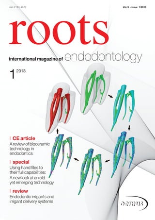
Roots International Magazine of Endodontology
1. issn 2193-4673 Vol. 9 • Issue 1/2013
roots
international magazine of endodontology
1 2013
| CE article
A review of bioceramic
technology in
endodontics
| special
Using hand files to
their full capabilities:
A new look at an old
yet emerging technology
| review
Endodontic irrigants and
irrigant delivery systems
2.
3. editorial _ roots I
Dear Reader,
_On 5 March, the Root Canal Anatomy Project (http://rootcanalanatomy.blogspot.com) will have
been online for two years. This project was conceived in the Laboratory of Endodontics of the University
of São Paulo, Brazil. During this time, the blog has registered over 210,000 visitors from 161 countries and
the videos have been watched more than 50,000 times. Considering that root-canal anatomy is a specific
subject in dentistry, we believe that our aim is being achieved.
The original goal of this project was the development and availability of non-commercial educational Prof. Marco Versiani, DDS, MS, PhD
resources in the endodontic field for educators, scholars, students, clinicians and the general public. The
main purpose is to demonstrate the complexity of the root-canal system in different groups of teeth and
the limitations of some procedures related to endodontic therapy. In a world where 3-D entertainment
rules, it is unthinkable that dentists, dental students and patients are still being educated using only
2-D models such as radiographs and photographs. The project emphasises the importance of animated
images of the internal anatomy of the teeth in the educational process.
People have asked me why the content of this project has not yet been commercialised. Basically, there
are two reasons for this. The first one is that the technology and training of our staff were only possible
because of a government sponsorship. So the government believed in our project and public money was
granted in order to develop our idea. It is thus only fair to make the project content available in the form
of free educational material.
The second reason has been guided by the following: dividing to multiply. Since the blog first went
online, the number of people who appreciate and respect our work has increased exponentially. I have
been invited to travel worldwide to talk about this project and had the unique opportunity to experience
other cultures and met amazing people I would otherwise not have met. Our images have been used on
invitation cards, personal web pages, educational flyers, and even on some covers of roots. Amazing!
It has been a wonderful experience to be a giver and a receiver at the same time. This is the most beautiful
of paradoxes. It is in the very act of giving of ourselves to others that we truly receive all for which we could
ever possibly wish.
While this editorial is not full of references to the newest innovations in endodontics or the answers
to your deep clinical questions, I am sure that you will be able to find such information in the pages of this
marvellous magazine. My purpose here is another one. Considering that this is the first issue of roots in
2013, I would like to wish you a year full of new friendships, happiness, peace, and unforgettable moments
with your family. I hope that you will keep providing the best of your skills in order to fulfil your patients’
needs and use our gift to provide pain release to make this world a better place. Keep giving! Giving is an
act of gratitude. Plant the seeds of generosity through your acts of giving, and you will grow the fruits of
abundance for yourself and those around you. Thank you for supporting us throughout these years.
My best wishes,
Prof. Marco Versiani, DDS, MS, PhD
Major Dental Officer (Brazilian Military Police)
Specialist in endodontics, didactics and bioethics
roots
1 _ 2013 I 03
4. I content _ roots
page 6 page 20 page 24
I editorial I industry
03 Dear Reader 38 Stropko Irrigator removes debris, making many
| Prof. Marco Versiani procedures easier
| Dr John J. Stropko
I CE article
39 Produits Dentaires presents PD MTA White
06 A review of bioceramic technology in endodontics
| Drs Ken Koch, Dennis Brave & Allen Ali Nasseh
I meetings
I special 40 International Events
14 Using hand files to their full capabilities:
A new look at an old yet emerging technology I about the publisher
| Dr Rich Mounce 41 | submission guidelines
20 Twisted Files changed the world of endodontics 42 | imprint
| Dr Sorin Sirbu
24 INITIAL®: The beginning of a new era for endodontic
instrumentation?
| Dr Matthieu Pérard, Dr Justine Le Clerc, Prof. Pierre Colon &
Prof. Jean-Marie Vulcain
I review
Cover image courtesy of Prof. Marco Versiani
30 Endodontic irrigants and irrigant delivery systems
3-D micro-CT models of a mandibular molar showing the changes of the
| Dr Gary Glassman
original root-canal anatomy (green) after preparation with a multiple-file rotary system.
Each colour represents preparation by one of five instruments. The last image in
the sequence represents the root canal after shaping (red) superimposed on the
original canal (green), demonstrating that most of the surface area was prepared
using the multiple-file system.
page 30 page 38 page 40
04 I roots 1_ 2013
5. FDI 2013 Istanbul
Annual World Dental Congress
28 to 31 August 2013 - Istanbul, Turkey
Bridging Continents for Global Oral Health
www.fdi2013istanbul.org
congress@fdi2013istanbul.org
6. I CE article _ bioceramic technology
A review of bioceramic
technology in endodontics
Authors_ Drs Ken Koch, Dennis Brave & Allen Ali Nasseh, USA
prognosis. The option of “saving the natural denti-
_ce credit roots
tion” is now back on the table.
By reading this article and then tak- However, before we investigate specific tech-
ing a short online quiz, you can gain niques, we must first ask ourselves is, “What are bio-
ADA CERP CE credits. To take the CE ceramics?” Bioceramics are ceramic materials specif-
quiz, visit www.dtstudyclub.com. ically designed for use in medicine and dentistry. They
The quiz is free for subscribers, include alumina and zirconia, bioactive glass, glass
who will be sent an access code. Please write support@ ceramics, coatings and composites, hydroxyapatite
dtstudyclub.com if you don’t receive it. Non subscribers and resorbable calcium phosphates.1, 2
may take the quiz for a $20 fee.
There are numerous bioceramics currently in use
in both dentistry and medicine, although more so in
_Since bioceramic technology was introduced medicine. Alumina and zirconia are among the bio-
to endodontics, the response has been exceptional. As inert ceramics used for prosthetic devices. Bioactive
more and more practitioners have thought through glasses and glass ceramics are available for use in
the process, they have been able to see not only the dentistry under various trade names. Additionally,
clear benefits of this technology in endodontics, but porous ceramics such as calcium phosphate-based
they are now asking how this technology can be materials have been used for filling bone defects. Even
applied to other aspects of dentistry. The application some basic calcium silicates such as ProRoot MTA
of bioceramic technology has not only changed (DENTSPLY) have been used in dentistry as root repair
Fig. 1_The particle size of BC Sealer endodontics both surgically and non surgically, it has materials and for apical retrofills.
is so fine (less than two microns), also begun to change the way we treatment plan our
it can actually be delivered with patients. As a result of bioceramic technology, we now Although employed in both medical and dental
a 0.012 capillary tip. (Photos/ have the ability to save more teeth in a predictable applications, it is important to understand the spe-
Provided by Ken Koch, DMD) fashion, while, in addition, improving their long-term cific advantages of bioceramics in dentistry and why
they have become so popular. Clearly the first answer
is related to physical properties. Bioceramics are
exceedingly biocompatible, non–toxic, do not shrink,
and are chemically stable within the biological envi-
ronment. Additionally, and this is very important in
endodontics, bioceramics will not result in a signifi-
cant inflammatory response if an over fill occurs
during the obturation process or in a root repair. A
further advantage of the material itself is its ability
(during the setting process) to form hydroxyapatite
and ultimately create a bond between dentin and the
filling material. A significant component of improv-
ing this adaptation to the canal wall is the hydrophilic
nature of the material. In essence, it is a bonded
restoration. However, to fully appreciate the proper-
ties associated with the use of bioceramic technology,
we must understand the hydration reactions involved
Fig. 1 in the setting of the material.
06 I roots 1_ 2013
7. CE article _ bioceramic technology I
_EndoSequence BC sealer setting reactions This material has been specifically designed as a
non-toxic calcium silicate cement that is easy to use
The calcium silicates in the powder hydrate to as an endodontic sealer. This is a key point. In addition
produce a calcium silicate hydrate gel and calcium to its excellent physical properties, the purpose of BC
hydroxide. The calcium hydroxide reacts with the Sealer is to improve the convenience and delivery
phosphate ions to precipitate hydroxyapatite and method of an excellent root canal sealer, while simul-
water. The water continues to react with the cal- taneously taking advantage of its bioactive charac-
cium silicates to precipitate additional gel-like cal- teristics (it utilizes the water inherent in the dentinal
cium silicate hydrate. The water supplied through tubules to drive the hydration reaction of the mate-
this reaction is an important factor in controlling rial, thereby shortening the setting time).
the hydration rate and the setting time as follow-
ing: As we know, dentin is composed of approximately
20 per cent (by volume) water, and it is this water that
The hydration reactions (A, B) of calcium silicates initiates the setting of the material and ultimately
can be approximated as follows: results in the formation of hydroxyapatite.4 Therefore,
if any residual moisture remains in the canal after
2[3CaO · SiO2] + 6H2O 3CaO · 2SiO2 · 3H2O + 3Ca(OH)2 (A) drying, it will not adversely affect the seal established
2[2CaO · SiO2] + 4H2O 3CaO · 2SiO2 · 3H2O + Ca(OH)2 (B) by the bioceramic cement. This is very important in
obturation and is a major improvement over previous
The precipitation reaction (C) of calcium phos- sealers. Furthermore, its hydrophilicity, small particle
phate apatite is as follows: size and chemical bonding to the canal walls also
contribute to its excellent hydraulics. But there is
7Ca(OH)2 + 3Ca(H2PO4 )2 Ca10(PO4 )6 (OH)2 + 12H2O (C) another aspect to sealer hydraulics. That is the shape
of the prepared canal itself.
For clinical purposes (in endodontics), the advan-
tages of a premixed sealer should be obvious. In Actually, it all begins with the file. To be more
addition to a significant saving of time and conven- specific, it all begins with the specific preparation
ience, one of the major issues associated with the created by the file—a constant taper preparation.
mixing of any cement, or sealer, is an insufficient and When using the EndoSequence technique, we can
non-homogenous mix. Such a mix may ultimately create either a 0.04 constant taper preparation or a
compromise the benefits associated with the mate- 0.06 taper. The real key is the constant taper prepara-
rial. Keeping this in mind, a new premixed bioceramic tion, because when accomplished it now gives us
sealer has been designed that hardens only when the ability to create predictable, reproducible shapes.
exposed to a moist environment, such as that pro- A variable taper preparation is not recommended
duced by the dentinal tubules.3 because its lack of shaping predictability (and its cor-
responding lack of reproducibility) will lead to a less
But, what is it specifically about bioceramics that than ideal master cone fit. This lack of endodontic Fig. 2a_This image shows the
make them so well suited to act as an endodontic synchronicity is why all variable taper preparations excellent adaption of the bioceramic
sealer? From our perspective as endodontists, some are associated with the overly expensive and more sealer (and gutta-percha) to the true
of the advantages are: high pH (12.8) during the ini- time consuming thermoplastic techniques. shape of the prepared canal.
tial 24 hours of the setting process (which is strongly
anti-bacterial); they are hydrophilic, not hydropho-
bic; they have enhanced biocompatibility; they do
not shrink or resorb (which is critical for a sealer-
based technique); they have excellent sealing ability;
they set quickly (three to four hours); and they are
easy to use (particle size is so small it can be used in a
syringe).
The introduction of a bioceramic sealer (Endo-
Sequence BC Sealer, Brasseler) allows us, for the first
time, to take advantage of all the benefits associated
with bioceramics but to not limit its use to merely
root repairs and apical retrofills. This is only possible
because of recent nanotechnology developments;
the particle size of BC Sealer is so fine (less than two
microns), it can actually be delivered with a 0.012
capillary tip (Fig. 1). Fig. 2a
roots
1 _ 2013 I 07
8. I CE article _ bioceramic technology
Knowing in advance what the final shape (constant est reported value was in Group IV, which employed
taper preparation) will be is a tremendous advantage ActiV GP sealer in combination with regular gutta-
in creating superior hydraulics. Then add in the feature percha cones. The conclusion of this study was that
of laser verified paper points and gutta-percha cones, employing a bioceramic sealer (such as BC Sealer) is
and we now start to develop a system where every- very promising in terms of strengthening the residual
thing matches (true endodontic synchronicity). root and increasing the in vitro fracture resistance of
endodontically treated teeth. This is a very significant
This concept of having everything match is so im- finding, especially regarding the long term retention
portant because it allows us, for the first time, to per- of an endodontically treated tooth.
form rotary endodontics in a truly conservative fash-
ion and to be able to use a hydraulic condensation In this particular study, the bioceramic sealer per-
technique. Furthermore, when used in conjunction formed best when combined with ActiV GP cones. In
with the EndoSequence filing system, this becomes a fact, bonding will occur between the bioceramic sealer
synchronized hydraulic condensation technique. This and the ceramic particles in the ActiV GP cones as
well as to the bioceramic particles present in the new
bioceramic coated cones (BC cones). The technique of
achieving a true bond between the root canal wall and
the master cone (as a result of creating endodontic
synchronicity and advanced material science) is
known as synchronized hydraulic condensation.
_Synchronized hydraulic condensation
The technique with this material is quite straight-
forward. Simply remove the syringe cap from the
EndoSequence BC Sealer syringe. Then attach an Intra
Fig. 2b Canal Tip of your choice to the hub of the syringe. The
Intra Canal Tip is flexible and can be bent to facilitate
Fig. 2b_A composite image has tremendous implications for the tooth as evi- access to the root canal. Also, because the particle size
demonstrating the true denced by a recent study published in the Journal of has been milled to such a fine size (less than 2 microns),
excellence of the technique. Endodontics.5 The purpose of this study was to evalu- a capillary tip (such as a 0.012) can be used to place
ate and compare the fracture resistance of roots obtu- the sealer.
rated with various contemporary-filling systems. The
investigators (Ghoneim, et. al.) instrumented 40 sin- Following this procedure, insert the tip of the sy-
gle-canal premolars using 0.06 taper EndoSequence ringe into the canal no deeper than the coronal one
files. The teeth were then obturated using four differ- third. Slowly and smoothly dispense a small amount
ent techniques. Group I used a bioceramic sealer iRoot of EndoSequence BC Sealer into the root canal. Then
SP (IRoot SP is BC Sealer in Europe) in combination with remove the disposable tip from the syringe and pro-
ActiV GP cones (Brasseler) while Group II used the ceed to coat the master gutta-percha cone with a thin
bioceramic sealer with regular gutta-percha. Group III layer of sealer. After the cone has been lightly coated,
utilized ActiV GP sealer plus ActiV GP cones and Group slowly insert it into the canal all the way to the final
IV employed ActiV G sealer with conventional gutta- working length. The synchronized master gutta-per-
percha cones. All four groups were obturated using a cha cone will carry sufficient material to seal the apex.6
single cone technique. Ten teeth were left unprepared
and these acted as a negative control for the study. The precise fit of the EndoSequence gutta-percha
master cone (in combination with a constant taper
Following preparation and obturation, all the teeth preparation) creates excellent hydraulics and, for
were embedded in acrylic molds and then subjected to that reason, it is recommended that the practitioner
a fracture resistance test in which a compressive load use only a small amount of sealer. Furthermore, as
(0.5mm/min) was applied until fracture. Subsequently, with all obturation techniques, it is important to in-
all data was statistically analyzed using the analysis of sert the master cone slowly to its final working
variance model and the Turkey post hoc test. length. Moreover, the EndoSequence System is now
available with bioceramic coated gutta-percha
Then results generated were quite remarkable. It cones. So in essence, what we can now achieve with
was demonstrated that the significantly highest frac- this technique is a chemical bond to the canal wall,
ture resistance was recorded for both the negative as a result of the hydroxyapatite that is created dur-
control and Group I (bioceramic sealer /Activ GP cone) ing the setting reaction of the bioceramic material
with no statistical difference between them. The low- and we also have a chemical bond between the
08 I roots 1_ 2013
9.
10. I CE article _ bioceramic technology
_Materials and methods
Sixteen recently extracted human molars were
mounted on individual stubs and underwent an ini-
tial high spatial resolution CT scan prior to any treat-
ment. Following biomechanical crown-down canal
preparation to an apical matrix of 35/0.04 and ultra-
sonic irrigation with 6 per cent sodium hypochlorite,
each sample was scanned a second time. Obturation
was completed using a single matched gutta-percha
Fig. 3a Fig. 3b cone and EndoSequence BC sealer. The coronal 4mm
of the gutta-percha was thermo-softened and com-
pacted vertically. Subsequent to canal obturation, a
third scan was made.
Scanning of the specimens was performed (Actis
150/130, Varian Medical Systems) with a 180-degree
rotation around the vertical axis and a single rotation
step of 0.9 degree with a cross-sectional pixel size of
approximately 24µm. All three backscatter projections
were aligned post-processing with sub-voxel accu-
Fig. 3c Fig. 3d
racy at 92 per cent CI in VG Studio Max 2.1 (Volume
Graphics GmbH) and manipulated to create regions of
Figs. 3a–5c_Cases treated with ceramic particles in the sealer and the ceramic par- interest for each of the scans.
bioceramics. (Clinical X-rays/ ticles on the bioceramic coated cone.
Provided by Allen Ali Nasseh, _Results
DDS, MMSc) Think about what we have just accomplished. We
are now doing root canals in a manner that truly is Analysis of volume occupied by sealer in relation to
easier, faster and better. As further evidence of this total original canal volumes was found to be extremely
technique, we asked Dr Adam Lloyd, the chairman of high with a mean of 97 per cent ± 2.8, much higher than
the Department of Endodontics at the University of reported previously using studies on canal surface
Tennessee, to share the results of a study recently area occupancy of material, with 75 per cent of sam-
conducted at the University of Tennessee.7 ples occupied at the ≥ 95 per cent level (Figs. 2a, 2b).
Fig. 4a Fig. 4b Fig. 4c
Fig. 5a Fig. 5b Fig. 5c
10 I roots 1_ 2013
11. CE article _ bioceramic technology I
While the properties associated with bioceramics
make them very attractive to dentistry, in general,
what would be their specific advantage if used as an
endodontic sealer? From our perspective as endo-
dontists, some of the advantages are: enhanced bio-
compatibility, possible increased strength of the root
following obturation, high pH (12.8) during the set-
ting process which is strongly anti-bacterial, sealing
ability related to its hydophilicity, and ease of use.8
Furthermore, the bioceramic sealer does not shrink
upon setting (it actually expands 0.002 per cent) and Fig. 6a Fig. 6b
once it is fully set, the material will not resorb.
The cases pictured in Figs. 3a through 5c demon-
strate the excellence of this technique.
_Retreatment of bioceramics
Bioceramic sealer cases are definitely retreatable
yet the issue of retreating these cases (and all the
associated misinformation) is not unlike that of glass
Fig. 6c Fig. 6d
ionomer. Historically there has been confusion about
retreating glass ionomer endodontic cases (glass
ionomer sealer is definitely retreatable when used as _Bioceramics as a root repair material Figs. 6a–6d_A case demonstrating
a sealer) and, similarly, there has been confusion retreatment of BC Sealer. (Clinical X-
concerning the retreatability of bioceramics.8 The key We are all familiar with the success of MTA (min- rays/Provided by Allen Ali Nasseh,
is using bioceramics as a sealer, not as a complete eral trioxide aggregate) as a root repair and apico DDS MMSc)
filler. This is why endodontic synchronicity is so im- retrofilling material. Furthermore, we realize that
portant and again, why the use of constant tapers because MTA is a modified Portland cement, it has
makes so much sense (it minimizes the amount of some limitations in terms of handling characteristics.
endodontic sealer thereby facilitating retreatment). It does not come premixed (and therefore must be
mixed by hand), is difficult to use on retrofills, and has
The technique itself is relatively straightforward. such a large particle size that it cannot be extruded
The key in retreating bioceramic cases is to use an through a small syringe. Yet it has a number of favor-
ultrasonic with a copious amount of water. This is able characteristics including a pH of 12.5, which is
particularly important at the start of the procedure in significantly anti-bacterial. However, in lieu of a
the coronal third of the tooth. Work the ultrasonic Portand cement-based material, we now have avail-
(with lots of water) down the canal to approximately able a medical grade bioceramic repair material.
half its length. At this point, add a solvent to the canal
(chloroform or xylol) and switch over to an Endo- This new repair material is, in fact, the Endo-
Sequence file (#30 or 35/0.04 taper) run at an in- Sequence Root Repair material, which comes either
creased rate of speed (1,000RPM). Proceed with this premixed in a syringe (just like BC Sealer) or as a pre-
file, all the way to the working length, using solvent mixed putty (Fig. 7). This is a tremendous help not just
when indicated. An alternative is to use hand files for in terms of assuring a proper mix but also in terms of
the final 2-3mm and then follow the gutta-percha ease of use. We now have a root repair material with
removal with a rotary file to ensure synchronicity. an easy and efficient delivery system. This is a key
development and a serious upgrade. This allows many
The case pictured in Figs. 6a and 6b demonstrates clinicians, not just specialists, to take advantage of
the retreatment of BC Sealer. its properties.
Fig. 7_EndoSequence Root Repair
Material. (Image courtesy of Real
World Endo)
Fig. 8_A section of material ready for
delievery.
Fig. 7 Fig. 8
roots
1 _ 2013 I 11
12. I CE article _ bioceramic technology
Fig. 9a Fig. 9b Fig. 9c Fig. 9d
Figs. 9a–10c_Cases demonstrate EndoSequence Root Repair material specifically has or as a syringeable paste possessed antibacterial proper-
healing and bone fill in less than six been created as a white premixed cement for both per- ties against a collection of Enterococcus faecalis strains.
months. (Clinical X-rays/Provided by manent root canal repairs and apico retrofillings. As a As a standard, they compared the ESRRM to MTA. Their
Allen Ali Nasseh, DDS MMSc) true bioceramic cement, the advantages of this new re- conclusion was, ESRRM, both putty and syringeable
pair material are its high pH (pH >12.5), high resistance forms and white ProRoot MTA demonstrated similar an-
to washout, no-shrinkage during setting, excellent bio- tibacterial efficacy against clinical strains ofE. faecalis.9
compatibility, and superb physical properties. In fact, it
has a compressive strength of 50–70MPa, which is sim- This research again validated earlier studies that
ilar to that of current root canal repair materials, ProRoot found ESRRM (Putty) and ESSRM (Paste) displayed sim-
MTA (DENTSPLY) and BioAggregate (Diadent). However, ilar in vitro biocompatiblity to MTA. Additionally, other
a significant upgrade with this material is its particle studies found that the ESRRM had cell viability similar to
size, which allows the premixed material to be extruded Gray and White MTA in both set and fresh conditions.10
through a syringe rather than inconsistent mixing by
hand and then placement with a hand instrument. Even more significant research was published
(January 2012) concerning bioceramics in general. In
The Clinicians Report (November 2011) published a comparison of endodontic sealers, it was demon-
findings on EndoSequence Root Repair Material. Some strated that in various moisture conditions within a
of its noted advantages as a root repair material were: root canal, iRoot SP (EndoSequence BC Sealer) out-
_easier to use and place than previous similar products, performed all the other sealers. The conclusion of
_good dispenser (tip/syringe) for easy dispensing, the study was, “Within the experimental conditions
_radiopaque, of this in vitrostudy, it can be concluded that the bond
_mulitple uses for a variety of clinical conditions, strength of iRoot SP to root dentin was higher than
_no mixing required. that of other sealers in all moisture conditions.”11
Furthermore, their final conclusion was that 95 As mentioned previously, the bioceramic material to
per cent of 19 CR Evaluators stated that they would use in surgical cases is the EndoSequence Root Repair
incorporate EndoSequence Root Repair Material into Material (RRM). The ESRRM is available in two different
their practice. Ninety-five percent rated it excellent or modes. There is a syringeable RRM (very similar to the
good and worthy of trial by colleagues. basic BC Sealer in its mode of delivery) and there is also
a RRM putty that is both stronger and malleable. The
Another significant piece of research was published consistency of the putty is similar to Cavit G. The RRM
in the Journal of Endodontics, where a research team in a syringe is obviously delivered by a syringe tip but the
investigated the antibacterial activity of EndoSequence technique associated with the putty is different.
Root Repair material against Enterococcus faecalis. The
aim of this study was to determine whether Endo- When using the putty, simply remove a small
Sequence Root Repair material either in its putty form amount from the room temperature jar and knead it for
Fig. 10a Fig. 10b Fig. 10c
12 I roots 1_ 2013
13. CE article _ bioceramic technology I
a few seconds with a spatula or in your gloved hands. introduction of Ceramir Crown & Bridge (Doxa Dental).
Then start to roll it into a hotdog shape. This is very sim- It is easy to predict that we will see more applications of
ilar to creating similar shapes with desiccated ZOE or this technology in different aspects of dental medicine.
SuperEBA (Bosworth). Once you have created an oblong
shape, you can pick up a section of it with a sterile in- In this article, we have introduced a new bioceramic
strument and use this to deliver it where needed (Fig. 8). sealer (EndoSequence BC Sealer) that when combined
This is an easy technique for apico retro fills, perforation with coated cones offers an exciting new obturation
repairs, and even for resorption defects. After placing technique (Synchronized Hydraulic Condensation). The
the putty into the apical preparation (or defect) simply properties associated with the new bioceramic sealer
wipe with a moist cotton ball and finish the procedure. also allow us to be more conservative in our endodon-
tic shaping which ultimately leads to the preservation
The cases pictured in Figs. 9a to 10c are evidence of of natural tooth structure. Surgical applications have
how beautifully this technique works. These cases are so also been introduced, and cases shown, which demon-
significant because they clearly demonstrate the ex- strate the remarkable ability of bioceramics. The future
traordinary healing capability of bioceramics, when used is bright for bioceramic technology and even more
as a repair material. The X-rays display amazing healing exciting for dental medicine._
and bone fill in less than six months, in the mandible.
Editorial note: A complete list of references is available from
_Pulp capping with bioceramics the publisher.
One of the other significant benefits of having bio-
ceramics come pre-mixed in a syringe (EndoSequence _about the authors roots
Root Repair Material) is the ability for all dentists to
now easily treat young patients in need of pulp caps or Dr Ken Koch, received both his DMD and certificate in endodontics
other pulpal therapies (e.g., pulpotomies). Previously, from the University of Pennsylvania School of Dental Medicine. He is
many specialists considered MTA to be the ideal mate- the founder and past director of the New Program in Postdoctoral
rial for a direct pulp cap because it did not seem to en- Endodontics at the Harvard School of Dental Medicine. Prior to his
gender a significant inflammatory response in the pulp. dndodontic career, Koch spent 10 years in the Air Force and held,
Unfortunately, due to price concerns and the difficulty among various positions, that of chief of prosthodontics at Osan Air
of placement, this methodology was not universally Force Base and chief of prosthodontics at McGuire Air Force Base.
accepted. However, we now have a true bioceramic In addition to having maintained a private practice, limited to endo-
material (ESRRM) that not only works well, but is eas- dontics, Koch has lectured extensively in both the United States and abroad. He is also the
ier to use. It is much easier. Hopefully, this will lead to an author of numerous articles on endodontics. Koch is a co-founder of Real World Endo.
increased use of bioceramics in our pediatric patients
and help these patients save their teeth. All dentists Dr Dennis Brave, a diplomate of the American Board of Endo-
can benefit from this upgrade in technique. dontics and a member of the College of Diplomates, received his
DDS degree from the Baltimore College of Dental Surgery, Uni-
The technique itself for a direct pulp cap with the versity of Maryland and his certificate in endodontics from the
bioceramic root repair material is as follows: Isolate the University of Pennsylvania. In endodontic practice for over 25
tooth under a rubber dam and disinfect the exposure years, he has lectured extensively throughout the world and
site with a cotton ball and NaOCl. Apply a small amount holds multiple patents, including the VisiFrame. Formerly an as-
of the RRM from the syringe or, take a small amount sociate clinical professor at the University of Pennsylvania, Brave
of the RRM putty from the jar, and place this over the currently holds a staff position at The Johns Hopkins Hospital. Along with having au-
exposure area. Then, cover the bioceramic repair mate- thored numerous articles on endodontics, Brave is a co-founder of Real World Endo.
rial with a compomer or glass ionomer restoration.
Following the placement of this material, proceed with Dr Allen Ali Nasseh, received his MMSc degree and Certificate
the final restoration, including etching if required. in Endodontics from the Harvard School of Dental Medicine in
Single visit direct pulp capping is now here. 1997. He received his DDS degree in 1994 from Northwestern
University Dental School. He maintains a private endodontic
_Future directions and prosthodontic practice in Boston (Microsurgicalendo.com) and holds a staff
applications position at the Harvard’s postdoctoral endodontic program.
Nasseh is the endodontic editor for several dental journals and
The future promises to be even more exciting in the periodicals and serves as the Alumni Editor of the “Harvard
world of bioceramics. There will be new fast set (8 to 10 Dental Bulletin.” He serves as the Clinical Director of Real World Endo.
minutes) repair materials introduced, as well as a spe-
cial bioceramic putty for pediatric use (primary teeth). The authors may be contacted via thier website, www.RealWorldEndo.com, or via
We have also seen the melding of bioceramic technol- email at info@realworldendo.com
ogy into the world of prosthodontic cements, with the
roots
1 _ 2013 I 13
14. I special _ instrumentation
Using hand files to their full
capabilities: A new look at an
old yet emerging technology
Author_ Dr Rich Mounce, USA
The endodontist is encouraged to compare their
treatment methods with those described here. The
Mani product line of files is described primarily
because these files are used daily by the author.
Examples of equivalent files are provided along-
side of Mani products throughout the article for
comparison.
There are myriad hand file designs, applications,
materials and manufacturing methods. In recent
years, multi axis grinding machines have provided
advancements of true clinical consequence, espe-
cially with regard to file flexibility and cutting abil-
ity. Given the wide diversity of available designs
and features, it is impossible to discuss the design,
clinical use or precautions required for every hand
file on the market. Neither barbed broaches nor
balanced force technique will be discussed.1
Fig. 1
_Introduction: Appreciating the unseen
Fig. 1_Mani D Finders. _Despite wide global acceptance of rotary nickel dimension
(Images provided by Dr Rich Mounce) titanium (RNT) canal enlargement, hand files remain
central to endodontic practice. It can be argued Hand files allow the clinician to manually “feel”
persuasively that proper canal negotiation and glide the unseen dimension in canal anatomy beyond
path creation are key ingredients to successful long- what radiographs alone can illustrate. Specifically,
term treatment, along with adequate and appro- by virtue of hand file resistance to apical advance-
priate irrigation, canal preparation, coronal seal, ment, the clinician can, by tactile feel, determine the
etc. Simply stated, after the preparatory steps of curvature, calcification, length, the anatomy of the
straight-line access and removal of the cervical MC, and if iatrogenic events may have occurred.
dentinal triangle with orifice openers, if the canal is Only cone beam technology comes close to provid-
not properly negotiated and a glide path prepared ing the tactile information provided by hand files
prior to RNT enlargement, cleaning and shaping (Planmeca).
procedures cannot be optimal.
Such tactile information helps determine treat-
This article was written primarily for the general ment strategies prior to shaping. Astute RNT use
dentist. It describes stainless steel (and, to a lesser has, as its foundation, intimate canal knowledge
degree, nickel titanium) hand files, reciprocation first by hand files. Forcing RNT files to length with-
and their clinical application. This article is in- out adequate hand file negotiation and a glide path
tended to be a clinical “how to” article, not a liter- is the harbinger of file fracture, canal transportation
ature review, hence a lack of extensive references. and inadequate cleaning and shaping.
14 I roots 1_ 2013
15. special _ instrumentation I
Fig. 2
_Hand file applications, differentiation _Principles for maximizing hand file Fig. 2_Mani K and H Files, and Mani
and general use principles effectiveness Reamers.
Hand files differ based on the following (among The use of hand files is based on several universal
other attributes): assumptions. These assumptions are:
1. Material of manufacture (carbon steel, stainless a) Optimal visualization of the access preparation,
steel, nickel titanium, among several other less ideally through the surgical microscope (Zeiss,
common materials). Global Surgical).
2. Taper (0.02 tapered, variable tapered, greater ta- b) Optimal radiographic evaluation of the tooth prior
pered). to access preparation including where necessary,
3. Initial cross sectional design before manufacture cone beam visualization. For those without CBCT
(triangular, square, rhomboid, among other initial technology, having two or optimally three different
shapes). pre-operative radiographic angles will provide the
4. Final cross sectional design. best possible visualization of canal anatomy short
5. Corrosion resistance. of a CBCT scan.
6. Handle design and material used for the hand file. c) Straight line access.
7. Tip sizes (of the individual instrument). d) Removal of the cervical dentinal triangle prior to
8. Progression of tip sizes across the spectrum of a hand file exploration.
given set of instruments. e) Copious irrigation at every stage in the procedure,
9. How the cutting flutes are produced (twisting, especially rinsing debris from the access prepara-
grinding, among other manufacturing methods). tion before hand files are inserted.
10. Tip design (active, non cutting, partially cutting). f) Pre-operative evaluation of the estimated and ex-
11. Whether the file is reciprocated, watch-wound pected true working length, final taper and master
(K files), rotated (K reamers), or used with a pull apical diameter.
stroke (H files). g) Curved files negotiate curved canals more effec-
12. Helix angle, rake angle, cutting angle (if different tively than straight ones. The EndoBender pliers
from the rake angle) number of flutes (as well as (Axis/Sybron) are an effective instrument to place
flute width, depth and number). the needed curvature onto hand files. Generally,
13. Possible variability of the cutting angle along the in canals that have been ledged or transported,
length of the file. placing an acute, 3- to 5-mm curve onto the apical
14. Linear length of the cutting flutes. portion of the hand file is beneficial. Multiple in-
15. In addition to the attributes above, hand files are sertions of curved hand files to bypass blocked and
designed to be stiff versus flexible, aggressive cut- transported canals (especially ledges) are the rule,
ting versus less aggressive, finishing files versus bulk not the exception. Alternatively, if no transporta-
shaping files, among other general classifications. tion has occurred (the canal is untouched or easily Fig. 3_Mani Flexile Files.
Fig. 3
roots
1 _ 2013 I 15
16. I special _ instrumentation
Fig. 4
Fig. 4_Mani RT Files. negotiable) the clinician can curve the file in their _General classes of hand files
fingers without an EndoBender.
h) Canals should always be negotiated with hand files Files primarily designed for canal negotiation
prior to using RNT files. Even if the clinician uses
a RNT glide path creator (PathFile, DENTSPLY Tulsa In calcified canals, hand file stiffness is an attrib-
or PreShapers, SpecializedEndo), the canal should ute. Mani D Finder files are representative of this
be first negotiated by hand to assure patency. class and are especially useful for early negotiation of
Clinician preference dictates whether a glide path calcified canals. The D finders have a D shaped cross
should be created by hand files or RNT files. section. Some files utilize carbon steel in manufac-
i) In the view of the author, hand files are single use ture and/or possess atypical tip sizes to facilitate
disposable instruments as they dull rapidly during negotiation. Stiffness can be attributed to either the
clinical function. files design (Mani D Finders) or the use of carbon steel
j) The use of nickel titanium hand files is a matter of and/or a combination of carbon steel and a modified
personal preference. While some clinicians desire design (Pathfinder CS, Axis/SybronEndo) (Fig. 1).
the flexibility and shape memory of nickel tita-
nium hand files, others do not. It should be noted K files
that nickel titanium hand files are available with
controlled memory, a proprietary thermo mechan- Generally, K files have a three or four-sided con-
ical process in which nickel titanium hand files lose figuration with more spirals than a K reamer. Mani
their shape memory yet retain their flexibility.2–4 K Files are four-sided. Overall, K files are the most
k) The principles of canal preparation must be ob- “universal” hand files covering the greatest number
served, irrespective of the methods utilized to of clinical indications.
achieve these principles (i.e., hand file canal en-
largement and/or RNT enlargement or a combi- K files are not as flexible as hand files designed
nation of these methods). These principles are to: specifically for flexibility (such as the Mani Flexile
_leave the canal in its original position (simply en- files discussed below) or nickel titanium hand files.
large it as described here); K files are used with a watch-winding hand motion
_leave the minor constriction (MC) of the apical and can be reciprocated (as described below). The
foramen at its original position and size; angle between the cutting flutes and long axis of a
_create a tapering funnel with narrowing cross K file is generally in the 25- to 40-degree range.5 Lex-
sectional diameters from orifice to apex; icon K Files are an additional example of another
_create a master apical taper that optimizes irriga- commercially available K file (DENTSPLY Tulsa).
tion and obturation hydraulics, and yet causes no
iatrogenic events (strip perforation, canal trans- K Reamers
portation unnecessary dentin removal—and does
not leave the tooth at risk of long term vertical Mani K Reamers are three-sided and contain fewer
Fig. 5_Mani SEC O K and H Files. fracture). spirals than K files. Smaller reamers are generally square
Fig. 5
16 I roots 1_ 2013
17. special _ instrumentation I
in cross section. Larger reamer sizes are generally trian- It is not advisable to use H files near the MC. The MC
gular. The angle between the cutting flutes and long axis can be transported easily if H files are used at or beyond
of a reamer is most often in the 10- to 30-degree range.5 the MC. Clinically, aside from transportation, such an
action lead to significant apical bleeding (Fig. 2).
Reamers are used in rotation, unlike K files. Hand
file rotation is associated with less canal transporta- Hand files of accentuated and variable taper
tion than K file watch winding.
Mani Flare Files are more tapered than standard
The use of K reamers versus K files is a matter of hand files—0.05 taper compared to 0.02 taper. They are
personal preference. K type instruments of both types used to prepare tapered canals for doctors who hand
(reamers versus K files) should be manipulated care- file the entire preparation among other more special-
fully when used counterclockwise due to the risk of ized uses such as verifying taper before cone fit.
instrument fracture. Lexicon K Reamers are an addi-
tional example of a commercially available K reamer Accentuated taper is also available with nickel ti-
(DENTSPLY Tulsa)—these are triangular in cross section. tanium GT Hand Files. ProFile 0.04 Hand Files are 0.04
tapered and come in a variety of tip sizes, again in
H files nickel titanium. ProTaper Universal Hand Files feature
the ProTaper variable taper design in shaping and
H files (Mani H Files as well) have conical spirals finishing files in various lengths (all of the above are
ground into them. They are used on the pull stroke for manufactured by DENTSPLY Tulsa).
gross removal of canal contents in the coronal third
and in retreatment. H files should not be rotated due Flexible Files
to fracture risk inherent in their design. The angle
between the cutting flutes and long axis of an H file is Mani Flexible Files are triangular in cross section.
generally in the 60- to 65-degree range.5 Files with a triangular cross section are more flexible
C
AD
I hereby agree to receive a free trail subscription of (4 issues per year).
I would like to subscribe to for € 44 including shipping and VAT
for German customers, € 46 including shipping and VAT for customers outside of Ger-
many, unless a written cancellation is sent within 14 days of the receipt of the trial sub-
scription. The subscription will be renewed automatically every year until a written
cancellation is sent to OEMUS MEDIA AG, Holbeinstr. 29, 04229 Leipzig, Germany, six
weeks prior to the renewal date.
Reply via Fax +49 341 48474-290 to OEMUS MEDIA AG or per E-mail to
grasse@oemus-media.de
Last Name, First Name
Company
Street
ZIP/City/Country
E-mail Signature
Notice of revocation: I am able to revoke the subscription within 14 days after my order by
sending a written cancellation to OEMUS MEDIA AG, Holbeinstr. 29, 04229 Leipzig, Germany.
roots 1/13
Signature
DHJ 1/10
OEMUS MEDIA AG Holbeinstraße 29, 04229 Leipzig, Germany
Tel.: +49 341 48474-0, Fax: +49 341 48474-290, E-Mail: grasse@oemus-media.de
18. I special _ instrumentation
than those with square cross sections. Flexible stain- article, the terms TWL and MC are synonymous.
less steel hand files are generally used in easily nego- The purpose of reciprocation is to save time, reduce
tiated canals. Clinician preference dictates whether hand fatigue and prepare a space into which RNT
to use flexible stainless steel files relative to nickel files can subsequently be inserted with minimal
titanium hand instruments (Fig. 3). torque stresses (prepare a glide path).
Additional files in this class are Lexicon FlexSSK Reciprocation is inherently safe. It is difficult to
Files (DENTSPLY Tulsa). These files are also available in fracture hand files when this technique is used
medium sizes (12, 17, 22, etc.). appropriately. Fracture or iatrogenic misadventure
generally occurs when the files are inappropriately
Aggressive cutting files placed (well beyond the MC), the wrong type of hand
file is reciprocated (H) and/or the speed is grossly
Mani RT files (possessing a parallelogram cross- exaggerated above the recommended levels.
section) and a 71-degree cutting angle, making them
more aggressive relative to many of the other files Reciprocating hand piece attach-
included here. RT files would be used primarily by ments fit onto an E-type coupling and
doctors who are hand filing the entire canal in can be powered at 900rpm, for example
conjunction with other hand files (Fig. 4). at the 18:1 setting on an electric endodon-
tic motor.
Nickel titanium files
Fig. 6
To initiate reciprocation, the file is left in the
GT Hand Files (made of nickel titanium) canal at the TWL and the reciprocating hand
are available in various tapers and tip sizes piece is placed over the file (the file is inserted into
(DENTSPLY Tulsa). Lexicon FlexNTK Files are the head of the reciprocating hand piece and is
made of nickel titanium and come in vari- held there while reciprocating). The attachment
ous tip sizes while maintaining a constant reciprocates the file clockwise and counter clock-
taper. As mentioned above, clinician wise—for example, with a 30-degree clockwise and
preference dictates whether a flexible 30-degree counterclockwise movement. These at-
stainless steel file is more desirable than tachments do not rotate the file a full 360 degrees—
a nickel titanium hand file. in contrast to how RNT files are powered. Different
reciprocating hand pieces may have variations on
Medium sizes, K, H and reamers the degree of clockwise or counterclockwise rota-
tion and possibly include a vertical amplitude.
Mani provides K Files, H Files and
stainless-steel reamers in medium sizes The Synea W&H-62A is an example of a recip-
(12, 17, 22, 27, etc.). ProFile Series 29 rocating hand piece (MounceEndo) attachment
Fig. 6_The Synea W&H WA-62. Stainless Steel 0.02 Hand Files have a with a 30-degree clockwise and 30-degree coun-
A reciprocating hand piece constant 29 per cent increase in tip size terclockwise motion. Reciprocation is the tech-
attachment. in 0.02 taper. Use of medium sizes avoids nique and file motion utilized in the Wave One
the dramatic increase in tip diameter canal preparation system (DENTSPLY Tulsa).
with increasing tip sizes, especially between a #10
an #15 hand file (a 50 per cent increase in size of the Clinically, using the SEC O K File as an example, the
#15 relative to the #10 hand file). SEC O K File is placed to the TWL, the attachment
placed over the file and reciprocation commences as
Safe-ended hand files and reciprocation described above. The file is reciprocated for 15 to 30
seconds, using a 1- to 3-mm vertical amplitude move-
Mani SEC O files are available in an H and K file ment. Clinically, the file will become less tightly bound
variety. Both are “safe-ended,” as they do not cut on as the canal is enlarged.
their tips. The Mani SEC O K File is ideal for recipro-
cation. SEC O H files (and H files in general) are not If, for example, a #08 SEC O K file is the first file that
reciprocated (Figs. 5 & 6). binds in the canal at TWL this file is reciprocated. Once
the #08 SEC O K File is reciprocated, the canal will now
Reciprocation is a very safe technique, whereby accept a #10 SEC O K File to TWL. The #10 SEC O K File
the clinician can use a reciprocating hand piece at- is reciprocated. Once reciprocation is complete, the
tachment to replicate manual hand file watch wind- canal will allow a #15 SEC O K File to reach the TWL.
ing. Clinically, reciprocation is used after the canal Once the canal is enlarged to approximately the size
has been negotiated to the TWL and reciprocation of a #15 or #20 hand file, the canal is ready for RNT
proceeds with the first file that binds at TWL. In this enlargement.
18 I roots 1_ 2013
20. I special _ instrumentation
Twisted Files changed
the world of endodontics
Case report
Author_ Dr Sorin Sirbu, Romania
Fig. 1_TF 25.12 to 25.04 files.
Fig. 2_The 25.12 file is beginning to
untwist near the tip because of
overworking. This file must be
replaced immediately.
Fig. 1 Fig. 2
_Introduction systems? Firstly, by its unique machining—which is a
SybronEndo patent.
There are many rotary systems on the dental
market at present. All of these systems are relatively The NiTi wire is brought into a special state
Fig. 3_Clinical examination of tooth similar, except for one. This system is called Twisted (called R-Phase) that allows the twisting of the file.
26 revealed a composite filling. Files (TF) and it was introduced to the dental market in This makes TF distinct from all the other systems, for
Fig. 4_The initial radiographic 2008. I am glad to have been among the first users of which the shape of the file is machined by milling,
examination showed a massive this system, which has changed the endodontic a mechanical process. This unique procedure lends
composite filling. world. How does this system differ from other rotary particular resistance to TF, as well as an extraordinary
Fig. 3 Fig. 4
20 I roots 1_ 2013
21. special _ instrumentation I
Fig. 5 Fig. 6
flexibility. Owing to this manufacturing technique, _The clinical procedure Fig. 5_After removing the composite
a TF untwists before breaking, warning the dentist filling, a secondary occlusal decay
in this way. In addition, being made by twisting and In this part, I will describe the TF technique. Treat- was observed.
not by polishing/milling, all the microcracks are elim- ment with TF always begins by creating a glide path Fig. 6_The opening of the pulp
inated, resulting in a more resistant, more robust file. in the canals with #6 to 20 K-files. After opening and chamber in tooth 26 and the
The manufacturing process is completed by applying access, treatment inside the canal begins. In the ab- identification of the canals.
an advanced surface conditioning treatment that sence of adequate access into the canal, there is the
makes the edges active (cutting). risk of overworking the file and its subsequent frac-
ture. By opening the canals with K-files, important
The tip of a TF is inactive, which allows it to follow information about the anatomy of the root canal is
the route of the canal easily and to minimise canal obtained, such as the existence of curves and the
transportation. The working sequence with this sys- diameter of the root canal.
tem is terribly easy and consequently working time is
reduced. Generally, the first TF that is introduced into the
canal is TF 25.08 (the apical diameter is 25mm and it
The files may be recognised and differentiated by has a taper of 8%), which in most cases will reach the Fig. 7_The shaped canals ready
the help of the practical system of codification. There working length previously detected by means of an for endodontic obturation using
are two coloured rings: the lower one (closer to the apex locator. The endodontic engine must be set at the warm vertical condensation
active part) shows the apical diameter (ISO standard; 500rpm and the torque at 2Ncm. The file is intro- technique.
for example: red = 25) and the upper one shows the duced into the canal in rotation and without pressure Fig. 8_The canals obturated by
taper size (Fig. 1). Two working lengths are available: applied. It is sufficient to advance 2 to 4mm when means of the warm vertical
23 and 27mm. introducing the file into the canal. If the file does not condensation technique.
Fig. 7 Fig. 8
roots
1 _ 2013 I 21
22. I special _ instrumentation
Fig. 9 Fig. 10 Fig. 11
Fig. 9_The sealing of the root canals advance, then a file with a smaller taper (TF 25.06) _Case 1
using RxFlow composite. must be used instead to achieve working length.
Fig. 10_The final composite The patient came to our clinic with acute apical
restoration. During preparation, there must be sodium hypo- periodontitis around tooth 26. When examined clini-
Fig. 11_The final X-ray showing all chlorite in the root canal at all times. The file is cleaned cally and radiographically, the tooth showed a large
four obturated canals. and examined to detect possible distortion before in- composite filling next to the distal pulp horn (Figs. 3
troduction to the canal and upon withdrawal. If the file & 4). The periodontal examination did not find any ir-
exhibits some distortion, it must be replaced (Fig. 2). If regularities; however, the tooth was extremely painful
TF 25.08 reaches working length easily, then a file with in vitality tests. Initially, I intended to replace the com-
a greater taper can be used (TF 25.10 or 25.12). posite filling. After removing the old composite filling,
I noticed secondary decay that reached up to the pulp
After reaching the desired taper, the final apical di- chamber (Fig. 5) and I subsequently decided to pursue
ameter is prepared. There are many studies in the en- endodontic treatment.
dodontic literature that have found that apical prepa-
ration up to a #25 K-file is insufficient. For this reason, The treatment was performed in one session.
after reaching the taper the TF 30.06 or 35.06 or both Four canals were identified (MB, MB2, DB and P;
are used. If greater apical diameters are desired, TF 40.04 Fig. 6). The main problem was in the MB2 canal,
or 50.04 can be used. The greater the apical diameter is, which had a 90-degree curvature. The treatment was
the greater the quantities of irrigation that reach the performed with TF 25.06 in the MB2 canal and with
apex will be and the cleaner the apex will be. It is gener- TF 25.08 in the other canals (Fig. 7). As a final irrigant,
ally known that apical preparation by means of rotary I used SmearClear (SybronEndo). After obturating
files with large diameters can create many problems the canals with warm vertical condensation using
because of the stiffness of the rotary files, such as trans- the Elements Obturation Unit (SybronEndo; Fig. 8),
Fig. 12_Tooth 37 at the initial clinic portation of the apex and changes to the root-canal the canals were sealed with a coloured composite
examination. anatomy. With TF, however, this does not occur, owing to (RxFlow, Dental Life Sciences; Fig. 9). Finally, the
Fig. 13_The initial radiographic the unique machining process, which ensures that the tooth was restored with a composite filling (Fig. 10)
examination of tooth 37. files are flexible, even those with large apical diameters. and the control X-ray was taken (Fig. 11).
Fig. 12 Fig. 13
22 I roots 1_ 2013
23. special _ instrumentation I
Fig. 14 Fig. 15 Fig. 16
_Case 2 _Conclusion Fig. 14_Shaped and cleaned canals.
Fig. 15_The impression left by the
The patient was referred to our clinic by another TF permits treatment even in the most difficult file on the gutta-percha cone attested
doctor who had come across difficulties when clinical situations and is essential to the dentist. to the merging of the MB and ML
identifying and working in the canals of tooth 37. Using TF, it is possible to widen the apex up to a canals.
The presence of a temporary filling done during #50 K-file without the risk of transporting the Fig. 16_Final endodontic obturation
previous treatment was observed during the clini- apex. In addition, owing to its unique machining, by means of the warm vertical
cal examination (Fig. 12). An initial X-ray was taken TF untwists before separating in the canal, there- condensation technique.
to identify any possible associated pathology, the by giving the dentist timely warning to replace
presence of canals, etc. (Fig. 13). the file and significantly decreasing the risk of
accidents while working with the rotary files.
After removing the temporary filling, three root Another major advantage is that this system aids
canals were identified, shaped and cleaned (Fig. 14). the maintenance of the root-canal anatomy ow-
The treatment was performed with TF 25.10 up to ing to the remarkable flexibility of the files._
40.04. The MB and ML canals merged, as shown by
the file impression from the MB canal on the gutta-
percha cone (Fig. 15). The final irrigation was done
with SmearClear. The tooth was obturated with _about the author roots
warm vertical condensation using the Elements
Obturation Unit (Fig. 16), and finally restored with Dr Sorin Sirbu graduated
composite material and a fibreglass post (Fig. 17). from the Carol Davila Univer-
sity of Medicine and Pharmacy
The control X-ray showed that the root canal and in Bucharest in Romania.
numerous accessory canals (Fig. 18) had been prop- At present, he works in a Fig. 17_Tooth 37 restored using
erly cleaned and obturated due to working with private dental practice in composite material and a fibreglass
TF rotary files and negative irrigation with EndoVac Bucharest. post.
(SybronEndo). Fig. 18_The final X-ray.
Fig. 17 Fig. 18
roots
1 _ 2013 I 23