2. tularemia-like disease in animal models, F. novicida provides
a safe and appropriate research alternative to virulent F. tu-
larensis strains.
Although research efforts have elucidated many of the mo-
lecular mechanisms of Francisella pathogenesis over the past
decade (9–12), the detailed mechanistic interplay between
bacterial factors and the host immune response remains in-
complete. One of the first steps during the bacteria-host
interaction is the phagocytosis or entry of bacteria into the
host cells. To date, there are five receptors described in the
phagocytosis of Francisella by macrophages: scavenger re-
ceptor A (SRA), mannose receptor (MR), Fc-gamma receptor
(Fc␥R), CR3 complement receptor (CR3) and surface-ex-
posed nucleolin (SEN) (13–16). After being recognized by one
of these receptors Francisella triggers a signaling cascade
that culminates in the rearrangement of actin filaments and
engulfment of the bacteria (17–19). This signaling cascade is
in part regulated by phosphorylation and dependent on ki-
nases such as Akt, Syk and Erk (20, 21). More recently,
Francisella was also shown to be endocytosed by liver cells in
a mechanism that involved cholesterol-rich domains on the
plasma membrane and clathrin-coated vesicles (22). In addi-
tion to phagocytosis, the host inflammatory response through
TLR signaling and the activation of the inflammasome are also
essential factors during the early stages of the infection (9–11,
23).
Francisella has quite a distinct inflammatory response com-
pared with other Gram-negative bacteria. Unlike E. coli or
Salmonella, the lipopolysaccharide (LPS) from Francisella is
not endotoxic and lacks the ability to stimulate toll-like recep-
tor 4 (TLR4), the pro-inflammatory receptor that recognizes
Gram-negative bacteria (24). The lack of TLR4 activation is
related to hypoacylation and the addition of very long chain
fatty acids to the diglucosamine backbone of the lipid A
portion of LPS (24). Typically, TLR4 is activated by lipid A
containing six short (12–14 carbon) fatty acids, whereas Fran-
cisella lipid A has four fatty acids with longer carbon chains
(16–18 carbon) (11, 24). On the other hand, Francisella has
been shown to activate TLR2 (25, 26) that recognizes lipopro-
teins, glycans, and glycophospholipids more frequently pres-
ent in Gram-positive bacteria, fungi, and protozoan parasites,
respectively (27). Francisella also activates another compo-
nent of innate immune system—the inflammasome (10). As-
sembly of the inflammasome is activated by Francisella infec-
tion and is responsible for triggering a pro-inflammatory
response through processing and maturing cytokines, such
as IL-1 and IL-18 (28–32), in addition to triggering a pro-
inflammatory response that leads to cell death by pyroptosis
(30–33). More recently, Peng et al. showed that several Fran-
cisella mutants defective in membrane proteins were hyper-
cytotoxic to host cells in a process that is dependent on the
inflammasome (29).
It has been proposed that Francisella actively suppresses
signaling by TLRs and the inflammasome to evade the host
immune system and effectively establish an infection (11). To
identify genes that are involved in host immune evasion, we
recently screened a comprehensive mutant library containing
over 3000 F. novicida strains for mutants that were hyper-
cytotoxic (34). Among the identified strains four F. novicida
genes were involved in LPS core synthesis and assembly.
These mutants are unable to synthesize the LPS core and
thus produce LPS with a very short glycan compared with the
long polysaccharide chains of the wild-type (WT) (34). Inter-
estingly, three mutants (⌬lpcC, ⌬manB, and ⌬manC) are hy-
per-cytotoxic to RAW 264.7 cells, a murine macrophage-like
cell line that lacks the production of ASC (apoptosis-associ-
ated speck-like protein containing a caspase recruiting do-
main), a key component of the inflammasome signaling. This
observation suggests that although ⌬lpcC, ⌬manB, and
⌬manC are also defective in membrane components, the
mechanism of cell killing may be different than the mutant
strains studied by Peng et al. (29). Also surprising was that
⌬lpcC, ⌬manB, and ⌬manC strains were phagocytosed at a
much higher rate than WT F. novicida via a mechanism that
does not require actin polymerization (34). Actin polymeriza-
tion has thus far been demonstrated to be involved in inter-
nalization of Francisella by either phagocytic or endocytic
pathways (21, 22, 35); therefore, the finding that ⌬lpcC,
⌬manB, and ⌬manC mutant strains are able to invade host
cells independent of actin filaments remains to be under-
stood. Deletion of lpcC from F. tularensis Schu S4 also results
in a similar phenotype, suggesting that this phenomenon acts
broadly among the different Francisella species rather than
specific to F. novicida (Tempel and Heffron, unpublished
observations).
To better understand the mechanism of infection and cell
killing by these hyper-cytotoxic F. novicida strains, we evalu-
ated cell signaling stimulated by the F. novicida ⌬lpcC strain
in comparison to its parent WT strain. Here we carried out a
comprehensive quantitative phosphoproteomic analysis us-
ing isobaric tags for relative and absolute quantification
(iTRAQ), which allowed us to analyze WT and ⌬lpcC F. novi-
cida infection with multiple replicates in the same experiment.
By combining network and function-enrichment analyses fol-
lowed by immunological and cellular experiments we identi-
fied new mechanistic aspects of host phagocytosis and cell
killing by F. novicida.
EXPERIMENTAL PROCEDURES
Ethics Statement—Mouse experiments were approved under the
protocol of the Oregon Health and Science University Institutional
Animal Care and Use Committee (IACUC B11220) and performed in
accordance with the guidelines for the Care and Use of Laboratory
Animals of the National Institutes of Health to minimize animal
suffering.
Bacterial Culture—Wild type and ⌬lpcC Francisella novicida were
grown on cysteine heart agar (CHA) from Ϫ80 °C freezer stocks
before culturing in liquid media. Bacteria were cultured overnight in
tryptic soy broth ϩ 0.1% cysteine (TSBC) with shaking (200 rpm), and
Phosphoproteome of Francisella-infected Cells
3298 Molecular & Cellular Proteomics 12.11
3. the optical density at 600 nm (OD600) was measured to determine
CFU/ml.
Cell Culture and Infection—RAW 264.7 or J774 murine macro-
phage-like cells (American Type Culture Collection, Manassas, VA)
were cultured in DMEM/High Glucose (HyClone, Logan, UT) supple-
mented with 10% fetal bovine serum (FBS; Invitrogen, Rockville, MD)
at 37°C in the presence of 5% CO2. For infection, bacteria were
added to 50–80% confluent cells in tissue culture plates at a multi-
plicity of infection (MOI) of 100. The cells were centrifuged at 1000 ϫ
g for 5 min at room temperature and incubated at 37°C with 5% CO2.
Thirty minutes to 1 h after infection, cells were washed three times
with PBS and either prepared for analysis (time ϭ 0) or overlaid with
DMEM containing 100 g/ml gentamicin to prevent growth of extra-
cellular bacteria and incubated at 37°C with 5% CO2. After one hour,
cells were washed three times with PBS and either prepared for
analysis or overlaid with 10 g/ml gentamicin and incubated at 37°C
with 5% CO2 for the duration of the infection. Each infection assay is
performed in triplicate unless otherwise stated.
Western Blot Analysis—RAW 264.7 or J774 murine macrophage-
like cell lines were cultured as described in 6-well plates and either
subjected to mock infections or acute 1 h infections with wild-type
(U112) or ⌬lpcC F. novicida strains, as indicated. Following infection,
cells were washed three times in PBS and incubated with media
containing 100 nM okadaic acid and 10 g/ml gentamycin. Four hours
after initial exposure to Francisella, cells were collected in PBS and
pellets stored at Ϫ80°C for 10 min. Cell pellets were then lysed in the
presence or absence of phosphatase inhibitors and EDTA depending
on whether they were to undergo lambda phosphatase treatment (20
mM Tris pH7.4, 150 mM NaCl, 0.5% Nonidet P-40, 1 mM dithiothreitol
(DTT), protease inhibitor mixture (Roche), Ϯ 5 mM EDTA, Ϯ 10 mM
NaF, Ϯ 10 mM Na3VO4, Ϯ 10 mM p-Nitrophenyl phosphate, Ϯ 100 nM
Okadaic Acid, and Ϯ 1ϫ HALT phosphatase inhibitor mixture (Pierce,
Rockford, IL)). Thirty g of cleared whole cell lysates (quantified using
the Bradford assay) was resolved on 10% Tris-Glycine SDS-PAGE.
Manganese and 5 U of lambda phosphatase (New England Biolabs,
Ipswich, MA) were added to whole cell lysates as indicated and the
reaction was incubated at 30°C for 30 min; stopped with Laemmli
protein loading buffer. Transferred proteins were subjected to West-
ern blot analysis using anti-TTP 1:1000 (Millipore ABE285), anti-RNA
Pol II Rpb1 1:1000 (SCBT sc-900), and anti-rabbit IgG-HRP second-
ary 1:10,000 (Promega, Madison, WI).
Cytokine Assay—RAW 264.7 cells were infected in triplicate with
⌬lpcC, parent, mock, or untreated in 24-well plates as described
above for 0, 2, or 6 h. At the end of each time point, 350 l of
supernatant was frozen at Ϫ80°C and submitted for analysis. The
samples were tested on Quansys Biosciences’ (Logan, UT) Q-Plex
ArrayTM
kits for expression of 21 unique cytokines. Q-PlexTM
tech-
nology is a multiplex approach that involves the micro-spotting of
individual groups of capture antibody in either a Cartesian or polar
coordinate system on the bottom of a 96-well plate, each spot being
its own micro ELISA. Each well was identically spotted. Standard
ELISA procedures were applied notably initial sample incubation,
washing, secondary antibody incubation, washing, incubation with
the label, and measurement are involved. The label and reporting
system used in a Q-Plex ArrayTM
is chemiluminescent.
Apoptosis and Necrosis Assay—RAW 264.7 cells were infected for
18 h in triplicate in an opaque white 96-well tissue culture plate (BD,
Billerica, MA) with ⌬lpcC, WT U112, mock, or uninfected. 10 mM
staurosporine or 100 mM ionomycin were added to triplicate wells as
controls for apoptosis or necrosis, respectively. Thirty minutes before
analysis, 5 l of 10% saponin was added to triplicate control wells as
a lysis control. The cells were analyzed for viability, cytotoxicity, and
apoptosis using the ApoTox-GloTM
Triplex Assay (Promega) accord-
ing to manufacturer’s instructions. The assay was read using a
SpectraMax® GeminiTM
XS plate reader (Molecular Devices, Sunny-
vale, CA).
Cytotoxicity (Cell Invasion) Assay—J774 cells were infected in trip-
licate in 24-well tissue culture plates (BD, Billerica, MA) with ⌬lpcC,
WT, mock, or uninfected, Ϯ cells invasion inhibitors (cytochalasin D,
5 g/ml; nocodazole, 10 g/ml; withaferin A, 5 M; wortmannin, 0.1
M). One hour after infection, the cells were washed and incubated
with DMEM containing 100 g/ml gentamicin, as above. After one
hour of incubation with gentamicin, macrophages were washed and
lysed with 0.5% saponin for 30 min at 37°C, and the lysates were
serially diluted and plated on CHA plates to determine F. novicida
CFU/ml.
Protein Extraction and Trypsin Digestion—In preparation for phos-
phorylation analyses, RAW 264.7 macrophage-like cells were infected
with either wild-type or ⌬lpcC Francisella strains as described above
for 30 min, washed with PBS and incubated for an additional 0, 1, 2,
and 4 h. The contents of each well of the 6-well plates were resus-
pended in 500 l of lysis buffer (8 M urea in 100 mM NH4HCO3 pH 8.0
containing Sigma phosphatase inhibitor cocktails 1 and 3). The con-
tents of 3 wells were combined and stored at Ϫ80°C. Proteins were
then thawed and quantified by BCA assay (Pierce). Protein disulfide
bonds were reduced by DTT (5 mM) for 1 h at 37°C, and the resulting
free thiol groups were alkylated by iodoacetamide (20 mM) for 1 h at
room temperature in the dark. After alkylation, samples were diluted
4-fold with 50 mM NH4HCO3 (pH 7.8) and digested with trypsin at ratio
of 1:50 (trypsin/protein) for 4 h at 37°C, diluted by another 2-fold with
the same buffer, and subjected to a second treatment with trypsin
and incubated at room temperature overnight. The reaction was
stopped by lowering the pH to 2.5 with trifluoroacetic acid. The
resulting peptides were desalted using C18 SPE (Discovery DSC-18,
Supelco) cartridges, concentrated down in SpeedVac SC250 Epress
(ThermoSavant) and their concentrations were measured by BCA
assay.
iTRAQ Labeling and Phosphopeptide Enrichment—Peptides (50
g) from each sample were subjected to 8-plex iTRAQ (Applied
Biosystems) labeling as the experimental design in Fig. 1, and ac-
cording to the manufacturer recommendations. Each iTRAQ experi-
ment had biological duplicate controls with mock infections and
biological triplicates of RAW 264.7 cells infected with either wild-type
or ⌬lpcC F. novicida strains, all collected at the same time point. After
labeling and mixing equal amounts of peptides, samples were de-
salted using C18 SPE cartridges, concentrated in a SpeedVac and
submitted to quantification by BCA assay. The phosphopeptide en-
richment was performed exactly as described by Nguyen et al., using
Fe3ϩ
-nitriloacetic acid functionalized magnetic beads (36). After en-
richment, phosphopeptides were concentrated by vacuum centrifu-
gation before mass spectrometry analysis.
Proteomic Analysis—Phosphopeptides were loaded to a trap col-
umn (150 m i.d. ϫ 4 cm; 5 m C18 particles, Phenomenex) and the
separation was achieved in a longer column (50 m i.d. ϫ 40 cm; 5
m C18 particles, Phenomenex) using an isobaric gradient of aceto-
nitrile over 200 min (37). Eluted peptides were directly analyzed by
electrospray ionization - mass spectrometer (Orbitrap Velos equipped
with ETD). Full scan - 400–200 m/z with resolution of 60,000 at 400
m/z. The five most intense ions were selected for HCD (40% normal-
ized collision energy) followed by decision tree ETD (z ϭ 3, m/zϽ650;
z ϭ 4, m/zϽ900; z ϭ 5, m/zϽ950) or CID scans (z ϭ 2; z ϭ 3,
m/zϾ650; z ϭ 4, m/zϾ900; z ϭ 5, m/zϾ950). MS/MS spectra were
converted to peak lists using DeconMSn (version 2.2.2.2, http://
omics.pnl.gov/software/DeconMSn.php) (38) using default parame-
ters and database searched using SEQUEST v27 against murine
protein sequences from Uniprot (downloaded on August 22nd, 2011)
and porcine trypsin both in forward and reverse orientations (32,780
total sequences). As searching parameters, precursor ion mass tol-
Phosphoproteome of Francisella-infected Cells
Molecular & Cellular Proteomics 12.11 3299
4. erance was 50 ppm, and fragmentation tolerances for CID, ETD,
and HCD were 0.5, 0.5, and 0.05 Da, respectively. Only fully tryptic
peptides were considered, with two missed cleavages allowed. Phos-
phorylation (ϩ79.9663 Da) of serine, threonine, or tyrosine was
searched as a dynamic modification. Cysteine carbamidomethylation
(ϩ57.0215 Da) and lysine and N terminus labeling with iTRAQ
(ϩ304.2022 Da) were searched as static modifications. The maximum
number of variable modifications was set to 3. Peptides were then
filtered using a cutoff of MSGF (39) Յ 1.0e-9, which resulted in ϳ1%
false-discovery rate. When peptides matched multiple sequences
only the first in the database was reported. Ascore (40) was used to
estimate the confidence of phosphorylation modification site assign-
ment, Sites with an A-Score Ն 19 were considered as unambiguous
sites (Ն 99% confidence). Sites with Ϫ1 Ascore indicate these sites
are the only choices for modification on the peptide identified.
Quantitative Data Analysis—iTRAQ reporter ion intensities were
extracted with MASIC (MS/MS Automated Selected Ion Chromato-
gram Generator, version v2.5.3923, http://omics.pnl.gov/software/
MASIC.php) (41) and the phosphopeptides fragmented multiple times
were summed together to remove redundancy in quantification and
improve signal to noise ratio followed by log transformation. To con-
vert log2 (ion intensities) to relative abundance measures we sub-
tracted the mean log intensity values for each individual peptide. The
normalization step was based on assumption that the majority of
phosphopeptides do not change in one direction, and the median
relative abundance should be equal between samples. This type of
normalization allows removing biases arising from pipetting error or
labeling efficiency. The missing data was imputed using K-NN ap-
proach similar to described previously (42), using Spearman correla-
tion as a distance measure. Because of the low number of replicates
(2–3), for testing of statistical significance of differential abundance of
phosphopeptides we applied a moderated ANOVA test available in
the “limma” BioConductor package (43). Pairwise differences were
inferred by applying post-hoc contrasts analysis (Mock-WT, Mock-
⌬lpcC, WT-⌬lpcC). The significance threshold was set at 0.05 of
Benjamin-Hochberg adjusted p value. All of the time points (0, 1, 2
and 4 h) were analyzed independently.
Sequence Analysis—Motif analysis for specific kinases was per-
formed online using motif-x (http://motif-x.med.harvard.edu/motif-
x.html) (44). For this analysis only confident phosphorylation sites with
Ascore Ն 19.0 or –1 were used, and results were filtered with an
enrichment significance Յ 0.01. The number of occurrences was
adjusted to 5 or 10 depending on the number of queried phospho-
sites. The protein-protein interaction network was built based on the
information deposited at InnateDB (innatebd.org, updated February
16th, 2012) (45). Proteins from experimentally verified interactions
were retrieved from InnateDB and the network was built in Cytoscape
v2.8.3 (cytoscape.org) (46). After building the network, only proteins
that directly interact with at least 2 phosphoproteins were kept. The
function-enrichment analysis was performed online using the Data-
base for Annotation, Visualization and Integrated Discovery (DAVID)
v6.7 (http://david.abcc.ncifcrf.gov/home.jsp) (47).
Murine Infection—Four- to six-week-old female C57BL/6J mice
were purchased from Jackson Laboratories (Bar Harbor, Maine) and
allowed to acclimatize for 1 week before infection. Groups of three
mice were infected intraperitoneally (intraperitoneal) with 2 ϫ 104
CFU
of WT or ⌬lpcC F. novicida, mock infected with PBS, or left untreated.
At 48 h postinfection, whole spleens were harvested, cut into 3 pieces
and placed in RNAlater (Ambion) at 4°C overnight before storing at
Ϫ80°C. All procedures and protocols were performed at OHSU in
compliance with Institutional Animal Care and Usage Committee
guidelines.
Generation of Probes for Microarray Experiments—DNA probes for
microarray experiments were generated by adding 2 g of total RNA
in a mixture containing 6 g of random hexamers (Invitrogen), 0.01 M
dithiothreitol, an aminoallyl-deoxynucleoside triphosphate mixture
containing 25 mM each dATP, dCTP, and dGTP, 15 mM dTTP, and 10
mM amino-allyl-dUTP (aa-dUTP) (Sigma), reaction buffer, and 400
units of SuperScript III reverse transcriptase (Invitrogen) and incubat-
ing at 42°C overnight. The RNA template then was hydrolyzed by
adding NaOH and EDTA to a final concentration of 0.2 and 0.1 M,
respectively, and incubating at 70°C for 15 min. Unincorporated
aa-dUTP was removed with a QIAquick column (Qiagen). The probe
was eluted with phosphate elution buffer (4 mM KH2PO4, pH 8.5, in
ultrapure water), dried, and resuspended in 0.1 M sodium carbonate
buffer (pH 9.0). To couple the amino-allyl cDNA with fluorescent
labels, Cy3 or Cy5 (Amersham Biosciences) was added for 1h. Un-
coupled label was removed using the Qiagen QIAquick PCR purifica-
tion kit (Valencia, CA).
Microarray Hybridization, Scanning, Image Analysis, Normalization,
and Analysis—Agilent 4 ϫ 44K mouse gene expression arrays were
used. Hybridization protocol was performed by manufacturer’s sug-
gestion. Equal volumes of the appropriate Cy3- and Cy5-labeled
probes were combined, dried, and then resuspended in the hybrid-
ization buffer and were heated to 95°C before hybridization. The
probe mixture then was added to the microarray slide and allowed to
hybridize overnight at 42°C with rotation in the Agilent hybridization
oven. Hybridized slides were washed sequentially in Agilent wash
buffer #1 and wash buffer #2. Individual TIFF images from each
channel were captured and analyzed with Agilent Feature Extractor.
Microarray data were normalized by LOWESS in TIGR MIDAS soft-
ware (available at http://www.jcvi.org/software/tm4) and data visual-
ized in TMEV.
mRNA Stability Assay—RAW 264.7 macrophage-like cells were
infected with either wild-type or ⌬lpcC Francisella strains in biological
triplicate for 1 h, followed by washes and addition of gentamicin as
described above. Cells were then treated with Actinomycin D (5
g/ml) and indicated time points were harvested directly in Trizol
(Invitrogen). From each replicate and time point, 500 ng of purified
total RNA was used for cDNA synthesis using oligoDT primers. Quan-
titative PCR was performed using gene-specific primers for rRNA 18S,
Gapdh, IL-1, and RANTES transcripts. (IL-1: TTGACAGTGATGAGA-
ATGACCTG, GAGATTTGAAGCTGGATGCTCT; RANTES: TTGCAGTC-
GTGTTTGTCACTC, TTCTTGAACCCACTTCTTCTCTG). Values were
normalized to rRNA 18S transcript as well as relative to their baseline
abundance at time 0.
RESULTS AND DISCUSSION
Changes in Host Cell Phosphoproteome Induced by F.
novicida Infection—In an effort to identify pathways that are
activated during cell invasion by Francisella we performed a
quantitative phosphoproteomic analysis of RAW 264.7 mac-
rophage-like cells infected with either wild-type or ⌬lpcC
Francisella strains. A total of 3008 phosphopeptides from
1256 phosphoproteins were identified with a false-discovery
rate of ϳ1% in phosphopeptide level (Fig. 1A, supplemental
Table S1).
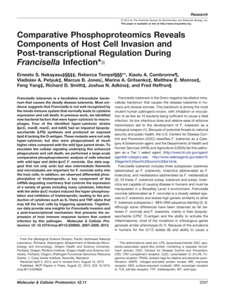
The quantitative analysis revealed 200, 69, 3, and 40 phos-
phopeptides that were significantly differentially abundant
comparing infected and control (mock) cells at 0, 1, 2, or 4 h,
respectively (Fig. 1B and supplemental Fig. S1, supplemental
Tables S2–S5). To provide insights on the kinase families
activated or repressed during Francisella infection, specific
kinase motifs were searched. Although protein kinase motifs
are often redundant, it is possible to assign these sequences
Phosphoproteome of Francisella-infected Cells
3300 Molecular & Cellular Proteomics 12.11
5. to enzyme families, thus providing insights about classes of
kinases differentially activated during the cell invasion proc-
ess (48). At the early time point (0 h postinfection) two motifs
(RxxS and SP, x being any amino acid residue) were found to
be enriched among the up-regulated phosphorylation sites,
and one motif (SP) was identified among down-regulated
phosphorylation sites (Fig. 2). At 1 h postinfection, both RxxS
and SP motifs were overrepresented among the up-regulated
phosphorylation sites, but no overrepresented motif was
found for the down-regulated sites, likely because of the low
number of down-regulated phosphopeptides. At the last time
point, only the RxxS motif was found to be overrepresented in
the up-regulated sites (Fig. 2). The motif RxxS is consistent
with either calmodulin kinase 2 (CaMK-II), PKA, PBK/AKT, or
PKC, whereas SP is consistent with ERK/MAPK, CDK, and
CDK-like motifs, thus suggesting that enzymes from these
families might be regulated during Francisella infection. In
agreement with these observations, Francisella infection is
known to activate TLR2, which triggers a cascade of MAPKs
(26).
To investigate potential pathways activated during Franci-
sella infection, we built a protein–protein interaction (PPI) net-
work and performed a function-enrichment analysis. For this
analysis we combined differentially phosphorylated proteins
found in all time points because proteins from the same
pathway are not necessarily simultaneously phosphorylated.
The PPI network was based on experimentally identified in-
teractions deposited in InnateDB. To have a more insightful
network, only proteins with significantly changing phosphory-
lation sites were used, resulting in a network of 222 proteins
(nodes) and 383 connections (edges). The network showed a
great number of proteins involved in transcription and trans-
lation regulation (RNA processing and post-transcription reg-
ulation), as well as degradation of polypeptides (proteasome)
(Fig. 3A). In support of the network analysis, these functions,
besides protein degradation, were found to be significantly
overrepresented by a DAVID analysis (Fig. 3B). Interestingly,
regulators of cytoskeleton organization and phagocytosis
were also enriched among the differentially phosphorylated
proteins (Figs. 3A and 3B), which may suggest the utilization
of these pathways in the host cell entry process by the bac-
teria. The protein network and function-enrichment analysis
were also consistent with motifs found to be overrepresented
in our analysis. For instance, the motif analyses suggested
that CDKs might be regulated during the infection process
and, in accordance with these data, cell cycle function was
suggested by the network and functional enrichment analysis
to be altered during infection (Figs. 1B, 3A, and 3B).
FIG. 1. Phosphoproteome analysis of cells infected with F. novi-
cida. A, Experimental design. RAW 264.7 macrophage-like cells were
infected for 30 min with wild-type (WT) Francisella or with ⌬lpcC,
followed by incubation for 0–4 h before harvesting. A negative control
was run in parallel using media alone (Mock). After infection, cells
were harvested, digested with trypsin and the resulting peptides were
labeled with iTRAQ. Equal parts of each iTRAQ sample were com-
bined and phosphopeptides were captured by immobilized metal
affinity chromatography (IMAC). Enriched phosphopeptide fractions
were analyzed by liquid chromatography-tandem mass spectrometry
(LC-MS/MS) leading to the identification of 3008 phosphopeptides. B,
Differentially abundant phosphopeptides from RAW 264.7 cells after
Francisella infection. Abbreviations: L, ⌬lpcC-infected; M, mock-in-
fected; n, number of phosphopeptides; W, wild type-infected.
FIG. 2. Analysis of phosphorylation motifs differentially abun-
dant in cells infected with F. novicida. Enrichment of phosphory-
lation site motifs was performed using Motif-x (http://motif-x.med.
harvard.edu/) and only the significantly different phosphopeptides in
the course of infection. Abbreviations: Up, up-regulated phosphory-
lation sites; Down, down-regulated phosphorylation sites.
Phosphoproteome of Francisella-infected Cells
Molecular & Cellular Proteomics 12.11 3301
6. Comparative Analysis of Wild-Type Versus ⌬lpcC Infected
Cells—To better understand the mechanism of host cell inva-
sion and killing by the ⌬lpcC mutant, we investigated the
differences in phosphorylation patterns between RAW 264.7
cells infected with WT or ⌬lpcC F. novicida strains. At the
earliest time point (0 h postinfection) phosphorylation signals,
seemed to be stronger, but not significant, in ⌬lpcC-infected
cells compared with WT-infected cells (Fig. 1B). At 1 and 2 h
postinfection the levels of protein phosphorylation in both
⌬lpcC- and WT-infected cells were almost identical (Fig. 1B).
The largest differences were observed at 4 h postinfection
(Fig. 1B). We speculate that the signaling observed 4 h postin-
fection is dependent on bacterial escape from the phagosome
into the host cell cytosol. Supporting this notion, the escape
from the phagosome occurs from 30 min to 4 h postinfection
(11). Furthermore, after the bacteria gain access to the host
cell cytosol some signaling occurs, such as inflammasome
activation by AIM2 (29).
To determine the significantly different phosphopeptides
from WT- and ⌬lpcC-infected cells a pairwise analysis was
done using the Benjamin-Hochberg test. A total of 36 phos-
phopeptides were significantly different in ⌬lpcC infection
compared with WT infection and were only observed at 4 h
postinfection (Fig. 4A, supplemental Table S6). Among the
enriched motifs, the RxxS motif was enriched within the up-
regulated phosphorylation sites (Fig. 4B), which suggests that
kinases with preference to basic motifs, such as PKB/Akt, are
active at this time point.
To identify pathways that were affected by proteins differ-
entially phosphorylated between WT and ⌬lpcC infections, we
searched for overrepresented functions. Only four gene on-
tology categories were enriched: cell leading edge, anchoring
junction, negative regulation of gene expression and RNA
processing (Fig. 4C). Indeed cell leading edge and anchoring
junction functions shared several proteins, thus we believe
that indeed they reflect the same pathway of cytoskeleton
FIG. 3. Analysis of functions over-
represented among differentially
phosphorylated proteins in F. novicida
infection. A, Network of differentially
phosphorylated proteins in RAW 264.7
cells during Francisella infection. A
protein–protein interaction map of the
differentially phosphorylated proteins
was retrieved from InnateDB and the
network was built using Cytoscape. Cir-
cles (nodes) represent each protein and
lines indicate physical interactions.
Nodes were colored according to the
phosphorylation abundance in infected
cells compared with the mock infection:
red, up-regulated; green, down-regula-
ted; blue, either unchanged or not
identified by LC-MS/MS. B, Enriched
functions among the differentially phos-
phorylated proteins in RAW 264.7 cells
during Francisella infection. Overrepre-
sented functions were annotated using
Database for Annotation, Visualization
and Integrated Discovery (DAVID).
Phosphoproteome of Francisella-infected Cells
3302 Molecular & Cellular Proteomics 12.11
7. rearrangement. The same was observed for negative regula-
tion of gene expression and RNA processing, which we inter-
pret as down-regulation of gene expression by RNA degra-
dation. Accordingly, we next performed a more complete
analysis of the proteins involved in these pathways and at-
tempted to correlate them with ⌬lpcC phenotypes.
Phosphorylation of Phagocytic and Cytoskeleton Pro-
teins—To identify phosphorylation events that could be in-
volved in the host cell invasion by ⌬lpcC, we examined the
phosphorylation quantification of proteins known to be in-
volved in Francisella entry in the host cells. Francisella entry
into host cells occurs mainly by two general mechanisms:
endocytosis and phagocytosis (11, 22). In the endocytic pro-
cess, Francisella enters host cells through cholesterol-rich
domains in the plasma membrane. This process depends on
clathrin and two adaptor proteins, Eps15 and AP2 (22). In the
phosphoproteomic data, phosphorylation of the heavy chain
of clathrin (Cltc) was shown to be reduced at the earliest time
point after infection (0 h) but unchanged at the later time
points (Fig. 5A). Cltc was also slightly less phosphorylated
during the infection with the WT compared with ⌬lpcC, but
this difference was not significant (supplemental Table S2).
Eps15 phosphorylation levels were unchanged over the two
time points when a peptide from this protein was detected,
and no phosphopeptides derived from AP2 were found.
These observations suggest that the endocytic process might
not be involved in the enhanced infectivity by ⌬lpcC or that
this process is not controlled by phosphorylation.
The phagocytic process of Francisella by host cells involves
at least five receptors: MR, SRA, Fc␥R, CR3, and SEN (17–
19). Of these known Francisella receptors, only phosphopep-
tides from Fc␥R were identified in our phosphoproteomic
dataset, and no changes in the phosphorylation levels were
observed (Fig. 5A). It is possible that ⌬lpcC is more easily
recognized by one or more of these receptors because of the
lack of LPS polysaccharides on the cell surface. Indeed, pre-
vious work has shown the importance of LPS O antigen
polysaccharide on the regulation of complement deposition
on Francisella surface (49). The deposition of the complement
protein C3 on Francisella surface enables the recognition of
the bacteria by CR3 promoting phagocytosis by opsonization
(13, 50); however, we inactivated complement proteins in our
experimental conditions.
After recognizing bacteria, the phagocytic receptors trigger
cell signaling that results in the rearrangement of cytoskeleton
and engulfment of the particle. In the case of Francisella, two
FIG. 4. Differentially phosphorylated proteins in cells infected with wild-type (W) or ⌬lpcC (L) F. novicida. A, Significantly different
phosphopeptides in WT or ⌬lpcC infection. Assigned phosphorylation sites are in parentheses and sites that were not confidently assigned to
a single amino acid residue (Ascore Ͻ 19.0 or Ϫ1) are represented by the number of phosphogroups and range of potential modification sites.
B, Enriched motif among the phosphosites that were differentially abundant comparing WT and ⌬lpcC F. novicida infection. C, Enriched
functions among the differentially phosphorylated proteins in RAW 264.7 cells comparing infection with WT and ⌬lpcC F. novicida strains.
Phosphoproteome of Francisella-infected Cells
Molecular & Cellular Proteomics 12.11 3303
8. pathways have been reported to be activated during the
phagocytosis process: phosphoinositol 3-kinase (PI3K)/Akt
and the tyrosine kinase Syk/mitogen-activated protein kinase
(MAPK) Erk2 pathways (20, 21). For the PI3K/Akt pathway,
phosphopeptides for PI3K and ribosomal protein S6 kinase
beta-1 (Rps6kb1) were identified. Although the phosphoryla-
tion on PI3K remained unchanged, Rps6kb1 phosphorylation
decreased in both WT and ⌬lpcC infection in early time points
(Fig. 5A). The lack of phosphorylation up-regulation suggests
that this pathway was not activated during the infection. In-
deed, another report has provided contradictory results re-
garding the role of PI3K/Akt pathway during Francisella phago-
cytosis (20), which could be potentially explained by their use
of different bacterial strains. On the other hand, two proteins
from the Syk/Erk2 pathway, kinase Erk2 and phospholipase
A2 (PLA2), were shown to be more phosphorylated in both WT
and ⌬lpcC Francisella infection at 0 h postinfection (Fig. 5A).
That only the last two enzymes of the cascade were found to
be overphosphorylated may indicate that the upstream sig-
naling had already faded. Interestingly, phosphorylation of
these proteins was up-regulated in ⌬lpcC-infected cells as
compared with WT infection at 4 h (Fig. 5A), which suggests
that this pathway could also be involved in the phagocytosis
of ⌬lpcC.
Next, we analyzed cytoskeletal proteins that can be in-
volved in the engulfment of bacteria. Although more proteins
involved in actin filament organization were found to be phos-
phorylated, only three were previously described to have a
role in phagocytic or endocytic pathways: mDia2, and formin-
like proteins 1 (Fmnl1) and 3 (Fmnl3) (51). Among these pro-
teins, only phosphorylation on mDia2 was found to be up-
regulated at the earliest infection time point, and no difference
between WT and ⌬lpcC infection was observed (Fig. 5A).
Among the microtubule proteins, tubulins were found to be
FIG. 5. Phosphorylation on endo-
cytic, phagocytic, and cytoskeleton
proteins, and the mechanism of cell
invasion. A, Phosphorylation on endo-
cytic, phagocytic and cytoskeleton pro-
teins. The endocytic, phagocytic and
cytoskeleton rearrangement pathways
were built based on literature search
(see text for references) and KEGG da-
tabase. B, Contribution of cytoskeleton
filaments to host cell entry of WT and
⌬lpcC F. novicida strains. J774 cells
were left untreated (Unt) or treated with 5
g/ml cytochalasin D (CytD or C), 10
g/ml nocodazole (Noc or N), or 5 M
withaferin A (WFA or W) before infection
with WT and ⌬lpcC F. novicida strains.
Data points significantly different to the
untreated control (t test, p Յ 0.05) are
marked the asterisks.
Phosphoproteome of Francisella-infected Cells
3304 Molecular & Cellular Proteomics 12.11
9. phosphorylated, but their modification levels did not change
with the F. novicida infection (Fig. 5A). On the other hand, the
intermediate filament protein vimentin was phosphorylated in
multiple sites (Table S1). Vimentin phosphorylation was higher
in the early time point in both WT and ⌬lpcC infections, but
was up-regulated only in ⌬lpcC at 4 h postinfection in a
pattern that was consistent with Erk2 and PLA2 phosphory-
lation (Fig. 5A). Indeed, vimentin has previously been shown
to interact with PLA2 (52); therefore, it is reasonable to believe
that they could be involved in similar cellular signaling path-
ways. Furthermore, previous studies indicate that vimentin
filaments are regulated by phosphorylation (53).
To test whether intermediate filaments and microtubules
have a function in Francisella entry into host cells, J774 cells
were treated with cytochalasin D, nocodazole and withaferin
A, which are inhibitors of actin, microtubules, and intermedi-
ate (vimentin) filaments, respectively. As previously shown
(34), ⌬lpcC F. novicida infected at a higher rate compared with
WT, and the inhibition of host cell actin polymerization with
cytochalasin D decreased the infectivity of the WT bacteria
(more than 2 logs) to a much greater extent than in the ⌬lpcC
infection (Fig. 5B). Blocking microtubule polymerization with
nocodazole also led to a similar phenotype of decreasing
bacterial entry in a greater extent in the WT (ϳ1 log) compared
with the ⌬lpcC (not significant) infection (Fig. 5B). Inhibition of
vimentin polymerization with withaferin A by itself had only a
small effect on the entry of WT Francisella into the host cells,
but in combination with nocodazole and cytochalasin D it
significantly reduced the number of internalized bacteria (Fig.
5B). The individual treatments with cytochalasin D, nocoda-
zole and withaferin A had no effect on the entry of ⌬lpcC into
host cells, but in combination these inhibitors significantly
reduced the number of internalized bacteria (up to ϳ1 log)
(Fig. 5B). It should be noted that the reduction in the number
of internalized bacteria was smaller in ⌬lpcC infection com-
pared with WT for all the treatments, suggesting that whereas
all these 3 cytoskeleton filaments play a role during Francisella
infection an additional factor may be required for entry of
⌬lpcC F. novicida uptake via the endocytic pathway.
Phosphorylation of RNA-processing Proteins, Post-tran-
scription Regulation, and Mechanism of Cell Killing by
⌬lpcC—We next performed an in-depth analysis of the pro-
teins involved in RNA degradation, as this group was over-
represented among the differentially abundant phosphopro-
teins in cells infected with ⌬lpcC compared with WT
Francisella. The phosphoprotein tristetraprolin (TTP), product
of the gene zfp36, drew our interest in particular because it
contained two phosphopeptides with the most pronounced
difference between ⌬lpcC versus WT Francisella infection
(Fig. 4A). Thus, we decided to validate TTP and investigate
possible functions for differential phosphorylation during
Francisella infection. Fig. 6A shows the MS/MS spectrum of a
peptide corresponding to the phosphorylation at serine 178,
and the magnified region details the signal intensity of the
iTRAQ report ions used for the quantitative analysis clearly
shows a difference between WT and ⌬lpcC infections. The
quantification at the 4 h time point exhibited 2.5- and 12.6-
fold increases in WT and ⌬lpcC Francisella infection com-
pared with the mock, respectively (Fig. 6B). This observation
was further validated by Western blot analysis. In independent
experiments using both J774 and RAW 264.7 macrophages,
we observed a dramatic increase in the phosphorylation sta-
tus of endogenous TTP, indicated by the appearance of mul-
tiple higher molecular weight bands, particularly following
infection with ⌬lpcC Francisella compared with WT (Fig. 6C).
Importantly, these higher molecular weight bands were dimin-
ished on treatment with -phosphatase, consistent with a
shift in electrophoretic mobility because of phosphorylation
(Fig. 6C). The Western blot against subunit 1 of RNA Poly-
merase II (Rbp1) served as a loading control, as well as a
positive control for the phosphatase treatment (Fig. 6C).
With this support for hyper-phosphorylation of TTP by
phosphoproteomic and Western blot analyses, we next inves-
tigated if this phenomenon could have consequences for in
FIG. 6. Dynamics of TTP phosphorylation in RAW 264. 7 cells
during Francisella infection. A, Tandem mass spectrum illustrating the
identification and quantification of the phosphopeptide QSISpFS-
GLPSGR (corresponding to phosphorylation at serine 178) from tris-
tetraprolin (Zfp36 or TTP) at 4 h postinfection. The expanded region
highlights the iTRAQ report ions used for quantification. B, Time
course quantification of TTP phosphorylation at serine 178. C, West-
ern blot analysis of J774 and RAW 264.7 cell lines 4 h post infection
with WT or ⌬lpcC F. novicida. The presence of phosphorylation on
TTP was determined by shift of electrophoretic mobility on treatment
with -phosphatase (PP).
Phosphoproteome of Francisella-infected Cells
Molecular & Cellular Proteomics 12.11 3305
10. vivo murine infections. TTP is a well-known component of the
mRNA-destabilizing machinery that binds to AU-rich ele-
ments (54, 55). TTP is also known to be hyper-phosphorylated
by p38 MAPK, which is associated with a decrease in the
RNA-binding capacity of this protein and results in the sub-
sequent inhibition of RNA degradation (55, 56). In addition, as
a negative feedback mechanism TTP phosphorylation was
shown to be regulated by protein phosphatase 2A (57). Be-
cause TTP hyper-phosphorylation was observed with a much
higher intensity in the ⌬lpcC infection (p Ͻ 0.01), we hypoth-
esized that Francisella could be down regulating TTP phos-
phorylation as a mechanism to shut down the translation of
important factors that control its infection. To test this hypoth-
esis we performed transcriptomic analyses using spleens of
mice infected with either ⌬lpcC or WT Francisella. We mea-
sured the abundance of ϳ30,000 genes by microarray, which
showed a more drastic change in expression profiles in mice
infected with WT compared with ⌬lpcC Francisella (supple-
mental Fig. S2A, supplemental Table S7). Next, we investi-
gated the levels of transcripts that were recently identified as
TTP substrates (54). Out of 310 mRNAs identified as targets of
TTP, 245 were present in our dataset (supplemental Figs. S2B
and S2C, supplemental Table S8). Although the TTP-regu-
lated RNAs were on average slightly more abundant in ⌬lpcC-
infected samples compared with mock infections, they were
slightly reduced in WT infections (supplemental Figs. S2B and
S2C). To test if this difference in transcript abundance among
the TTP-regulated RNAs was significant, we compared the
transcripts found during ⌬lpcC infections to those observed in
WT infections (t test, p Յ 0.05). Although 25% of the tran-
scripts were more abundant in ⌬lpcC compared with WT
infections, this proportion was increased to 45% when only
the TTP-regulated RNAs were considered, showing a clear
enrichment (Chi-square contingency test, p ϭ 0.0001). These
FIG. 7. Cytokine profile and killing mechanism of cells infected with F. novicida. A, Cytokine profile. Cytokines were measured in culture
supernatants 6 h postinfection using the Q-Plex ArrayTM
kit. B, mRNA stability for IL-1 and RANTES was determined with qPCR following
infection and Actinomycin D treatment. (n ϭ 3, ϮS.E., 2-way ANOVA, p Ͻ 0.001 for IL-1, Bonferroni post tests, *p Ͻ 0.05, **p Ͻ 0.01) Viability
(C), cytotoxicity (D), and apoptosis/necrosis (E) assays. Cells were infected with WT and ⌬lpcC F. novicida strains and assayed for viability,
cytotoxicity and apoptosis/necrosis using the ApoTox-GloTM
Triplex Assay kit. As positive controls for lysis, necrosis and apoptosis, cells were
treated with saponin, ionomycin (Ionom.), staurosporine (Stauros.), respectively.
Phosphoproteome of Francisella-infected Cells
3306 Molecular & Cellular Proteomics 12.11
11. data suggest that hyper-phosphorylation on TTP has an im-
pact on the transcript levels during Francisella infection.
These results may help explain the down-regulation of many
immune response genes observed in monocytes infected with
either F. novicida or F. tularensis Schu S4 (58).
TTP has been shown to be a key player on the regulation of
cytokine production (54, 55, 59). Thus, we speculate that
Francisella may inhibit TTP phosphorylation and consequently
targeting the mRNA of selected cytokines to degradation and
preventing their translation. To test this hypothesis, we in-
fected RAW 264.7 cells and measured the cytokines by a
multiplex ELISA array 6 h after infection. Six cytokines were
shown to be produced in higher amounts during infection with
⌬lpcC compared with WT: IL-1, MDC, TARC, MCP-1, TNF-␣
and RANTES (Fig. 7A). Out of these six, IL-1, MDC, MCP-1
and TNF-␣, have been previously shown to be regulated by
TTP. These results suggest that Francisella impairs the pro-
duction of cytokines, likely by a mechanism that involves
TTP-mediated mRNA degradation. To directly investigate this
mechanism in RAW 264.7 cells, we used qPCR to monitor
mRNA stability of IL-1 and RANTES cytokines after infection
and in the presence of actinomycin D (Fig. 7B). IL-1 and
RANTES were selected as representative pro-inflammatory
cytokines and chemokines, respectively. With IL-1, we ob-
served a deficiency in its mRNA degradation following infec-
tion with ⌬lpcC compared with WT; the most dramatic delin-
eation appeared within the first hour. With RANTES, there was
a trending delay in mRNA degradation with ⌬lpcC within the
first 2 h but it caught up with WT by 4 h postinfection.
Importantly, there were no statistically significant differences
in control Gapdh mRNA stability between WT and ⌬lpcC
infection. Together, our data suggest that Francisella can
regulate the mRNA stability of particular cytokines, such as
IL-1, but for others it may only represent a contributing
mechanism.
Considering that several cytokines have cytotoxic effects,
we analyzed the relationship of cytokine production and the
hyper-cytotoxic phenotype of the ⌬lpcC mutant. Because
some of these cytokines, such as IL-1 and TNF-␣, kill cells
by triggering apoptosis (60, 61), we aimed to identify the
mechanism of host cell death by ⌬lpcC using the ApoTox-
Glo™ triplex assay, which analyzes viability, necrosis, and
apoptosis with the same assay well. Consistent with previous
results (34), ⌬lpcC is more cytotoxic to host cells than WT
(Fig. 7C). Furthermore, ⌬lpcC infection led to an increase in
apoptosis, similar to the positive control with the apoptotic
inducer staurosporine (Figs. 7D and 7E). These results sug-
gest that Francisella avoids triggering the host apoptotic path-
way potentially by inhibiting the production of select cyto-
kines. In support of our results, Francisella has been shown to
inhibit apoptotic signaling and to prolong the lifespan of in-
fected neutrophils (62). Indeed, Francisella targeting of IL-1-
and TNF-␣-mediated signaling has been proposed as poten-
tial mechanisms for anti-apoptotic activity in infected neutro-
phils (63).
CONCLUSIONS
We performed a comparative phosphoproteomic analysis
of murine cells infected with F. novicida. Our data showed the
participation of different cytoskeleton filaments in the process
of bacterial entry into the host cells. Furthermore, by a com-
bination of phosphoproteomics, transcriptomics, and cellular
and immunological assays, we showed a deregulation of the
signaling that controls TTP activity, a key component of post-
transcriptional regulation in innate immune response (54, 57,
59). These findings suggest that Francisella targets host post-
transcriptional regulatory machinery to repress the expression
of innate immune factors that controls the infection.
Acknowledgments—We thank our colleagues Charles Ansong,
Therese Clauss, Ron Moore, Penny Colton, and Brooke Kaiser for
insightful comments, input, and suggestions.
* This work was supported by the National Institute of Allergy and
Infectious Diseases (NIH/DHHS through interagency agreement Y1-
AI-4894-01; project website www.SysBEP.org) and the National In-
stitute for General Medicine (GM094623). This work used instrumen-
tation and capabilities developed with support from the NIH grant
5P41RR018522-10, the National Institute of General Medical Sci-
ences grant 8 P41 GM103493-10, and the U. S. Department of Energy
Office of Biological and Environmental Research (DOE/BER). Signifi-
cant portions of this work were performed in the EMSL, a DOE/BER
national scientific user facility located at Pacific Northwest National
Laboratory. The Pacific Northwest National Laboratory is operated for
the DOE by Battelle under Contract DE-AC05-76RLO1830. This work
was also supported by the NIH award NS076094 to X.A.C.
□S This article contains supplemental Figs. S1 and S2 and Tables
S1 to S8.
** To whom correspondence should be addressed: Department of
Molecular Microbiology and Immunology, L220, Oregon Health and
Science University, 3181 SW Sam Jackson Park Road, Portland, OR
97239. Tel.: (503) 494-6841; Fax: (503) 494-6862; E-mail: tempelr@
ohsu.edu.
‡‡Current address: Bindley Bioscience Center, Purdue University,
West Lafayette, IN, USA.
§§Both authors contributed equally to this work.
Data availability: The LC-MS/MS runs and annotated spectra are
available at PeptideAtlas under the accession number PASS00297.
REFERENCES
1. Sjostedt, A. (2007) Tularemia: history, epidemiology, pathogen physiology,
and clinical manifestations. Ann. N. Y. Acad. Sci. 1105, 1–29
2. Sjostedt, A. (2005) Family III. Francisellaceae. in Bergey’s Manual® of
Systematic Bacteriology (Brenner, D. J., Krieg, N. R., Staley, J. T., and
Garrity, G. M. eds.), Springer, New York, NY. pp 199–209
3. Champion, M. D., Zeng, Q., Nix, E. B., Nano, F. E., Keim, P., Kodira, C. D.,
Borowsky, M., Young, S., Koehrsen, M., Engels, R., Pearson, M., How-
arth, C., Larson, L., White, J., Alvarado, L., Forsman, M., Bearden, S. W.,
Sjostedt, A., Titball, R., Michell, S. L., Birren, B., and Galagan, J. (2009)
Comparative genomic characterization of Francisella tularensis strains
belonging to low and high virulence subspecies. PLoS Pathogens 5,
e1000459
4. Rohmer, L., Fong, C., Abmayr, S., Wasnick, M., Larson Freeman, T. J.,
Radey, M., Guina, T., Svensson, K., Hayden, H. S., Jacobs, M., Gal-
lagher, L. A., Manoil, C., Ernst, R. K., Drees, B., Buckley, D., Haugen, E.,
Bovee, D., Zhou, Y., Chang, J., Levy, R., Lim, R., Gillett, W., Guenthener,
Phosphoproteome of Francisella-infected Cells
Molecular & Cellular Proteomics 12.11 3307
12. D., Kang, A., Shaffer, S. A., Taylor, G., Chen, J., Gallis, B., D’Argenio,
D. A., Forsman, M., Olson, M. V., Goodlett, D. R., Kaul, R., Miller, S. I.,
and Brittnacher, M. J. (2007) Comparison of Francisella tularensis ge-
nomes reveals evolutionary events associated with the emergence of
human pathogenic strains. Genome Biol. 8, R102
5. Thomas, R. M., Titball, R. W., Oyston, P. C., Griffin, K., Waters, E., Hitchen,
P. G., Michell, S. L., Grice, I. D., Wilson, J. C., and Prior, J. L. (2007) The
immunologically distinct O antigens from Francisella tularensis subspe-
cies tularensis and Francisella novicida are both virulence determinants
and protective antigens. Infection Immunity 75, 371–378
6. Atianand, M. K., Duffy, E. B., Shah, A., Kar, S., Malik, M., and Harton, J. A.
(2011) Francisella tularensis reveals a disparity between human and
mouse NLRP3 inflammasome activation. J. Biol. Chem. 286,
39033–39042
7. Dotson, R. J., Rabadi, S. M., Westcott, E. L., Bradley, S., Catlett, S. V.,
Banik, S., Harton, J. A., Bakshi, C. S., and Malik, M. (2013) Repression of
inflammasome by Francisella tularensis during early stages of infection.
J. Biol. Chem. in press 288, 23844–23857
8. Shen, H., Chen, W., and Conlan, J. W. (2004) Mice sublethally infected with
Francisella novicida U112 develop only marginal protective immunity
against systemic or aerosol challenge with virulent type A or B strains of
F. tularensis. Microb. Pathogenesis 37, 107–110
9. Asare, R., and Kwaik, Y. A. (2010) Exploitation of host cell biology and
evasion of immunity by francisella tularensis. Front. Microbiol. 1, 145
10. Henry, T., and Monack, D. M. (2007) Activation of the inflammasome upon
Francisella tularensis infection: interplay of innate immune pathways and
virulence factors. Cell. Microbiol. 9, 2543–2551
11. Jones, C. L., Napier, B. A., Sampson, T. R., Llewellyn, A. C., Schroeder,
M. R., and Weiss, D. S. (2012) Subversion of host recognition and
defense systems by Francisella spp. Microbiol. Mol. Biol. Rev. 76,
383–404
12. Bosio, C. M. (2011) The subversion of the immune system by francisella
tularensis. Front. Microbiol. 2, 9
13. Balagopal, A., MacFarlane, A. S., Mohapatra, N., Soni, S., Gunn, J. S., and
Schlesinger, L. S. (2006) Characterization of the receptor-ligand path-
ways important for entry and survival of Francisella tularensis in human
macrophages. Infection Immunity 74, 5114–5125
14. Barel, M., Hovanessian, A. G., Meibom, K., Briand, J. P., Dupuis, M., and
Charbit, A. (2008) A novel receptor - ligand pathway for entry of Franci-
sella tularensis in monocyte-like THP-1 cells: interaction between sur-
face nucleolin and bacterial elongation factor Tu. BMC Microbiol. 8, 145
15. Geier, H., and Celli, J. (2011) Phagocytic receptors dictate phagosomal
escape and intracellular proliferation of Francisella tularensis. Infection
Immunity 79, 2204–2214
16. Schulert, G. S., and Allen, L. A. (2006) Differential infection of mononuclear
phagocytes by Francisella tularensis: role of the macrophage mannose
receptor. J. Leukocyte Biol. 80, 563–571
17. Clemens, D. L., Lee, B. Y., and Horwitz, M. A. (2005) Francisella tularensis
enters macrophages via a novel process involving pseudopod loops.
Infection Immunity 73, 5892–5902
18. Lai, X. H., Golovliov, I., and Sjostedt, A. (2001) Francisella tularensis in-
duces cytopathogenicity and apoptosis in murine macrophages via a
mechanism that requires intracellular bacterial multiplication. Infection
Immunity 69, 4691–4694
19. Lindemann, S. R., McLendon, M. K., Apicella, M. A., and Jones, B. D. (2007)
An in vitro model system used to study adherence and invasion of
Francisella tularensis live vaccine strain in nonphagocytic cells. Infection
Immunity 75, 3178–3182
20. Parsa, K. V., Butchar, J. P., Rajaram, M. V., Cremer, T. J., and Tridandapani,
S. (2008) The tyrosine kinase Syk promotes phagocytosis of Francisella
through the activation of Erk. Mol. Immunol. 45, 3012–3021
21. Tamilselvam, B., and Daefler, S. (2008) Francisella targets cholesterol-rich
host cell membrane domains for entry into macrophages. J. Immunol.
180, 8262–8271
22. Law, H. T., Lin, A. E. J., Kim, Y., Quach, B., Nano, F. E., and Guttman, J. A.
(2011) Francisella tularensis uses cholesterol and clathrin-based endo-
cytic mechanisms to invade hepatocytes. Sci Rep-Uk 1
23. Cremer, T. J., Butchar, J. P., and Tridandapani, S. (2011) Francisella sub-
verts innate immune signaling: Focus On PI3K/Akt. Front. Microbiol. 5,
13
24. Hajjar, A. M., Harvey, M. D., Shaffer, S. A., Goodlett, D. R., Sjostedt, A.,
Edebro, H., Forsman, M., Bystrom, M., Pelletier, M., Wilson, C. B., Miller,
S. I., Skerrett, S. J., and Ernst, R. K. (2006) Lack of in vitro and in vivo
recognition of Francisella tularensis subspecies lipopolysaccharide by
Toll-like receptors. Infection Immunity 74, 6730–6738
25. Katz, J., Zhang, P., Martin, M., Vogel, S. N., and Michalek, S. M. (2006)
Toll-like receptor 2 is required for inflammatory responses to Francisella
tularensis LVS. Infection Immunity 74, 2809–2816
26. Cole, L. E., Shirey, K. A., Barry, E., Santiago, A., Rallabhandi, P., Elkins,
K. L., Puche, A. C., Michalek, S. M., and Vogel, S. N. (2007) Toll-like
receptor 2-mediated signaling requirements for Francisella tularensis live
vaccine strain infection of murine macrophages. Infection Immunity 75,
4127–4137
27. Kawai, T., and Akira, S. (2011) Toll-like receptors and their crosstalk with
other innate receptors in infection and immunity. Immunity 34, 637–650
28. Belhocine, K., and Monack, D. M. (2012) Francisella infection triggers
activation of the AIM2 inflammasome in murine dendritic cells. Cell.
Microbiol. 14, 71–80
29. Peng, K., Broz, P., Jones, J., Joubert, L. M., and Monack, D. (2011)
Elevated AIM2-mediated pyroptosis triggered by hypercytotoxic Franci-
sella mutant strains is attributed to increased intracellular bacteriolysis.
Cell. Microbiol. 13, 1586–1600
30. Jones, J. W., Kayagaki, N., Broz, P., Henry, T., Newton, K., O’Rourke, K.,
Chan, S., Dong, J., Qu, Y., Roose-Girma, M., Dixit, V. M., and Monack,
D. M. (2010) Absent in melanoma 2 is required for innate immune
recognition of Francisella tularensis. Proc. Natl. Acad. Sci. U. S. A.107,
9771–9776
31. Fernandes-Alnemri, T., Yu, J. W., Juliana, C., Solorzano, L., Kang, S., Wu,
J., Datta, P., McCormick, M., Huang, L., McDermott, E., Eisenlohr, L.,
Landel, C. P., and Alnemri, E. S. (2010) The AIM2 inflammasome is
critical for innate immunity to Francisella tularensis. Nat. Immunol. 11,
385–393
32. Rathinam, V. A., Jiang, Z., Waggoner, S. N., Sharma, S., Cole, L. E.,
Waggoner, L., Vanaja, S. K., Monks, B. G., Ganesan, S., Latz, E., Hor-
nung, V., Vogel, S. N., Szomolanyi-Tsuda, E., and Fitzgerald, K. A. (2010)
The AIM2 inflammasome is essential for host defense against cytosolic
bacteria and DNA viruses. Nat. Immunol. 11, 395–402
33. Broz, P., von Moltke, J., Jones, J. W., Vance, R. E., and Monack, D. M.
(2010) Differential requirement for Caspase-1 autoproteolysis in patho-
gen-induced cell death and cytokine processing. Cell Host Microbe 8,
471–483
34. Lai, X. H., Shirley, R. L., Crosa, L., Kanistanon, D., Tempel, R., Ernst, R. K.,
Gallagher, L. A., Manoil, C., and Heffron, F. (2010) Mutations of Franci-
sella novicida that alter the mechanism of its phagocytosis by murine
macrophages. PloS One 5, e11857
35. Craven, R. R., Hall, J. D., Fuller, J. R., Taft-Benz, S., and Kawula, T. H.
(2008) Francisella tularensis invasion of lung epithelial cells. Infection
Immunity 76, 2833–2842
36. Nguyen, T. H., Brechenmacher, L., Aldrich, J. T., Clauss, T. R., Gritsenko,
M. A., Hixson, K. K., Libault, M., Tanaka, K., Yang, F., Yao, Q., Pasa-
Tolic, L., Xu, D., Nguyen, H. T., and Stacey, G. (2012) Quantitative
Phosphoproteomic Analysis of Soybean Root Hairs Inoculated with Bra-
dyrhizobium japonicum. Mol. Cell. Proteomics 11, 1140–1155
37. Zhao, R., Ding, S. J., Shen, Y., Camp, D. G., 2nd, Livesay, E. A., Udseth, H.,
and Smith, R. D. (2009) Automated metal-free multiple-column nanoLC
for improved phosphopeptide analysis sensitivity and throughput.
J. Chromatog. 877, 663–670
38. Mayampurath, A. M., Jaitly, N., Purvine, S. O., Monroe, M. E., Auberry, K. J.,
Adkins, J. N., and Smith, R. D. (2008) DeconMSn: a software tool for
accurate parent ion monoisotopic mass determination for tandem mass
spectra. Bioinformatics 24, 1021–1023
39. Kim, S., Gupta, N., and Pevzner, P. A. (2008) Spectral probabilities and
generating functions of tandem mass spectra: a strike against decoy
databases. J. Proteome Res. 7, 3354–3363
40. Beausoleil, S. A., Villen, J., Gerber, S. A., Rush, J., and Gygi, S. P. (2006) A
probability-based approach for high-throughput protein phosphorylation
analysis and site localization. Nat. Biotechnol. 24, 1285–1292
41. Monroe, M. E., Shaw, J. L., Daly, D. S., Adkins, J. N., and Smith, R. D.
(2008) MASIC: a software program for fast quantitation and flexible
visualization of chromatographic profiles from detected LC-MS(/MS)
features. Comput. Biol. Chem. 32, 215–217
42. Troyanskaya, O., Cantor, M., Sherlock, G., Brown, P., Hastie, T., Tibshirani,
Phosphoproteome of Francisella-infected Cells
3308 Molecular & Cellular Proteomics 12.11
13. R., Botstein, D., and Altman, R. B. (2001) Missing value estimation
methods for DNA microarrays. Bioinformatics 17, 520–525
43. Smyth, G. K. (2005) Limma: linear models for microarray data. in Bioinfor-
matics and Computational Biology Solutions using R and Bioconductor
(Gentleman, R., Carey, V., Dudoit, S., Irizarry, R., and Huber, W. eds.),
Springer, New York, NY. pp 397–420
44. Schwartz, D., and Gygi, S. P. (2005) An iterative statistical approach to the
identification of protein phosphorylation motifs from large-scale data
sets. Nat. Biotechnol. 23, 1391–1398
45. Lynn, D. J., Winsor, G. L., Chan, C., Richard, N., Laird, M. R., Barsky, A.,
Gardy, J. L., Roche, F. M., Chan, T. H., Shah, N., Lo, R., Naseer, M., Que,
J., Yau, M., Acab, M., Tulpan, D., Whiteside, M. D., Chikatamarla, A.,
Mah, B., Munzner, T., Hokamp, K., Hancock, R. E., and Brinkman, F. S.
(2008) InnateDB: facilitating systems-level analyses of the mammalian
innate immune response. Mol. Sys. Biol. 4, 218
46. Smoot, M. E., Ono, K., Ruscheinski, J., Wang, P. L., and Ideker, T. (2011)
Cytoscape 2.8: new features for data integration and network visualiza-
tion. Bioinformatics 27, 431–432
47. Huang da, W., Sherman, B. T., and Lempicki, R. A. (2009) Systematic and
integrative analysis of large gene lists using DAVID bioinformatics re-
sources. Nature protocols 4, 44–57
48. Trost, M., English, L., Lemieux, S., Courcelles, M., Desjardins, M., and
Thibault, P. (2009) The phagosomal proteome in interferon-gamma-ac-
tivated macrophages. Immunity 30, 143–154
49. Clay, C. D., Soni, S., Gunn, J. S., and Schlesinger, L. S. (2008) Evasion of
complement-mediated lysis and complement C3 deposition are regu-
lated by Francisella tularensis lipopolysaccharide O antigen. Journal of
immunology 181, 5568–5578
50. Schwartz, J. T., Barker, J. H., Long, M. E., Kaufman, J., McCracken, J., and
Allen, L. A. (2012) Natural IgM mediates complement-dependent uptake
of Francisella tularensis by human neutrophils via complement receptors
1 and 3 in nonimmune serum. J. Immunol. 189, 3064–3077
51. Campellone, K. G., and Welch, M. D. (2010) A nucleator arms race: cellular
control of actin assembly. Nat. Rev. 11, 237–251
52. Kuwata, H., Yamamoto, S., Miyazaki, Y., Shimbara, S., Nakatani, Y., Su-
zuki, H., Ueda, N., Yamamoto, S., Murakami, M., and Kudo, I. (2000)
Studies on a mechanism by which cytosolic phospholipase A2 regulates
the expression and function of type IIA secretory phospholipase A2.
J. Immunol. 165, 4024–4031
53. Eriksson, J. E., He, T., Trejo-Skalli, A. V., Harmala-Brasken, A. S., Hellman,
J., Chou, Y. H., and Goldman, R. D. (2004) Specific in vivo phosphory-
lation sites determine the assembly dynamics of vimentin intermediate
filaments. J. Cell Sci. 117, 919–932
54. Kratochvill, F., Machacek, C., Vogl, C., Ebner, F., Sedlyarov, V., Gruber,
A. R., Hartweger, H., Vielnascher, R., Karaghiosoff, M., Rulicke, T.,
Muller, M., Hofacker, I., Lang, R., and Kovarik, P. (2011) Tristetraprolin-
driven regulatory circuit controls quality and timing of mRNA decay in
inflammation. Mol. Sys. Biol. 7, 560
55. Ronkina, N., Menon, M. B., Schwermann, J., Tiedje, C., Hitti, E., Kotlyarov,
A., and Gaestel, M. (2010) MAPKAP kinases MK2 and MK3 in inflamma-
tion: complex regulation of TNF biosynthesis via expression and phos-
phorylation of tristetraprolin. Biochem. Pharmacol. 80, 1915–1920
56. Brooks, S. A., and Blackshear, P. J. (2013) Tristetraprolin (TTP): interactions
with mRNA and proteins, and current thoughts on mechanisms of action.
Biochim. Biophys. Acta 1829, 666–679
57. Sun, L., Stoecklin, G., Van Way, S., Hinkovska-Galcheva, V., Guo, R. F.,
Anderson, P., and Shanley, T. P. (2007) Tristetraprolin (TTP)-14-3-3
complex formation protects TTP from dephosphorylation by protein
phosphatase 2a and stabilizes tumor necrosis factor-alpha mRNA.
J. Biol. Chem. 282, 3766–3777
58. Butchar, J. P., Cremer, T. J., Clay, C. D., Gavrilin, M. A., Wewers, M. D.,
Marsh, C. B., Schlesinger, L. S., and Tridandapani, S. (2008) Microarray
analysis of human monocytes infected with Francisella tularensis iden-
tifies new targets of host response subversion. PloS One 3, e2924
59. Qiu, L. Q., Stumpo, D. J., and Blackshear, P. J. (2012) Myeloid-specific
tristetraprolin deficiency in mice results in extreme lipopolysaccharide
sensitivity in an otherwise minimal phenotype. J. Immunol. 188,
5150–5159
60. Hogquist, K. A., Nett, M. A., Unanue, E. R., and Chaplin, D. D. (1991)
Interleukin 1 is processed and released during apoptosis. Proc. Natl.
Acad. Sci. U. S. A. 88, 8485–8489
61. Rath, P. C., and Aggarwal, B. B. (1999) TNF-induced signaling in apoptosis.
J. Clin. Immunol. 19, 350–364
62. Schwartz, J. T., Barker, J. H., Kaufman, J., Fayram, D. C., McCracken,
J. M., and Allen, L. A. (2012) Francisella tularensis inhibits the intrinsic
and extrinsic pathways to delay constitutive apoptosis and prolong
human neutrophil lifespan. J. Immunol. 188, 3351–3363
63. Schwartz, J. T., Bandyopadhyay, S., Kobayashi, S. D., McCracken, J.,
Whitney, A. R., Deleo, F. R., and Allen, L. A. (2013) Francisella tularensis
alters human neutrophil gene expression: insights into the molecular
basis of delayed neutrophil apoptosis. J. Innate Immun. 5, 124–136
Phosphoproteome of Francisella-infected Cells
Molecular & Cellular Proteomics 12.11 3309