SlideShare une entreprise Scribd logo
1  sur  12
Télécharger pour lire hors ligne
Evaluation of the Patient with Hip Pain
JOHN J. WILSON, MD, MS, and MASARU FURUKAWA, MD, MS, University of Wisconsin School of Medicine
and Public Health, Madison, Wisconsin

Hip pain is a common and disabling condition that affects patients of all ages. The differential diagnosis of hip pain
is broad, presenting a diagnostic challenge. Patients often express that their hip pain is localized to one of three anatomic regions: the anterior hip and groin, the posterior hip and buttock, or the lateral hip. Anterior hip and groin
pain is commonly associated with intra-articular pathology, such as osteoarthritis and hip labral tears. Posterior hip
pain is associated with piriformis syndrome, sacroiliac joint dysfunction, lumbar radiculopathy, and less commonly
ischiofemoral impingement and vascular claudication. Lateral hip pain occurs with greater trochanteric pain syndrome. Clinical examination tests, although helpful, are not highly sensitive or specific for most diagnoses; however,
a rational approach to the hip examination can be used. Radiography should be performed if acute fracture, dislocations, or stress fractures are suspected. Initial plain radiography of the hip should include an anteroposterior view of
the pelvis and frog-leg lateral view of the symptomatic hip. Magnetic resonance imaging should be performed if the
history and plain radiograph results are not diagnostic. Magnetic resonance imaging is valuable for the detection of
occult traumatic fractures, stress fractures, and osteonecrosis of the femoral head. Magnetic resonance arthrography
is the diagnostic test of choice for labral tears. (Am Fam Physician. 2014;89(1):27-34. Copyright © 2014 American
Academy of Family Physicians.)
▲

Patient information:
A handout on this topic,
written by the authors
of this article, is available at http://www.
aafp.org/afp/2014/0101/
p27-s1.html. Access to
this handout is free and
unrestricted.
More online
at http://www.
aafp.org/afp.

CME This clinical content
conforms to AAFP criteria
for continuing medical
education (CME). See CME
Quiz questions on page 6.

Author disclosure: No relevant financial affiliations.

H

ip pain is a common presentation in primary care and can
affect patients of all ages. In one
study, 14.3% of adults 60 years
and older reported significant hip pain on
most days over the previous six weeks.1 Hip
pain often presents a diagnostic and therapeutic challenge. The differential diagnosis
of hip pain (eTable A) is broad, including
both intra-articular and extra-articular
pathology, and varies by age. A history and
physical examination are essential to accurately diagnose the cause of hip pain.
Anatomy
The hip joint is a ball-and-socket synovial
joint designed to allow multiaxial motion
while transferring loads between the upper
and lower body. The acetabular rim is lined
by fibrocartilage (labrum), which adds depth
and stability to the femoroacetabular joint.
The articular surfaces are covered by hyaline cartilage that dissipates shear and compressive forces during load bearing and hip
motion. The hip’s major innervating nerves
originate in the lumbosacral region, which
can make it difficult to distinguish between
primary hip pain and radicular lumbar pain.
The hip joint’s wide range of motion is second only to that of the glenohumeral joint

and is enabled by the large number of muscle groups that surround the hip. The flexor
muscles include the iliopsoas, rectus femoris,
pectineus, and sartorius muscles. The gluteus maximus and hamstring muscle groups
allow for hip extension. Smaller muscles, such
as gluteus medius and minimus, piriformis,
obturator externus and internus, and quadratus femoris muscles, insert around the greater
trochanter, allowing for abduction, adduction, and internal and external rotation.
In persons who are skeletally immature,
there are several growth centers of the pelvis
and femur where injuries can occur. Potential sites of apophyseal injury in the hip
region include the ischium, anterior superior
iliac spine, anterior inferior iliac spine, iliac
crest, lesser trochanter, and greater trochanter. The apophysis of the superior iliac spine
matures last and is susceptible to injury up
to 25 years of age.2
Evaluation of Hip Pain
HISTORY

Age alone can narrow the differential diagnosis of hip pain. In prepubescent and adolescent patients, congenital malformations
of the femoroacetabular joint, avulsion fractures, and apophyseal or epiphyseal injuries should be considered. In those who are

January 1, 2014 ◆ American Family Physician
www.aafp.org/afp
Downloaded from the Volume 89, Number 1 website at www.aafp.org/afp. Copyright © 2014 American Academy of FamilyAmerican For the private, nonPhysicians. Family Physician
commercial use of one individual user of the website. All other rights reserved. Contact copyrights@aafp.org for copyright questions and/or permission requests.

27
Hip Pain
SORT: KEY RECOMMENDATIONS FOR PRACTICE
Evidence
rating

References

Initial plain radiography of the hip should include an anteroposterior view of the pelvis and a frog-leg
lateral view of the symptomatic hip.

C

4

Magnetic resonance imaging should be used for detection of occult hip fractures, stress fractures,
and osteonecrosis of the femoral head.

C

23, 30, 33

Magnetic resonance arthrography is the diagnostic test of choice for labral tears.

C

6, 19

Ultrasonography is a helpful diagnostic modality for patients with suspected bursitis, joint effusion,
or functional causes of hip pain (e.g., snapping hip), and can be employed for therapeutic imagingguided injections and aspirations around the hip.

C

8, 9

Clinical recommendation

A = consistent, good-quality patient-oriented evidence; B = inconsistent or limited-quality patient-oriented evidence; C = consensus, diseaseoriented evidence, usual practice, expert opinion, or case series. For information about the SORT evidence rating system, go to http://www.aafp.
org/afpsort.

A

B

C

Figure 1. Gait testing. (A) C sign. Patients often localize pain by cupping the anterolateral hip with the thumb and forefinger in the shape of a “C.” (B) Gait analysis. The patient is observed while walking to evaluate for limp or antalgic
gait characteristics. (C) Modified Trendelenburg test (single leg stance phase). The patient stands with feet shoulder
width apart and lifts one leg. The examiner observes for a drop in the level of the iliac crest on the side of the lifted leg.

skeletally mature, hip pain is often a result of musculotendinous strain, ligamentous sprain, contusion, or
bursitis. In older adults, degenerative osteoarthritis and
fractures should be considered first.
Patients with hip pain should be asked about antecedent trauma or inciting activity, factors that increase
or decrease the pain, mechanism of injury, and time of
28 American Family Physician

onset. Questions related to hip function, such as the ease
of getting in and out of a car, putting on shoes, running,
walking, and going up and down stairs, can be helpful.3
Location of the pain is informative because hip pain
often localizes to one of three basic anatomic regions:
the anterior hip and groin, posterior hip and buttock,
and lateral hip (eFigure A).

www.aafp.org/afp

Volume 89, Number 1

◆

January 1, 2014
Hip Pain
Table 1. Physical Examination Tests for the Evaluation of Hip Pain
Test

Other names

Positioning

Positive findings

Differential diagnosis

Gait testing (C sign,
Figure 1A; gait analysis,
Figure 1B)

—

Standing

Antalgic gait, Trendelenburg gait,
pelvic wink (rotation of more
than 40 degrees in the axial plane
toward the affected hip when
terminally extending the hip),
excessive pronation or supination
of the ankles, and limps caused
by differing leg lengths

Hip labral tear, transient synovitis,
Legg-Calvé-Perthes disease,
SCFE

Modified Trendelenburg
test (Figure 1C)

Single leg
stance phase

Standing

2-cm drop in the level of the iliac
crest, indicating weakness on the
contralateral side

Hip labral tear, transient synovitis,
Legg-Calvé-Perthes disease,
SCFE

ROM testing (Figure 2)

—

Supine,
lateral, or
sitting

Pain with passive ROM, limited
ROM

Pain with passive ROM: Transient
synovitis, septic arthritis
Limited ROM: Loose bodies,
chondral lesions, osteoarthritis,
Legg-Calvé-Perthes disease,
osteonecrosis

FABER test (Figure 3)

Patrick test

Supine

Posterior pain localized to the
sacroiliac joint, lumbar spine, or
posterior hip; groin pain with the
test is sensitive for intra-articular
pathology

Hip labral tear, loose bodies,
chondral lesions, femoral
acetabular impingement,
osteoarthritis, sacroiliac joint
dysfunction, iliopsoas bursitis

FADIR test (Figure 4)

Impingement
test

Supine

Pain

Hip labral tear, loose bodies,
chondral lesions, femoral
acetabular impingement

Log roll test (Figure 5)

Passive supine
rotation,
Freiberg test

Supine

Restricted movement, pain

Piriformis syndrome, SCFE

Straight leg raise against
resistance test (Figure 6)

Stinchfield test

Supine

Weakness to resistance, pain

Athletic pubalgia (sports hernia),
SCFE, femoral acetabular
impingement

Ober test (eFigure B)

Passive
adduction

Lateral

Passive adduction past midline
cannot be achieved

External snapping hip, greater
trochanteric pain syndrome

FABER = flexion, abduction, external rotation; FADIR = flexion, adduction, internal rotation; ROM = range of motion; SCFE = slipped capital femoral
epiphysis.

PHYSICAL EXAMINATION

The hip examination should evaluate the hip, back,
abdomen, and vascular and neurologic systems. It
should start with a gait analysis and stance assessment
(Figure 1), followed by evaluation of the patient in seated,
supine, lateral, and prone positions (Figures 2 through 6,
and eFigure B). Physical examination tests for the evaluation of hip pain are summarized in Table 1.
IMAGING

Radiography. Radiography of the hip should be performed if there is any suspicion of acute fracture, dislocation, or stress fracture. Initial plain radiography of the
hip should include an anteroposterior view of the pelvis
and a frog-leg lateral view of the symptomatic hip.4
January 1, 2014

◆

Volume 89, Number 1

Magnetic Resonance Imaging and Arthrography. Conventional magnetic resonance imaging (MRI) of the hip
can detect many soft tissue abnormalities, and is the
preferred imaging modality if plain radiography does
not identify specific pathology in a patient with persistent pain.5 Conventional MRI has a sensitivity of 30%
and an accuracy of 36% for diagnosing hip labral tears,
whereas magnetic resonance arthrography provides
added sensitivity of 90% and accuracy of 91% for the
detection of labral tears.6,7
Ultrasonography. Ultrasonography is a useful technique for evaluating individual tendons, confirming suspected bursitis, and identifying joint effusions
and functional causes of hip pain.8 Ultrasonography is
especially useful for safely and accurately performing

www.aafp.org/afp

American Family Physician 29
Hip Pain

45°

10°

A

C

20-30°

20-35°
30-70°

B

D

Figure 2. Hip range-of-motion testing (photos demonstrate normal range of motion). (A) Abduction. (B) Adduction.
(C) Extension. (D) Internal and external rotation.

30 American Family Physician

www.aafp.org/afp

Volume 89, Number 1

◆

January 1, 2014
Hip Pain

A

B

Figure 3. FABER test (flexion, abduction, external rotation; Patrick test). The examiner moves
the leg into 45 degrees of flexion, then (A) externally rotates and (B) abducts the leg so that
the ankle rests proximal to the knee of the contralateral leg.

A

B

Figure 4. FADIR test (flexion, adduction, internal rotation; impingement test). The examiner
passively moves the leg into (A) full flexion, then into (B) adduction and internal rotation.
January 1, 2014

◆

Volume 89, Number 1

www.aafp.org/afp

American Family Physician 31
Hip Pain

imaging-guided injections and aspirations
around the hip.9 It is ideal for an experienced
ultrasonographer to perform the diagnostic
study; however, emerging evidence suggests
that less experienced clinicians with appropriate training can make diagnoses with
reliability similar to that of an experienced
musculoskeletal ultrasonographer.10,11
Differential Diagnosis
of Anterior Hip Pain
Anterior hip or groin pain suggests involvement of the hip joint itself. Patients often
localize pain by cupping the anterolateral hip
with the thumb and forefinger in the shape of
a “C.” This is known as the C sign (Figure 1A).

Figure 5. Log roll test (passive supine rotation; Freiberg test). Patient’s
leg is extended and relaxed on examination table as the examiner
internally and externally rotates the leg (log roll).

OSTEOARTHRITIS

Osteoarthritis is the most likely diagnosis in
older adults with limited motion and gradual
onset of symptoms. Patients have a constant,
deep, aching pain and stiffness that are worse
with prolonged standing and weight bearing. Examination reveals decreased range of
motion, and extremes of hip motion often
cause pain. Plain radiographs demonstrate
the presence of asymmetrical joint-space
narrowing, osteophytosis, and subchondral
sclerosis and cyst formation.12
FEMOROACETABULAR IMPINGEMENT

Figure 6. Straight leg raise against resistance test (Stinchfield test).

Patients with femoroacetabular impinge- The patient lifts the straight leg to 45 degrees while the examiner
ment are often young and physically active. applies downward force on the thigh.
They describe insidious onset of pain that is
worse with sitting, rising from a seat, getting in or out of pain usually has an insidious onset, but occasionally
a car, or leaning forward.13 The pain is located primarily begins acutely after a traumatic event. About one-half
in the groin with occasional radiation to the lateral hip of patients with this injury also have mechanical sympand anterior thigh.14 The FABER test (flexion, abduction, toms, such as catching or painful clicking with activity.17
external rotation; Figure 3) has a sensitivity of 96% to The FADIR and FABER tests are effective for detect99%. The FADIR test (flexion, adduction, internal rota- ing intra-articular pathology (the sensitivity is 96% to
tion; Figure 4), log roll test (Figure 5), and straight leg 75% for the FADIR test and is 88% for the FABER test),
raise against resistance test (Figure 6) are also effective, although neither test has high specificity.14,15,18 Magnetic
with sensitivities of 88%, 56%, and 30%, respectively.14,15 resonance arthrography is considered the diagnostic test
In addition to the anteroposterior and lateral radiograph of choice for labral tears.6,19 However, if a labral tear is not
views, a Dunn view should be obtained to help detect suspected, other less invasive imaging modalities, such
subtle lesions.16
as plain radiography and conventional MRI, should be
used first to rule out other causes of hip and groin pain.
HIP LABRAL TEAR

Hip labral tears cause dull or sharp groin pain, and onehalf of patients with a labral tear have pain that radiates to the lateral hip, anterior thigh, and buttock. The
32 American Family Physician

ILIOPSOAS BURSITIS (INTERNAL SNAPPING HIP)

Patients with this condition have anterior hip pain when
extending the hip from a flexed position, often associated

www.aafp.org/afp

Volume 89, Number 1

◆

January 1, 2014
Hip Pain

with intermittent catching, snapping, or popping of the
hip.20 Dynamic real-time ultrasonography is particularly
useful in evaluating the various forms of snapping hip.8
OCCULT OR STRESS FRACTURE

Occult or stress fracture of the hip should be considered if trauma or repetitive weight-bearing exercise is
involved, even if plain radiograph results are negative.21
Clinically, these injuries cause anterior hip or groin pain
that is worse with activity.21 Pain may be present with
extremes of motion, active straight leg raise, the log roll
test, or hopping.22 MRI is useful for the detection of
occult traumatic fractures and stress fractures not seen
on plain radiographs.23
TRANSIENT SYNOVITIS AND SEPTIC ARTHRITIS

Acute onset of atraumatic anterior hip pain that results
in impaired weight bearing should raise suspicion for
transient synovitis and septic arthritis. Risk factors for
septic arthritis in adults include age older than 80 years,
diabetes mellitus, rheumatoid arthritis, recent joint surgery, and hip or knee prostheses.24 Fever, complete blood
count, erythrocyte sedimentation rate, and C-reactive
protein level should be used to evaluate the risk of septic arthritis.25,26 MRI is useful for differentiating septic
arthritis from transient synovitis.27,28 However, hip aspiration using guided imaging such as fluoroscopy, computed tomography, or ultrasonography is recommended
if a septic joint is suspected.29
OSTEONECROSIS

Legg-Calvé-Perthes disease is an idiopathic osteonecrosis of the femoral head in children two to 12 years of
age, with a male-to-female ratio of 4:1.4 In adults, risk
factors for osteonecrosis include systemic lupus erythematosus, sickle cell disease, human immunodeficiency
virus infection, smoking, alcoholism, and corticosteroid
use.30,31 Pain is the presenting symptom and is usually
insidious. Range of motion is initially preserved but can
become limited and painful as the disease progresses.32
MRI is valuable in the diagnosis and prognostication of
osteonecrosis of the femoral head.30,33
Differential Diagnosis of Posterior Hip
and Buttock Pain

Piriformis syndrome causes buttock pain that is aggravated by sitting or walking, with or without ipsilateral
radiation down the posterior thigh from sciatic nerve
compression.34,35 Pain with the log roll test is the most
◆

Volume 89, Number 1

OTHER

Other causes of posterior hip pain include sacroiliac joint
dysfunction,39 lumbar radiculopathy,40 and vascular claudication.41 The presence of a limp, groin pain, and limited internal rotation of the hip is more predictive of hip
disorders than disorders originating from the low back.42
Differential Diagnosis of Lateral Hip Pain
GREATER TROCHANTERIC PAIN SYNDROME

Lateral hip pain affects 10% to 25% of the general population.43 Greater trochanteric pain syndrome refers to
pain over the greater trochanter. Several disorders of the
lateral hip can lead to this type of pain, including iliotibial band thickening, bursitis, and tears of the gluteus
medius and minimus muscle attachment.43-45 Patients
may have mild morning stiffness and may be unable to
sleep on the affected side. Gluteus minimus and medius
injuries present with pain in the posterior lateral aspect
of the hip as a result of partial or full-thickness tearing at
the gluteal insertion. Most patients have an atraumatic,
insidious onset of symptoms from repetitive use.43,45,46
Data Sources: We searched articles on hip pathology in American Family Physician, along with their references. We also searched the Agency
for Healthcare Research and Quality Evidence Reports, Clinical Evidence,
Institute for Clinical Systems Improvement, the U.S. Preventive Services
Task Force guidelines, the National Guideline Clearinghouse, and UpToDate. We performed a PubMed search using the keywords greater trochanteric pain syndrome, hip pain physical examination, imaging femoral
hip stress fractures, imaging hip labral tear, imaging osteomyelitis,
ischiofemoral impingement syndrome, meralgia paresthetica review, MRI
arthrogram hip labrum, septic arthritis systematic review, and ultrasound
hip pain. Search dates: March and April 2011, and August 15, 2013.
The authors thank Kristen Prewitt, DO, (model examiner in the figures)
and Grace Trabulsi (model patient) for their assistance.

PIRIFORMIS SYNDROME
AND ISCHIOFEMORAL IMPINGEMENT

January 1, 2014

sensitive test, but tenderness with palpation of the sciatic
notch can help with the diagnosis.35
Ischiofemoral impingement is a less well-understood
condition that can lead to nonspecific buttock pain with
radiation to the posterior thigh.36,37 This condition is
thought to be a result of impingement of the quadratus
femoris muscle between the lesser trochanter and the
ischium.
Unlike sciatica from disc herniation, piriformis syndrome and ischiofemoral impingement are exacerbated
by active external hip rotation. MRI is useful for diagnosing these conditions.38

The Authors
JOHN J. WILSON, MD, MS, is an assistant professor in the Department of
Family Medicine at the University of Wisconsin School of Medicine and
Public Health in Madison. He is also a team physician for the University of
Wisconsin Intercollegiate Athletics.

www.aafp.org/afp

American Family Physician 33
Hip Pain

MASARU FURUKAWA, MD, MS, is a postgraduate trainee in the Department of Family Medicine at the University of Wisconsin School of Medicine
and Public Health.
Address correspondence to John J. Wilson, MD, MS, University of
Wisconsin–Madison, 1685 Highland Ave., Madison, WI 53705 (e-mail:
Wilson@Ortho.wisc.edu). Reprints are not available from the authors.
REFERENCES
1. Christmas C, Crespo CJ, Franckowiak SC, et al. How common is hip pain
among older adults? Results from the Third National Health and Nutrition Examination Survey. J Fam Pract. 2002;51(4):345-348.
2. Rossi F, Dragoni S. Acute avulsion fractures of the pelvis in adolescent
competitive athletes. Skeletal Radiol. 2001;30(3):127-131.
3. Martin HD, Shears SA, Palmer IJ. Evaluation of the hip. Sports Med
Arthrosc. 2010;18(2):63-75.
4. Gough-Palmer A, McHugh K. Investigating hip pain in a well child. BMJ.
2007;334(7605):1216-1217.

21. Egol KA, Koval KJ, Kummer F, et al. Stress fractures of the femoral neck.
Clin Orthop Relat Res. 1998;(348):72-78.
22. Fullerton LR Jr, Snowdy HA. Femoral neck stress fractures. Am J Sports
Med. 1988;16(4):365-377.
23. Newberg AH, Newman JS. Imaging the painful hip. Clin Orthop Relat
Res. 2003;(406):19-28.
24. Margaretten ME, Kohlwes J, Moore D, et al. Does this adult patient have
septic arthritis? JAMA. 2007;297(13):1478-1488.
25. Eich GF, Superti-Furga A, Umbricht FS, et al. The painful hip: evaluation of criteria for clinical decision-making. Eur J Pediatr. 1999;158(11):
923-928.
26. Kocher MS, Zurakowski D, Kasser JR. Differentiating between septic
arthritis and transient synovitis of the hip in children. J Bone Joint Surg
Am. 1999;81(12):1662-1670.
27. Learch TJ, Farooki S. Magnetic resonance imaging of septic arthritis. Clin
Imaging. 2000;24(4):236-242.
28. Lee SK, Suh KJ, Kim YW, et al. Septic arthritis versus transient synovitis
at MR imaging. Radiology. 1999;211(2):459-465.

5. Bencardino JT, Palmer WE. Imaging of hip disorders in athletes. Radiol
Clin North Am. 2002;40(2):267-287.

29. Leopold SS, Battista V, Oliverio JA. Safety and efficacy of intraarticular
hip injection using anatomic landmarks. Clin Orthop Relat Res. 2001;
(391):192-197.

6. Czerny C, Hofmann S, Neuhold A, et al. Lesions of the acetabular
labrum: accuracy of MR imaging and MR arthrography in detection and
staging. Radiology. 1996;200(1):225-230.

30. Mitchell DG, Rao VM, Dalinka MK, et al. Femoral head avascular necrosis: correlation of MR imaging, radiographic staging, radionuclide imaging, and clinical findings. Radiology. 1987;162(3):709-715.

7. Czerny C, Hofmann S, Urban M, et al. MR arthrography of the adult
acetabular capsular-labral complex. AJR Am J Roentgenol. 1999;173(2):
345-349.

31. Mont MA, Zywiel MG, Marker DR, et al. The natural history of untreated
asymptomatic osteonecrosis of the femoral head. J Bone Joint Surg Am.
2010;92(12):2165-2170.

8. Deslandes M, Guillin R, Cardinal E, et al. The snapping iliopsoas tendon:
new mechanisms using dynamic sonography. AJR Am J Roentgenol.
2008;190(3):576-581.

32. Assouline-Dayan Y, Chang C, Greenspan A, et al. Pathogenesis and natural history of osteonecrosis. Semin Arthritis Rheum. 2002;32(2):94-124.

9. Blankenbaker DG, De Smet AA. Hip injuries in athletes. Radiol Clin
North Am. 2010;48(6):1155-1178.

33. Totty WG, Murphy WA, Ganz WI, et al. Magnetic resonance imaging of the normal and ischemic femoral head. AJR Am J Roentgenol.
1984;143(6):1273-1280.

10. Balint PV, Sturrock RD. Intraobserver repeatability and interobserver
reproducibility in musculoskeletal ultrasound imaging measurements.
Clin Exp Rheumatol. 2001;19(1):89-92.

34. Kirschner JS, Foye PM, Cole JL. Piriformis syndrome, diagnosis and treatment. Muscle Nerve. 2009;40(1):10-18.

11. Ramwadhdoebe S, Sakkers RJ, Uiterwaal CS, et al. Evaluation of a
training program for general ultrasound screening for developmental dysplasia of the hip in preventive child health care. Pediatr Radiol.
2010;40(10):1634-1639.

35. Hopayian K, Song F, Riera R, et al. The clinical features of the piriformis
syndrome. Eur Spine J. 2010;19(12):2095-2109.
36. Torriani M, Souto SC, Thomas BJ, et al. Ischiofemoral impingement syndrome. AJR Am J Roentgenol. 2009;193(1):186-190.

12. Altman R, Alarcón G, Appelrouth D, et al. The American College of
Rheumatology criteria for the classification and reporting of osteoarthritis of the hip. Arthritis Rheum. 1991;34(5):505-514.

37. Ali AM, Whitwell D, Ostlere SJ. Case report: imaging and surgical treatment of a snapping hip due to ischiofemoral impingement. Skeletal
Radiol. 2011;40(5):653-656.

13. Banerjee P, McLean CR. Femoroacetabular impingement. Curr Rev Musculoskelet Med. 2011;4(1):23-32.

38. Lee EY, Margherita AJ, Gierada DS, et al. MRI of piriformis syndrome.
AJR Am J Roentgenol. 2004;183(1):63-64.

14. Clohisy JC, Knaus ER, Hunt DM, et al. Clinical presentation of patients
with symptomatic anterior hip impingement. Clin Orthop Relat Res.
2009;467(3):638-644.

39. Slipman CW, Jackson HB, Lipetz JS, et al. Sacroiliac joint pain referral
zones. Arch Phys Med Rehabil. 2000;81(3):334-338.

15. Ito K, Leunig M, Ganz R. Histopathologic features of the acetabular
labrum in femoroacetabular impingement. Clin Orthop Relat Res.
2004;(429):262-271.
16. Beall DP, Sweet CF, Martin HD, et al. Imaging findings of femoroacetabular impingement syndrome. Skeletal Radiol. 2005;34(11):691-701.
17. Burnett RS, Della Rocca GJ, Prather H, et al. Clinical presentation of
patients with tears of the acetabular labrum. J Bone Joint Surg Am.
2006;88(7):1448-1457.
18. Leunig M, Werlen S, Ungersböck A, et al. Evaluation of the acetabular
labrum by MR arthrography [published correction appears in J Bone Joint
Surg Br. 1997;79(4):693]. J Bone Joint Surg Br. 1997;79(2):230-234.
19. Groh MM, Herrera J. A comprehensive review of hip labral tears. Curr
Rev Musculoskelet Med. 2009;2(2):105-117.
20. Blankenbaker DG, De Smet AA, Keene JS. Sonography of the iliopsoas
tendon and injection of the iliopsoas bursa for diagnosis and management of the painful snapping hip. Skeletal Radiol. 2006;35(8):565-571.

34 American Family Physician

40. Moore KL, Dalley AF, Agur AM. Clinically Oriented Anatomy. 6th ed.
Philadelphia, Pa.: Lippincott Williams & Wilkins; 2010.
41. Adlakha S, Burket M, Cooper C. Percutaneous intervention for chronic
total occlusion of the internal iliac artery for unrelenting buttock claudication. Catheter Cardiovasc Interv. 2009;74(2):257-259.
42. Brown MD, Gomez-Marin O, Brookfield KF, et al. Differential diagnosis of hip disease versus spine disease. Clin Orthop Relat Res. 2004;
(419):280-284.
43. Segal NA, Felson DT, Torner JC, et al.; Multicenter Osteoarthritis Study
Group. Greater trochanteric pain syndrome. Arch Phys Med Rehabil.
2007;88(8):988-992.
44. Strauss EJ, Nho SJ, Kelly BT. Greater trochanteric pain syndrome. Sports
Med Arthrosc. 2010;18(2):113-119.
45. Williams BS, Cohen SP. Greater trochanteric pain syndrome. Anesth
Analg. 2009;108(5):1662-1670.
46. Tibor LM, Sekiya JK. Differential diagnosis of pain around the hip joint.
Arthroscopy. 2008;24(12):1407-1421.

www.aafp.org/afp

Volume 89, Number 1

◆

January 1, 2014
Downloaded from the American Family Physician website at www.aafp.org/afp. Copyright © 2013 American Academy of Family Physicians. For the private, noncommercial use of one individual user of the website. All other rights reserved. Contact copyrights@aafp.org for copyright questions and/or permission requests.

Dull, diffuse pain radiating to
inner thigh; pain with direct
pressure, sneezing, sit-ups,
kicking, Valsalva maneuver

Paresthesia, hypesthesia

Pain characteristics

Deep, referred pain; pain with
standing after prolonged
sitting

Dull or sharp, referred pain;
pain with weight bearing

Deep, referred pain;
intermittent catching,
snapping, or popping

Deep, referred pain; pain with
weight bearing

Deep, referred pain; painful
clicking

Deep, aching pain and
stiffness; pain with weight
bearing

Femoroacetabular
impingement

Hip labral tear

Iliopsoas bursitis
(internal snapping
hip)

Legg-Calvé-Perthes
disease

Loose bodies and
chondral lesions

Osteoarthritis
of the hip

Older than 50 years, pain with activity
that is relieved with rest

Mechanical symptoms, history of hip
dislocation or low-energy trauma,
history of Legg-Calvé-Perthes disease

2 to 12 years of age, male
predominance

Ballet dancers, runners

Mechanical symptoms, such as
catching or painful clicking; history
of hip dislocation

Pain with getting in and out of a car

Females (especially with female athlete
triad), endurance athletes, low
aerobic fitness, steroid use, smokers

Soccer, rugby, football, hockey players

Obesity, pregnancy, tight pants or belt,
conditions with increased intraabdominal pressure

History/risk factors

Internal rotation < 15 degrees, flexion
< 115 degrees

Limited ROM, catching and grinding
with provocative maneuvers, positive
FADIR and FABER tests

Antalgic gait, limited ROM or stiffness

Snap with FABER to extension,
adduction, and internal rotation;
reproduction of snapping with
extension of hip from flexed position

Trendelenburg or antalgic gait, loss of
internal rotation, positive FADIR and
FABER tests

FADIR and FABER tests are sensitive

Painful ROM, pain on palpation of
greater trochanter

No hernia, tenderness of the inguinal
canal or pubic tubercle, adductor
origin, pain with resisted sit-up or hip
flexion

Anterior thigh hypesthesia, dysesthesia

Examination findings

FABER = flexion, abduction, external rotation; FADIR = flexion, adduction, internal rotation; MRI = magnetic resonance imaging; ROM = range of motion.

Deep, referred pain; pain with
weight bearing

Femoral neck
fracture/stress
fracture

Anterolateral hip and groin pain (C sign)

Athletic pubalgia
(sports hernia)

Anterior groin pain

Meralgia
paresthetica

Anterior thigh pain

Diagnosis

eTable A. Differential Diagnosis of Hip Pain

continued

Radiography: Presence of osteophytes at the
acetabular joint margin, asymmetrical joint-space
narrowing, subchondral sclerosis and cyst formation

MRI: Can detect chondral and fibrous loose bodies

Radiography: Can show ossified or osteochondral
loose bodies

Radiography: Early small femoral epiphysis, sclerosis
and flattening of the femoral head

Dynamic ultrasonography: Snapping of iliopsoas or
iliotibial band over greater trochanter

Ultrasonography: Tendinopathy, bursitis, fluid around
tendon

MRI: Bursitis and edema of the iliotibial band

Radiography: No bony involvement

Magnetic resonance arthrography: offers added
sensitivity and specificity

MRI: Can show a labral tear

Radiography: Cam or pincer deformity, acetabular
retroversion, coxa profunda

MRI: Early bony edema

Radiography: Cortical disruption

MRI: Can show tear or detachment of the rectus
abdominis or adductor longus

Radiography: No bony involvement

None

Additional testing
34BDownloaded from the American Family Physician website at www.aafp.org/afp. Copyright © 2013 American Academy of Family Physicians. For the private, nonAmerican Family Physician
www.aafp.org/afp
Volume 89, Number 1 January 1, 2014

commercial use of one individual user of the website. All other rights reserved. Contact copyrights@aafp.org for copyright questions and/or permission requests.

◆

Pain characteristics

Refusal to bear weight, pain
with leg movement

Refusal to bear weight

Septic arthritis

Transient synovitis

Pain with direct pressure,
radiation down lateral
thigh, snapping or popping

Deep, referred pain; pain with
weight bearing

Slipped capital
femoral epiphysis

Greater trochanteric
pain syndrome

Pain with direct pressure,
radiation down lateral thigh
and buttock

Pain with direct pressure,
radiation down lateral thigh

Greater trochanteric
bursitis*

Iliac crest apophysis
avulsion

History of direct trauma, skeletal
immaturity (younger than 25 years)

Middle-aged women

Associated with knee osteoarthritis,
increased body mass index, low back
pain; female predominance

Runners, middle-aged women

All age groups, audible snap with
ambulation

Children: 3 to 8 years of age,
sometimes fever and ill appearance

Adults: Older than 80 years, diabetes
mellitus, rheumatoid arthritis, recent
joint surgery, hip or knee prostheses

Children: 3 to 8 years of age, fever, ill
appearance

11 to 14 years of age, overweight
(80th to 100th percentile)

Adults: Lupus, sickle cell disease,
human immunodeficiency virus
infection, corticosteroid use, smoking,
and alcohol use; insidious onset, but
can be acute with history of trauma

History/risk factors

Iliac crest tenderness and/or ecchymosis

Weak hip abduction, pain with resisted
external rotation, Trendelenburg gait is
sensitive and specific

Proximal iliotibial band tenderness,
Trendelenburg gait is sensitive and
specific

Pain over greater trochanter

Positive Ober test, snap with Ober test,
pain over greater trochanter

Pain with extremes of ROM

Guarding against any ROM; pain with
passive ROM

Antalgic gait with foot externally rotated
on occasion, positive log roll and
straight leg raise against resistance
tests, pain with hip internal rotation
relieved with external rotation

Pain on ambulation, positive log roll test,
gradual limitation of ROM

Examination findings

*—Conditions associated with greater trochanteric pain syndrome.

FABER = flexion, abduction, external rotation; FADIR = flexion, adduction, internal rotation; MRI = magnetic resonance imaging; ROM = range of motion.

Tenderness to direct
palpation

Gluteal muscle tear
or avulsion*

Posterolateral pain

Pain with direct pressure,
radiation down lateral thigh

External snapping
hip*

Lateral pain

Deep, referred pain; pain with
weight bearing

Osteonecrosis
of the hip

Anterolateral hip and groin pain (C sign) (continued)

Diagnosis

eTable A. Differential Diagnosis of Hip Pain (continued)

Radiography: Apophysis widening, soft tissue swelling
around iliac crest
continued

MRI: Gluteal muscle edema or tears

Dynamic ultrasonography: Snapping of iliopsoas or
iliotibial band over greater trochanter

Ultrasonography: Tendinopathy, bursitis, fluid around
tendon

MRI: Bursitis and edema of the iliotibial band

Radiography: No bony involvement

MRI: Useful for differentiating septic arthritis from
transient synovitis

Hip aspiration guided by fluoroscopy, computed
tomography, or ultrasonography; Gram stain and
culture of joint aspirate

Radiography: Widened epiphysis early, slippage of
femur under epiphysis later

MRI: Bony edema, subchondral collapse

Radiography: Femoral head lucency and subchondral
sclerosis, subchondral collapse (i.e., crescent sign),
flattening of the femoral head

Additional testing
Buttock pain, pain with direct
pressure

Pain characteristics

Buttock or back pain with
posterior thigh radiation,
sciatica symptoms

Buttock pain with posterior
thigh radiation, sciatica
symptoms

Pain radiates to lumbar back,
buttock, and groin

Ischial apophysis
avulsion

Ischiofemoral
impingement

Piriformis syndrome

Sacroiliac joint
dysfunction

Female predominance, common in
pregnancy, history of minor trauma

History of direct trauma to buttock
or pain with sitting, weakness and
numbness are rare compared with
lumbar radicular symptoms

Groin and/or buttock pain that may
radiate distally

Skeletal immaturity, eccentric muscle
contraction (cutting, kicking,
jumping)

Eccentric muscle contraction while hip
flexed and leg extended

History/risk factors

FABER test elicits posterior pain localized
to the sacroiliac joint, sacroiliac joint
line tenderness

Positive log roll test, tenderness over the
sciatic notch

None established

Ischial tuberosity tenderness,
ecchymosis, weakness to leg flexion,
palpable gap in hamstring

Examination findings

*—Conditions associated with greater trochanteric pain syndrome.

FABER = flexion, abduction, external rotation; FADIR = flexion, adduction, internal rotation; MRI = magnetic resonance imaging; ROM = range of motion.

Buttock pain, pain with direct
pressure

Hamstring muscle
strain or avulsion

Posterior pain

Diagnosis

eTable A. Differential Diagnosis of Hip Pain (continued)

Radiography: Possibly no findings, narrowing and
sclerotic changes of the sacroiliac joint space

MRI: Lumbar spine has no disk herniation, piriformis
muscle atrophy or hypertrophy, edema surrounding
the sciatic nerve

MRI: Soft tissue edema around quadratus femoris
muscle

MRI: Hamstring edema and retraction

Radiography: Avulsion or strain of hamstring
attachment to ischium

Additional testing

Hip Pain

January 1, 2014 ◆ American Family Physician
www.aafp.org/afp
American Family private, nonDownloaded from the Volume 89, Number 1 website at www.aafp.org/afp. Copyright © 2013 American Academy of Family Physicians. For thePhysician 34C
commercial use of one individual user of the website. All other rights reserved. Contact copyrights@aafp.org for copyright questions and/or permission requests.
Hip Pain

The rights holder did not grant the American Academy of Family Physicians the right to sublicense this material to a third party. For the missing
item, see the original print version of this publication.

eFigure A. Localization of hip pain. (A) Posterior view. (B) Anterior view.

A

B

C

eFigure B. Ober test (passive adduction). The patient is positioned on his or her side, with the unaffected hip on the
examination table. The examiner stands behind the patient with one hand on the patient’s hip, and the other hand
supporting the lower leg. (A) To evaluate the tensor fasciae latae: The hip and knee are held at 0 degrees of extension
and allowed to passively adduct with gravity. (B) The gluteus medius: The hip is held at 0 degrees of extension and
45 to 90 degrees of knee flexion. (C) The gluteus maximus: The shoulders are rotated back toward the table, with the
hip in flexion and knee in extension.

34DDownloaded from the American Family Physician website at www.aafp.org/afp. Copyright © 2013 American Academy of Family Physicians. For the private, nonAmerican Family Physician
www.aafp.org/afp
Volume 89, Number 1 January 1, 2014
◆

commercial use of one individual user of the website. All other rights reserved. Contact copyrights@aafp.org for copyright questions and/or permission requests.

Contenu connexe

Tendances

Genu varum valgus ,coxa varum
Genu varum valgus ,coxa varumGenu varum valgus ,coxa varum
Genu varum valgus ,coxa varumKishore Vemula
 
Sporting Hip and Groin
Sporting Hip and Groin Sporting Hip and Groin
Sporting Hip and Groin Tony Tompos
 
An approach to limping child
An approach to limping childAn approach to limping child
An approach to limping childmanoj das
 
Genu varum orthotic management
Genu varum orthotic managementGenu varum orthotic management
Genu varum orthotic managementBindubala15
 
Hip Injuries in Athletes 2015
Hip Injuries in Athletes 2015Hip Injuries in Athletes 2015
Hip Injuries in Athletes 2015washingtonortho
 
The limping child dr. ibrahim rakha
The limping child   dr. ibrahim rakhaThe limping child   dr. ibrahim rakha
The limping child dr. ibrahim rakhaMls Xresidentess
 
Flexible flatfoot (pes planovalgus)
Flexible flatfoot (pes planovalgus)Flexible flatfoot (pes planovalgus)
Flexible flatfoot (pes planovalgus)Hamid Hejrati
 
Crimson Publishers-Ankle Joint, Knee, Hip Distortion Syndrome Connected with...
 Crimson Publishers-Ankle Joint, Knee, Hip Distortion Syndrome Connected with... Crimson Publishers-Ankle Joint, Knee, Hip Distortion Syndrome Connected with...
Crimson Publishers-Ankle Joint, Knee, Hip Distortion Syndrome Connected with...CrimsonPublishersOPROJ
 
Labral Reconstruction: Newport Beach, CA 2014
Labral Reconstruction: Newport Beach, CA 2014Labral Reconstruction: Newport Beach, CA 2014
Labral Reconstruction: Newport Beach, CA 2014washingtonortho
 
Current Concepts in Patellofemoral Pain Syndrome: Treatment and Rehabilitatio...
Current Concepts in Patellofemoral Pain Syndrome: Treatment and Rehabilitatio...Current Concepts in Patellofemoral Pain Syndrome: Treatment and Rehabilitatio...
Current Concepts in Patellofemoral Pain Syndrome: Treatment and Rehabilitatio...National University of Health Sciences
 
Sporting Hip and Groin IST 2
Sporting Hip and Groin IST 2Sporting Hip and Groin IST 2
Sporting Hip and Groin IST 2Darren Finnegan
 
Athletic pubalgia - Κήλη αθλητή
Athletic pubalgia - Κήλη αθλητήAthletic pubalgia - Κήλη αθλητή
Athletic pubalgia - Κήλη αθλητήStavros Alevrogiannis
 
Pes planus
Pes planusPes planus
Pes planusRK Dahal
 
Foot orthoses for the treatment of patellofemoral pain
Foot orthoses for the treatment of patellofemoral painFoot orthoses for the treatment of patellofemoral pain
Foot orthoses for the treatment of patellofemoral painIsaac Knott
 
Cote_InservicePP_CEI
Cote_InservicePP_CEICote_InservicePP_CEI
Cote_InservicePP_CEIWilliam Cote
 
Deformities of the Foot
Deformities of the FootDeformities of the Foot
Deformities of the FootEneutron
 

Tendances (20)

Genu varum valgus ,coxa varum
Genu varum valgus ,coxa varumGenu varum valgus ,coxa varum
Genu varum valgus ,coxa varum
 
Hemiepiphysiodesis
HemiepiphysiodesisHemiepiphysiodesis
Hemiepiphysiodesis
 
Sporting Hip and Groin
Sporting Hip and Groin Sporting Hip and Groin
Sporting Hip and Groin
 
An approach to limping child
An approach to limping childAn approach to limping child
An approach to limping child
 
Genu varum orthotic management
Genu varum orthotic managementGenu varum orthotic management
Genu varum orthotic management
 
Hip Injuries in Athletes 2015
Hip Injuries in Athletes 2015Hip Injuries in Athletes 2015
Hip Injuries in Athletes 2015
 
The limping child dr. ibrahim rakha
The limping child   dr. ibrahim rakhaThe limping child   dr. ibrahim rakha
The limping child dr. ibrahim rakha
 
Flexible flatfoot (pes planovalgus)
Flexible flatfoot (pes planovalgus)Flexible flatfoot (pes planovalgus)
Flexible flatfoot (pes planovalgus)
 
Polousky ortho
Polousky orthoPolousky ortho
Polousky ortho
 
Crimson Publishers-Ankle Joint, Knee, Hip Distortion Syndrome Connected with...
 Crimson Publishers-Ankle Joint, Knee, Hip Distortion Syndrome Connected with... Crimson Publishers-Ankle Joint, Knee, Hip Distortion Syndrome Connected with...
Crimson Publishers-Ankle Joint, Knee, Hip Distortion Syndrome Connected with...
 
Labral Reconstruction: Newport Beach, CA 2014
Labral Reconstruction: Newport Beach, CA 2014Labral Reconstruction: Newport Beach, CA 2014
Labral Reconstruction: Newport Beach, CA 2014
 
Current Concepts in Patellofemoral Pain Syndrome: Treatment and Rehabilitatio...
Current Concepts in Patellofemoral Pain Syndrome: Treatment and Rehabilitatio...Current Concepts in Patellofemoral Pain Syndrome: Treatment and Rehabilitatio...
Current Concepts in Patellofemoral Pain Syndrome: Treatment and Rehabilitatio...
 
Sporting Hip and Groin IST 2
Sporting Hip and Groin IST 2Sporting Hip and Groin IST 2
Sporting Hip and Groin IST 2
 
Patellofemoral Mystery: Differential Diagnosis
Patellofemoral Mystery: Differential DiagnosisPatellofemoral Mystery: Differential Diagnosis
Patellofemoral Mystery: Differential Diagnosis
 
Athletic pubalgia - Κήλη αθλητή
Athletic pubalgia - Κήλη αθλητήAthletic pubalgia - Κήλη αθλητή
Athletic pubalgia - Κήλη αθλητή
 
Pes planus
Pes planusPes planus
Pes planus
 
Foot orthoses for the treatment of patellofemoral pain
Foot orthoses for the treatment of patellofemoral painFoot orthoses for the treatment of patellofemoral pain
Foot orthoses for the treatment of patellofemoral pain
 
Cote_InservicePP_CEI
Cote_InservicePP_CEICote_InservicePP_CEI
Cote_InservicePP_CEI
 
AZK(HOORISH BALOACH)
AZK(HOORISH BALOACH)AZK(HOORISH BALOACH)
AZK(HOORISH BALOACH)
 
Deformities of the Foot
Deformities of the FootDeformities of the Foot
Deformities of the Foot
 

En vedette (12)

Pie diabetico3 clasificacion
Pie diabetico3 clasificacionPie diabetico3 clasificacion
Pie diabetico3 clasificacion
 
Avaliação Radiologica do Quadril Doloroso
Avaliação Radiologica do Quadril DolorosoAvaliação Radiologica do Quadril Doloroso
Avaliação Radiologica do Quadril Doloroso
 
Lumbalgia
LumbalgiaLumbalgia
Lumbalgia
 
Internal causes of hip pain - Itamar Botser, MD
Internal causes of hip pain - Itamar Botser, MDInternal causes of hip pain - Itamar Botser, MD
Internal causes of hip pain - Itamar Botser, MD
 
Atlas de cadera
Atlas de caderaAtlas de cadera
Atlas de cadera
 
Pelvis
PelvisPelvis
Pelvis
 
Semiologia del miembro superior
Semiologia del miembro superiorSemiologia del miembro superior
Semiologia del miembro superior
 
Luxacion de codo
Luxacion de codoLuxacion de codo
Luxacion de codo
 
Presentacion de un caso clinico
Presentacion de un caso clinicoPresentacion de un caso clinico
Presentacion de un caso clinico
 
Semiología de pelvis y cadera
Semiología de pelvis y caderaSemiología de pelvis y cadera
Semiología de pelvis y cadera
 
Fracturas y luxaciones codo
Fracturas y luxaciones codoFracturas y luxaciones codo
Fracturas y luxaciones codo
 
Lesiones de Rodilla y Exploración
Lesiones de Rodilla y ExploraciónLesiones de Rodilla y Exploración
Lesiones de Rodilla y Exploración
 

Similaire à Evaluation of Hip Pain: A Guide to Differential Diagnosis and Management

Kangen Kamu Jelek
Kangen Kamu JelekKangen Kamu Jelek
Kangen Kamu Jeleknyctzerro
 
Hip and spine syndrome (PMR)
Hip and spine syndrome (PMR)Hip and spine syndrome (PMR)
Hip and spine syndrome (PMR)mrinal joshi
 
Pediatric knee copy
Pediatric knee   copyPediatric knee   copy
Pediatric knee copyluay hassan
 
Picture quiz - chronic knee pain in an adolescent
Picture quiz - chronic knee pain in an adolescentPicture quiz - chronic knee pain in an adolescent
Picture quiz - chronic knee pain in an adolescentmeducationdotnet
 
Differential diagnosis of hip
Differential diagnosis of hipDifferential diagnosis of hip
Differential diagnosis of hipRutuja Patharkar
 
Hip joint clinical Examination power point
Hip joint clinical Examination power pointHip joint clinical Examination power point
Hip joint clinical Examination power pointmohjaelbadawy
 
Benefits of Mechanical Manipulation of the Sacroiliac Joint: A Transient Syno...
Benefits of Mechanical Manipulation of the Sacroiliac Joint: A Transient Syno...Benefits of Mechanical Manipulation of the Sacroiliac Joint: A Transient Syno...
Benefits of Mechanical Manipulation of the Sacroiliac Joint: A Transient Syno...CrimsonPublishersOPROJ
 
Orthopedic surgery 9th pediatric orthopedic ( 1 )
Orthopedic surgery 9th pediatric orthopedic ( 1 )Orthopedic surgery 9th pediatric orthopedic ( 1 )
Orthopedic surgery 9th pediatric orthopedic ( 1 )RamiAboali
 
Guided Growth for Angular Knee Deformities in Nutritional Rickets Children
Guided Growth for Angular Knee Deformities in Nutritional Rickets ChildrenGuided Growth for Angular Knee Deformities in Nutritional Rickets Children
Guided Growth for Angular Knee Deformities in Nutritional Rickets ChildrenTamer El-Sobky
 
painfull accessory navicular bone(1).pptx
painfull accessory navicular bone(1).pptxpainfull accessory navicular bone(1).pptx
painfull accessory navicular bone(1).pptxAmmar1212009
 
approach to knee pain 2.pptx
approach to knee pain 2.pptxapproach to knee pain 2.pptx
approach to knee pain 2.pptxBertoltMoh
 
Sequential Ipsilateral Avulsion of the Anterior Inferior Iliac Spine and the ...
Sequential Ipsilateral Avulsion of the Anterior Inferior Iliac Spine and the ...Sequential Ipsilateral Avulsion of the Anterior Inferior Iliac Spine and the ...
Sequential Ipsilateral Avulsion of the Anterior Inferior Iliac Spine and the ...abdelaziz ahmed
 
Adult Orthopedic Imaging Mastery Project - Pelvic Ring Fractures
Adult Orthopedic Imaging Mastery Project - Pelvic Ring FracturesAdult Orthopedic Imaging Mastery Project - Pelvic Ring Fractures
Adult Orthopedic Imaging Mastery Project - Pelvic Ring FracturesSean M. Fox
 
Approach to hip joint pain in childhood
Approach to hip joint pain in childhoodApproach to hip joint pain in childhood
Approach to hip joint pain in childhoodaminpakdaman
 

Similaire à Evaluation of Hip Pain: A Guide to Differential Diagnosis and Management (20)

Kangen Kamu Jelek
Kangen Kamu JelekKangen Kamu Jelek
Kangen Kamu Jelek
 
Hip and spine syndrome (PMR)
Hip and spine syndrome (PMR)Hip and spine syndrome (PMR)
Hip and spine syndrome (PMR)
 
Pediatric knee copy
Pediatric knee   copyPediatric knee   copy
Pediatric knee copy
 
Picture quiz - chronic knee pain in an adolescent
Picture quiz - chronic knee pain in an adolescentPicture quiz - chronic knee pain in an adolescent
Picture quiz - chronic knee pain in an adolescent
 
Femoroacetabular impingement
Femoroacetabular impingementFemoroacetabular impingement
Femoroacetabular impingement
 
Differential diagnosis of hip
Differential diagnosis of hipDifferential diagnosis of hip
Differential diagnosis of hip
 
Hip joint clinical Examination power point
Hip joint clinical Examination power pointHip joint clinical Examination power point
Hip joint clinical Examination power point
 
Approach knee pain
Approach knee painApproach knee pain
Approach knee pain
 
Benefits of Mechanical Manipulation of the Sacroiliac Joint: A Transient Syno...
Benefits of Mechanical Manipulation of the Sacroiliac Joint: A Transient Syno...Benefits of Mechanical Manipulation of the Sacroiliac Joint: A Transient Syno...
Benefits of Mechanical Manipulation of the Sacroiliac Joint: A Transient Syno...
 
Msk assessment level 6
Msk assessment level 6Msk assessment level 6
Msk assessment level 6
 
Athletic pubalgia
Athletic pubalgiaAthletic pubalgia
Athletic pubalgia
 
Orthopedic surgery 9th pediatric orthopedic ( 1 )
Orthopedic surgery 9th pediatric orthopedic ( 1 )Orthopedic surgery 9th pediatric orthopedic ( 1 )
Orthopedic surgery 9th pediatric orthopedic ( 1 )
 
Guided Growth for Angular Knee Deformities in Nutritional Rickets Children
Guided Growth for Angular Knee Deformities in Nutritional Rickets ChildrenGuided Growth for Angular Knee Deformities in Nutritional Rickets Children
Guided Growth for Angular Knee Deformities in Nutritional Rickets Children
 
painfull accessory navicular bone(1).pptx
painfull accessory navicular bone(1).pptxpainfull accessory navicular bone(1).pptx
painfull accessory navicular bone(1).pptx
 
approach to knee pain 2.pptx
approach to knee pain 2.pptxapproach to knee pain 2.pptx
approach to knee pain 2.pptx
 
Sequential Ipsilateral Avulsion of the Anterior Inferior Iliac Spine and the ...
Sequential Ipsilateral Avulsion of the Anterior Inferior Iliac Spine and the ...Sequential Ipsilateral Avulsion of the Anterior Inferior Iliac Spine and the ...
Sequential Ipsilateral Avulsion of the Anterior Inferior Iliac Spine and the ...
 
Adult Orthopedic Imaging Mastery Project - Pelvic Ring Fractures
Adult Orthopedic Imaging Mastery Project - Pelvic Ring FracturesAdult Orthopedic Imaging Mastery Project - Pelvic Ring Fractures
Adult Orthopedic Imaging Mastery Project - Pelvic Ring Fractures
 
Approach to hip joint pain in childhood
Approach to hip joint pain in childhoodApproach to hip joint pain in childhood
Approach to hip joint pain in childhood
 
Nof fracture
Nof fractureNof fracture
Nof fracture
 
Coxa vara
Coxa varaCoxa vara
Coxa vara
 

Plus de Residentes1hun

Resolucion 5521 de 27 dic de 2013 actualizacion del pos (1)
Resolucion 5521 de 27 dic de 2013   actualizacion del pos (1)Resolucion 5521 de 27 dic de 2013   actualizacion del pos (1)
Resolucion 5521 de 27 dic de 2013 actualizacion del pos (1)Residentes1hun
 
Esclerodermia localizada
Esclerodermia localizadaEsclerodermia localizada
Esclerodermia localizadaResidentes1hun
 
miopatia y debilidad, criterios de les,esclerosis sistemica y E.mixta del tej...
miopatia y debilidad, criterios de les,esclerosis sistemica y E.mixta del tej...miopatia y debilidad, criterios de les,esclerosis sistemica y E.mixta del tej...
miopatia y debilidad, criterios de les,esclerosis sistemica y E.mixta del tej...Residentes1hun
 
Lupus neuropsiquiatria
Lupus neuropsiquiatriaLupus neuropsiquiatria
Lupus neuropsiquiatriaResidentes1hun
 
Presentacion clinico radiopatologia
Presentacion clinico radiopatologiaPresentacion clinico radiopatologia
Presentacion clinico radiopatologiaResidentes1hun
 
Comorbilidad en geriatria
Comorbilidad en geriatriaComorbilidad en geriatria
Comorbilidad en geriatriaResidentes1hun
 
Valvulopatias mitral y tricuspidea
Valvulopatias  mitral y tricuspideaValvulopatias  mitral y tricuspidea
Valvulopatias mitral y tricuspideaResidentes1hun
 
Valoracion preanestesica en paciente cardiaco en cx no fci
Valoracion preanestesica en paciente cardiaco en cx no fciValoracion preanestesica en paciente cardiaco en cx no fci
Valoracion preanestesica en paciente cardiaco en cx no fciResidentes1hun
 
Guias hta 2013 europeas
Guias hta 2013 europeasGuias hta 2013 europeas
Guias hta 2013 europeasResidentes1hun
 
Copia de aminoglicosidos en uci
Copia de aminoglicosidos en uciCopia de aminoglicosidos en uci
Copia de aminoglicosidos en uciResidentes1hun
 
Evidencia en el tratamiento 2013
Evidencia en el tratamiento 2013Evidencia en el tratamiento 2013
Evidencia en el tratamiento 2013Residentes1hun
 
Aspectos neuroqx de infeccion del snc 2012
Aspectos neuroqx de infeccion del snc 2012Aspectos neuroqx de infeccion del snc 2012
Aspectos neuroqx de infeccion del snc 2012Residentes1hun
 
Empiema subdural en meningitis bacteriana aguda 2012
Empiema subdural en meningitis bacteriana aguda 2012Empiema subdural en meningitis bacteriana aguda 2012
Empiema subdural en meningitis bacteriana aguda 2012Residentes1hun
 
Terapia de resincronizacion cardiaca
Terapia de resincronizacion cardiacaTerapia de resincronizacion cardiaca
Terapia de resincronizacion cardiacaResidentes1hun
 
Pie diabetico guia 2012
Pie diabetico guia 2012Pie diabetico guia 2012
Pie diabetico guia 2012Residentes1hun
 
Vasopresores a dosis altas
Vasopresores a dosis altasVasopresores a dosis altas
Vasopresores a dosis altasResidentes1hun
 

Plus de Residentes1hun (20)

Artropatia de jaccoud
Artropatia de jaccoudArtropatia de jaccoud
Artropatia de jaccoud
 
Resolucion 5521 de 27 dic de 2013 actualizacion del pos (1)
Resolucion 5521 de 27 dic de 2013   actualizacion del pos (1)Resolucion 5521 de 27 dic de 2013   actualizacion del pos (1)
Resolucion 5521 de 27 dic de 2013 actualizacion del pos (1)
 
Esclerodermia localizada
Esclerodermia localizadaEsclerodermia localizada
Esclerodermia localizada
 
miopatia y debilidad, criterios de les,esclerosis sistemica y E.mixta del tej...
miopatia y debilidad, criterios de les,esclerosis sistemica y E.mixta del tej...miopatia y debilidad, criterios de les,esclerosis sistemica y E.mixta del tej...
miopatia y debilidad, criterios de les,esclerosis sistemica y E.mixta del tej...
 
Lupus neuropsiquiatria
Lupus neuropsiquiatriaLupus neuropsiquiatria
Lupus neuropsiquiatria
 
Presentacion clinico radiopatologia
Presentacion clinico radiopatologiaPresentacion clinico radiopatologia
Presentacion clinico radiopatologia
 
Comorbilidad en geriatria
Comorbilidad en geriatriaComorbilidad en geriatria
Comorbilidad en geriatria
 
Valvulopatias mitral y tricuspidea
Valvulopatias  mitral y tricuspideaValvulopatias  mitral y tricuspidea
Valvulopatias mitral y tricuspidea
 
Complicaciones de iam
Complicaciones de iamComplicaciones de iam
Complicaciones de iam
 
Valoracion preanestesica en paciente cardiaco en cx no fci
Valoracion preanestesica en paciente cardiaco en cx no fciValoracion preanestesica en paciente cardiaco en cx no fci
Valoracion preanestesica en paciente cardiaco en cx no fci
 
Guias hta 2013 europeas
Guias hta 2013 europeasGuias hta 2013 europeas
Guias hta 2013 europeas
 
Copia de aminoglicosidos en uci
Copia de aminoglicosidos en uciCopia de aminoglicosidos en uci
Copia de aminoglicosidos en uci
 
Evidencia en el tratamiento 2013
Evidencia en el tratamiento 2013Evidencia en el tratamiento 2013
Evidencia en el tratamiento 2013
 
Aspectos neuroqx de infeccion del snc 2012
Aspectos neuroqx de infeccion del snc 2012Aspectos neuroqx de infeccion del snc 2012
Aspectos neuroqx de infeccion del snc 2012
 
Empiema subdural en meningitis bacteriana aguda 2012
Empiema subdural en meningitis bacteriana aguda 2012Empiema subdural en meningitis bacteriana aguda 2012
Empiema subdural en meningitis bacteriana aguda 2012
 
Nimodipina 2006
Nimodipina 2006Nimodipina 2006
Nimodipina 2006
 
Terapia de resincronizacion cardiaca
Terapia de resincronizacion cardiacaTerapia de resincronizacion cardiaca
Terapia de resincronizacion cardiaca
 
Pedis 2004
Pedis 2004Pedis 2004
Pedis 2004
 
Pie diabetico guia 2012
Pie diabetico guia 2012Pie diabetico guia 2012
Pie diabetico guia 2012
 
Vasopresores a dosis altas
Vasopresores a dosis altasVasopresores a dosis altas
Vasopresores a dosis altas
 

Dernier

Statistical modeling in pharmaceutical research and development.
Statistical modeling in pharmaceutical research and development.Statistical modeling in pharmaceutical research and development.
Statistical modeling in pharmaceutical research and development.ANJALI
 
Call Girls Jp Nagar Just Call 7001305949 Top Class Call Girl Service Available
Call Girls Jp Nagar Just Call 7001305949 Top Class Call Girl Service AvailableCall Girls Jp Nagar Just Call 7001305949 Top Class Call Girl Service Available
Call Girls Jp Nagar Just Call 7001305949 Top Class Call Girl Service Availablenarwatsonia7
 
Russian Call Girls Chickpet - 7001305949 Booking and charges genuine rate for...
Russian Call Girls Chickpet - 7001305949 Booking and charges genuine rate for...Russian Call Girls Chickpet - 7001305949 Booking and charges genuine rate for...
Russian Call Girls Chickpet - 7001305949 Booking and charges genuine rate for...narwatsonia7
 
Housewife Call Girls Hsr Layout - Call 7001305949 Rs-3500 with A/C Room Cash ...
Housewife Call Girls Hsr Layout - Call 7001305949 Rs-3500 with A/C Room Cash ...Housewife Call Girls Hsr Layout - Call 7001305949 Rs-3500 with A/C Room Cash ...
Housewife Call Girls Hsr Layout - Call 7001305949 Rs-3500 with A/C Room Cash ...narwatsonia7
 
Call Girls Electronic City Just Call 7001305949 Top Class Call Girl Service A...
Call Girls Electronic City Just Call 7001305949 Top Class Call Girl Service A...Call Girls Electronic City Just Call 7001305949 Top Class Call Girl Service A...
Call Girls Electronic City Just Call 7001305949 Top Class Call Girl Service A...narwatsonia7
 
Call Girls Service in Virugambakkam - 7001305949 | 24x7 Service Available Nea...
Call Girls Service in Virugambakkam - 7001305949 | 24x7 Service Available Nea...Call Girls Service in Virugambakkam - 7001305949 | 24x7 Service Available Nea...
Call Girls Service in Virugambakkam - 7001305949 | 24x7 Service Available Nea...Nehru place Escorts
 
High Profile Call Girls Jaipur Vani 8445551418 Independent Escort Service Jaipur
High Profile Call Girls Jaipur Vani 8445551418 Independent Escort Service JaipurHigh Profile Call Girls Jaipur Vani 8445551418 Independent Escort Service Jaipur
High Profile Call Girls Jaipur Vani 8445551418 Independent Escort Service Jaipurparulsinha
 
High Profile Call Girls Mavalli - 7001305949 | 24x7 Service Available Near Me
High Profile Call Girls Mavalli - 7001305949 | 24x7 Service Available Near MeHigh Profile Call Girls Mavalli - 7001305949 | 24x7 Service Available Near Me
High Profile Call Girls Mavalli - 7001305949 | 24x7 Service Available Near Menarwatsonia7
 
Call Girls ITPL Just Call 7001305949 Top Class Call Girl Service Available
Call Girls ITPL Just Call 7001305949 Top Class Call Girl Service AvailableCall Girls ITPL Just Call 7001305949 Top Class Call Girl Service Available
Call Girls ITPL Just Call 7001305949 Top Class Call Girl Service Availablenarwatsonia7
 
Call Girls Service Noida Maya 9711199012 Independent Escort Service Noida
Call Girls Service Noida Maya 9711199012 Independent Escort Service NoidaCall Girls Service Noida Maya 9711199012 Independent Escort Service Noida
Call Girls Service Noida Maya 9711199012 Independent Escort Service NoidaPooja Gupta
 
Call Girls Hsr Layout Just Call 7001305949 Top Class Call Girl Service Available
Call Girls Hsr Layout Just Call 7001305949 Top Class Call Girl Service AvailableCall Girls Hsr Layout Just Call 7001305949 Top Class Call Girl Service Available
Call Girls Hsr Layout Just Call 7001305949 Top Class Call Girl Service Availablenarwatsonia7
 
Hematology and Immunology - Leukocytes Functions
Hematology and Immunology - Leukocytes FunctionsHematology and Immunology - Leukocytes Functions
Hematology and Immunology - Leukocytes FunctionsMedicoseAcademics
 
Call Girls Hebbal Just Call 7001305949 Top Class Call Girl Service Available
Call Girls Hebbal Just Call 7001305949 Top Class Call Girl Service AvailableCall Girls Hebbal Just Call 7001305949 Top Class Call Girl Service Available
Call Girls Hebbal Just Call 7001305949 Top Class Call Girl Service Availablenarwatsonia7
 
Noida Sector 135 Call Girls ( 9873940964 ) Book Hot And Sexy Girls In A Few C...
Noida Sector 135 Call Girls ( 9873940964 ) Book Hot And Sexy Girls In A Few C...Noida Sector 135 Call Girls ( 9873940964 ) Book Hot And Sexy Girls In A Few C...
Noida Sector 135 Call Girls ( 9873940964 ) Book Hot And Sexy Girls In A Few C...rajnisinghkjn
 
Hemostasis Physiology and Clinical correlations by Dr Faiza.pdf
Hemostasis Physiology and Clinical correlations by Dr Faiza.pdfHemostasis Physiology and Clinical correlations by Dr Faiza.pdf
Hemostasis Physiology and Clinical correlations by Dr Faiza.pdfMedicoseAcademics
 
Call Girls Jayanagar Just Call 7001305949 Top Class Call Girl Service Available
Call Girls Jayanagar Just Call 7001305949 Top Class Call Girl Service AvailableCall Girls Jayanagar Just Call 7001305949 Top Class Call Girl Service Available
Call Girls Jayanagar Just Call 7001305949 Top Class Call Girl Service Availablenarwatsonia7
 
Book Call Girls in Kasavanahalli - 7001305949 with real photos and phone numbers
Book Call Girls in Kasavanahalli - 7001305949 with real photos and phone numbersBook Call Girls in Kasavanahalli - 7001305949 with real photos and phone numbers
Book Call Girls in Kasavanahalli - 7001305949 with real photos and phone numbersnarwatsonia7
 
Russian Call Girls Gunjur Mugalur Road : 7001305949 High Profile Model Escort...
Russian Call Girls Gunjur Mugalur Road : 7001305949 High Profile Model Escort...Russian Call Girls Gunjur Mugalur Road : 7001305949 High Profile Model Escort...
Russian Call Girls Gunjur Mugalur Road : 7001305949 High Profile Model Escort...narwatsonia7
 
Call Girls Thane Just Call 9910780858 Get High Class Call Girls Service
Call Girls Thane Just Call 9910780858 Get High Class Call Girls ServiceCall Girls Thane Just Call 9910780858 Get High Class Call Girls Service
Call Girls Thane Just Call 9910780858 Get High Class Call Girls Servicesonalikaur4
 
College Call Girls Vyasarpadi Whatsapp 7001305949 Independent Escort Service
College Call Girls Vyasarpadi Whatsapp 7001305949 Independent Escort ServiceCollege Call Girls Vyasarpadi Whatsapp 7001305949 Independent Escort Service
College Call Girls Vyasarpadi Whatsapp 7001305949 Independent Escort ServiceNehru place Escorts
 

Dernier (20)

Statistical modeling in pharmaceutical research and development.
Statistical modeling in pharmaceutical research and development.Statistical modeling in pharmaceutical research and development.
Statistical modeling in pharmaceutical research and development.
 
Call Girls Jp Nagar Just Call 7001305949 Top Class Call Girl Service Available
Call Girls Jp Nagar Just Call 7001305949 Top Class Call Girl Service AvailableCall Girls Jp Nagar Just Call 7001305949 Top Class Call Girl Service Available
Call Girls Jp Nagar Just Call 7001305949 Top Class Call Girl Service Available
 
Russian Call Girls Chickpet - 7001305949 Booking and charges genuine rate for...
Russian Call Girls Chickpet - 7001305949 Booking and charges genuine rate for...Russian Call Girls Chickpet - 7001305949 Booking and charges genuine rate for...
Russian Call Girls Chickpet - 7001305949 Booking and charges genuine rate for...
 
Housewife Call Girls Hsr Layout - Call 7001305949 Rs-3500 with A/C Room Cash ...
Housewife Call Girls Hsr Layout - Call 7001305949 Rs-3500 with A/C Room Cash ...Housewife Call Girls Hsr Layout - Call 7001305949 Rs-3500 with A/C Room Cash ...
Housewife Call Girls Hsr Layout - Call 7001305949 Rs-3500 with A/C Room Cash ...
 
Call Girls Electronic City Just Call 7001305949 Top Class Call Girl Service A...
Call Girls Electronic City Just Call 7001305949 Top Class Call Girl Service A...Call Girls Electronic City Just Call 7001305949 Top Class Call Girl Service A...
Call Girls Electronic City Just Call 7001305949 Top Class Call Girl Service A...
 
Call Girls Service in Virugambakkam - 7001305949 | 24x7 Service Available Nea...
Call Girls Service in Virugambakkam - 7001305949 | 24x7 Service Available Nea...Call Girls Service in Virugambakkam - 7001305949 | 24x7 Service Available Nea...
Call Girls Service in Virugambakkam - 7001305949 | 24x7 Service Available Nea...
 
High Profile Call Girls Jaipur Vani 8445551418 Independent Escort Service Jaipur
High Profile Call Girls Jaipur Vani 8445551418 Independent Escort Service JaipurHigh Profile Call Girls Jaipur Vani 8445551418 Independent Escort Service Jaipur
High Profile Call Girls Jaipur Vani 8445551418 Independent Escort Service Jaipur
 
High Profile Call Girls Mavalli - 7001305949 | 24x7 Service Available Near Me
High Profile Call Girls Mavalli - 7001305949 | 24x7 Service Available Near MeHigh Profile Call Girls Mavalli - 7001305949 | 24x7 Service Available Near Me
High Profile Call Girls Mavalli - 7001305949 | 24x7 Service Available Near Me
 
Call Girls ITPL Just Call 7001305949 Top Class Call Girl Service Available
Call Girls ITPL Just Call 7001305949 Top Class Call Girl Service AvailableCall Girls ITPL Just Call 7001305949 Top Class Call Girl Service Available
Call Girls ITPL Just Call 7001305949 Top Class Call Girl Service Available
 
Call Girls Service Noida Maya 9711199012 Independent Escort Service Noida
Call Girls Service Noida Maya 9711199012 Independent Escort Service NoidaCall Girls Service Noida Maya 9711199012 Independent Escort Service Noida
Call Girls Service Noida Maya 9711199012 Independent Escort Service Noida
 
Call Girls Hsr Layout Just Call 7001305949 Top Class Call Girl Service Available
Call Girls Hsr Layout Just Call 7001305949 Top Class Call Girl Service AvailableCall Girls Hsr Layout Just Call 7001305949 Top Class Call Girl Service Available
Call Girls Hsr Layout Just Call 7001305949 Top Class Call Girl Service Available
 
Hematology and Immunology - Leukocytes Functions
Hematology and Immunology - Leukocytes FunctionsHematology and Immunology - Leukocytes Functions
Hematology and Immunology - Leukocytes Functions
 
Call Girls Hebbal Just Call 7001305949 Top Class Call Girl Service Available
Call Girls Hebbal Just Call 7001305949 Top Class Call Girl Service AvailableCall Girls Hebbal Just Call 7001305949 Top Class Call Girl Service Available
Call Girls Hebbal Just Call 7001305949 Top Class Call Girl Service Available
 
Noida Sector 135 Call Girls ( 9873940964 ) Book Hot And Sexy Girls In A Few C...
Noida Sector 135 Call Girls ( 9873940964 ) Book Hot And Sexy Girls In A Few C...Noida Sector 135 Call Girls ( 9873940964 ) Book Hot And Sexy Girls In A Few C...
Noida Sector 135 Call Girls ( 9873940964 ) Book Hot And Sexy Girls In A Few C...
 
Hemostasis Physiology and Clinical correlations by Dr Faiza.pdf
Hemostasis Physiology and Clinical correlations by Dr Faiza.pdfHemostasis Physiology and Clinical correlations by Dr Faiza.pdf
Hemostasis Physiology and Clinical correlations by Dr Faiza.pdf
 
Call Girls Jayanagar Just Call 7001305949 Top Class Call Girl Service Available
Call Girls Jayanagar Just Call 7001305949 Top Class Call Girl Service AvailableCall Girls Jayanagar Just Call 7001305949 Top Class Call Girl Service Available
Call Girls Jayanagar Just Call 7001305949 Top Class Call Girl Service Available
 
Book Call Girls in Kasavanahalli - 7001305949 with real photos and phone numbers
Book Call Girls in Kasavanahalli - 7001305949 with real photos and phone numbersBook Call Girls in Kasavanahalli - 7001305949 with real photos and phone numbers
Book Call Girls in Kasavanahalli - 7001305949 with real photos and phone numbers
 
Russian Call Girls Gunjur Mugalur Road : 7001305949 High Profile Model Escort...
Russian Call Girls Gunjur Mugalur Road : 7001305949 High Profile Model Escort...Russian Call Girls Gunjur Mugalur Road : 7001305949 High Profile Model Escort...
Russian Call Girls Gunjur Mugalur Road : 7001305949 High Profile Model Escort...
 
Call Girls Thane Just Call 9910780858 Get High Class Call Girls Service
Call Girls Thane Just Call 9910780858 Get High Class Call Girls ServiceCall Girls Thane Just Call 9910780858 Get High Class Call Girls Service
Call Girls Thane Just Call 9910780858 Get High Class Call Girls Service
 
College Call Girls Vyasarpadi Whatsapp 7001305949 Independent Escort Service
College Call Girls Vyasarpadi Whatsapp 7001305949 Independent Escort ServiceCollege Call Girls Vyasarpadi Whatsapp 7001305949 Independent Escort Service
College Call Girls Vyasarpadi Whatsapp 7001305949 Independent Escort Service
 

Evaluation of Hip Pain: A Guide to Differential Diagnosis and Management

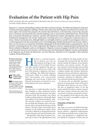
  • 1. Evaluation of the Patient with Hip Pain JOHN J. WILSON, MD, MS, and MASARU FURUKAWA, MD, MS, University of Wisconsin School of Medicine and Public Health, Madison, Wisconsin Hip pain is a common and disabling condition that affects patients of all ages. The differential diagnosis of hip pain is broad, presenting a diagnostic challenge. Patients often express that their hip pain is localized to one of three anatomic regions: the anterior hip and groin, the posterior hip and buttock, or the lateral hip. Anterior hip and groin pain is commonly associated with intra-articular pathology, such as osteoarthritis and hip labral tears. Posterior hip pain is associated with piriformis syndrome, sacroiliac joint dysfunction, lumbar radiculopathy, and less commonly ischiofemoral impingement and vascular claudication. Lateral hip pain occurs with greater trochanteric pain syndrome. Clinical examination tests, although helpful, are not highly sensitive or specific for most diagnoses; however, a rational approach to the hip examination can be used. Radiography should be performed if acute fracture, dislocations, or stress fractures are suspected. Initial plain radiography of the hip should include an anteroposterior view of the pelvis and frog-leg lateral view of the symptomatic hip. Magnetic resonance imaging should be performed if the history and plain radiograph results are not diagnostic. Magnetic resonance imaging is valuable for the detection of occult traumatic fractures, stress fractures, and osteonecrosis of the femoral head. Magnetic resonance arthrography is the diagnostic test of choice for labral tears. (Am Fam Physician. 2014;89(1):27-34. Copyright © 2014 American Academy of Family Physicians.) ▲ Patient information: A handout on this topic, written by the authors of this article, is available at http://www. aafp.org/afp/2014/0101/ p27-s1.html. Access to this handout is free and unrestricted. More online at http://www. aafp.org/afp. CME This clinical content conforms to AAFP criteria for continuing medical education (CME). See CME Quiz questions on page 6. Author disclosure: No relevant financial affiliations. H ip pain is a common presentation in primary care and can affect patients of all ages. In one study, 14.3% of adults 60 years and older reported significant hip pain on most days over the previous six weeks.1 Hip pain often presents a diagnostic and therapeutic challenge. The differential diagnosis of hip pain (eTable A) is broad, including both intra-articular and extra-articular pathology, and varies by age. A history and physical examination are essential to accurately diagnose the cause of hip pain. Anatomy The hip joint is a ball-and-socket synovial joint designed to allow multiaxial motion while transferring loads between the upper and lower body. The acetabular rim is lined by fibrocartilage (labrum), which adds depth and stability to the femoroacetabular joint. The articular surfaces are covered by hyaline cartilage that dissipates shear and compressive forces during load bearing and hip motion. The hip’s major innervating nerves originate in the lumbosacral region, which can make it difficult to distinguish between primary hip pain and radicular lumbar pain. The hip joint’s wide range of motion is second only to that of the glenohumeral joint and is enabled by the large number of muscle groups that surround the hip. The flexor muscles include the iliopsoas, rectus femoris, pectineus, and sartorius muscles. The gluteus maximus and hamstring muscle groups allow for hip extension. Smaller muscles, such as gluteus medius and minimus, piriformis, obturator externus and internus, and quadratus femoris muscles, insert around the greater trochanter, allowing for abduction, adduction, and internal and external rotation. In persons who are skeletally immature, there are several growth centers of the pelvis and femur where injuries can occur. Potential sites of apophyseal injury in the hip region include the ischium, anterior superior iliac spine, anterior inferior iliac spine, iliac crest, lesser trochanter, and greater trochanter. The apophysis of the superior iliac spine matures last and is susceptible to injury up to 25 years of age.2 Evaluation of Hip Pain HISTORY Age alone can narrow the differential diagnosis of hip pain. In prepubescent and adolescent patients, congenital malformations of the femoroacetabular joint, avulsion fractures, and apophyseal or epiphyseal injuries should be considered. In those who are January 1, 2014 ◆ American Family Physician www.aafp.org/afp Downloaded from the Volume 89, Number 1 website at www.aafp.org/afp. Copyright © 2014 American Academy of FamilyAmerican For the private, nonPhysicians. Family Physician commercial use of one individual user of the website. All other rights reserved. Contact copyrights@aafp.org for copyright questions and/or permission requests. 27
  • 2. Hip Pain SORT: KEY RECOMMENDATIONS FOR PRACTICE Evidence rating References Initial plain radiography of the hip should include an anteroposterior view of the pelvis and a frog-leg lateral view of the symptomatic hip. C 4 Magnetic resonance imaging should be used for detection of occult hip fractures, stress fractures, and osteonecrosis of the femoral head. C 23, 30, 33 Magnetic resonance arthrography is the diagnostic test of choice for labral tears. C 6, 19 Ultrasonography is a helpful diagnostic modality for patients with suspected bursitis, joint effusion, or functional causes of hip pain (e.g., snapping hip), and can be employed for therapeutic imagingguided injections and aspirations around the hip. C 8, 9 Clinical recommendation A = consistent, good-quality patient-oriented evidence; B = inconsistent or limited-quality patient-oriented evidence; C = consensus, diseaseoriented evidence, usual practice, expert opinion, or case series. For information about the SORT evidence rating system, go to http://www.aafp. org/afpsort. A B C Figure 1. Gait testing. (A) C sign. Patients often localize pain by cupping the anterolateral hip with the thumb and forefinger in the shape of a “C.” (B) Gait analysis. The patient is observed while walking to evaluate for limp or antalgic gait characteristics. (C) Modified Trendelenburg test (single leg stance phase). The patient stands with feet shoulder width apart and lifts one leg. The examiner observes for a drop in the level of the iliac crest on the side of the lifted leg. skeletally mature, hip pain is often a result of musculotendinous strain, ligamentous sprain, contusion, or bursitis. In older adults, degenerative osteoarthritis and fractures should be considered first. Patients with hip pain should be asked about antecedent trauma or inciting activity, factors that increase or decrease the pain, mechanism of injury, and time of 28 American Family Physician onset. Questions related to hip function, such as the ease of getting in and out of a car, putting on shoes, running, walking, and going up and down stairs, can be helpful.3 Location of the pain is informative because hip pain often localizes to one of three basic anatomic regions: the anterior hip and groin, posterior hip and buttock, and lateral hip (eFigure A). www.aafp.org/afp Volume 89, Number 1 ◆ January 1, 2014
  • 3. Hip Pain Table 1. Physical Examination Tests for the Evaluation of Hip Pain Test Other names Positioning Positive findings Differential diagnosis Gait testing (C sign, Figure 1A; gait analysis, Figure 1B) — Standing Antalgic gait, Trendelenburg gait, pelvic wink (rotation of more than 40 degrees in the axial plane toward the affected hip when terminally extending the hip), excessive pronation or supination of the ankles, and limps caused by differing leg lengths Hip labral tear, transient synovitis, Legg-Calvé-Perthes disease, SCFE Modified Trendelenburg test (Figure 1C) Single leg stance phase Standing 2-cm drop in the level of the iliac crest, indicating weakness on the contralateral side Hip labral tear, transient synovitis, Legg-Calvé-Perthes disease, SCFE ROM testing (Figure 2) — Supine, lateral, or sitting Pain with passive ROM, limited ROM Pain with passive ROM: Transient synovitis, septic arthritis Limited ROM: Loose bodies, chondral lesions, osteoarthritis, Legg-Calvé-Perthes disease, osteonecrosis FABER test (Figure 3) Patrick test Supine Posterior pain localized to the sacroiliac joint, lumbar spine, or posterior hip; groin pain with the test is sensitive for intra-articular pathology Hip labral tear, loose bodies, chondral lesions, femoral acetabular impingement, osteoarthritis, sacroiliac joint dysfunction, iliopsoas bursitis FADIR test (Figure 4) Impingement test Supine Pain Hip labral tear, loose bodies, chondral lesions, femoral acetabular impingement Log roll test (Figure 5) Passive supine rotation, Freiberg test Supine Restricted movement, pain Piriformis syndrome, SCFE Straight leg raise against resistance test (Figure 6) Stinchfield test Supine Weakness to resistance, pain Athletic pubalgia (sports hernia), SCFE, femoral acetabular impingement Ober test (eFigure B) Passive adduction Lateral Passive adduction past midline cannot be achieved External snapping hip, greater trochanteric pain syndrome FABER = flexion, abduction, external rotation; FADIR = flexion, adduction, internal rotation; ROM = range of motion; SCFE = slipped capital femoral epiphysis. PHYSICAL EXAMINATION The hip examination should evaluate the hip, back, abdomen, and vascular and neurologic systems. It should start with a gait analysis and stance assessment (Figure 1), followed by evaluation of the patient in seated, supine, lateral, and prone positions (Figures 2 through 6, and eFigure B). Physical examination tests for the evaluation of hip pain are summarized in Table 1. IMAGING Radiography. Radiography of the hip should be performed if there is any suspicion of acute fracture, dislocation, or stress fracture. Initial plain radiography of the hip should include an anteroposterior view of the pelvis and a frog-leg lateral view of the symptomatic hip.4 January 1, 2014 ◆ Volume 89, Number 1 Magnetic Resonance Imaging and Arthrography. Conventional magnetic resonance imaging (MRI) of the hip can detect many soft tissue abnormalities, and is the preferred imaging modality if plain radiography does not identify specific pathology in a patient with persistent pain.5 Conventional MRI has a sensitivity of 30% and an accuracy of 36% for diagnosing hip labral tears, whereas magnetic resonance arthrography provides added sensitivity of 90% and accuracy of 91% for the detection of labral tears.6,7 Ultrasonography. Ultrasonography is a useful technique for evaluating individual tendons, confirming suspected bursitis, and identifying joint effusions and functional causes of hip pain.8 Ultrasonography is especially useful for safely and accurately performing www.aafp.org/afp American Family Physician 29
  • 4. Hip Pain 45° 10° A C 20-30° 20-35° 30-70° B D Figure 2. Hip range-of-motion testing (photos demonstrate normal range of motion). (A) Abduction. (B) Adduction. (C) Extension. (D) Internal and external rotation. 30 American Family Physician www.aafp.org/afp Volume 89, Number 1 ◆ January 1, 2014
  • 5. Hip Pain A B Figure 3. FABER test (flexion, abduction, external rotation; Patrick test). The examiner moves the leg into 45 degrees of flexion, then (A) externally rotates and (B) abducts the leg so that the ankle rests proximal to the knee of the contralateral leg. A B Figure 4. FADIR test (flexion, adduction, internal rotation; impingement test). The examiner passively moves the leg into (A) full flexion, then into (B) adduction and internal rotation. January 1, 2014 ◆ Volume 89, Number 1 www.aafp.org/afp American Family Physician 31
  • 6. Hip Pain imaging-guided injections and aspirations around the hip.9 It is ideal for an experienced ultrasonographer to perform the diagnostic study; however, emerging evidence suggests that less experienced clinicians with appropriate training can make diagnoses with reliability similar to that of an experienced musculoskeletal ultrasonographer.10,11 Differential Diagnosis of Anterior Hip Pain Anterior hip or groin pain suggests involvement of the hip joint itself. Patients often localize pain by cupping the anterolateral hip with the thumb and forefinger in the shape of a “C.” This is known as the C sign (Figure 1A). Figure 5. Log roll test (passive supine rotation; Freiberg test). Patient’s leg is extended and relaxed on examination table as the examiner internally and externally rotates the leg (log roll). OSTEOARTHRITIS Osteoarthritis is the most likely diagnosis in older adults with limited motion and gradual onset of symptoms. Patients have a constant, deep, aching pain and stiffness that are worse with prolonged standing and weight bearing. Examination reveals decreased range of motion, and extremes of hip motion often cause pain. Plain radiographs demonstrate the presence of asymmetrical joint-space narrowing, osteophytosis, and subchondral sclerosis and cyst formation.12 FEMOROACETABULAR IMPINGEMENT Figure 6. Straight leg raise against resistance test (Stinchfield test). Patients with femoroacetabular impinge- The patient lifts the straight leg to 45 degrees while the examiner ment are often young and physically active. applies downward force on the thigh. They describe insidious onset of pain that is worse with sitting, rising from a seat, getting in or out of pain usually has an insidious onset, but occasionally a car, or leaning forward.13 The pain is located primarily begins acutely after a traumatic event. About one-half in the groin with occasional radiation to the lateral hip of patients with this injury also have mechanical sympand anterior thigh.14 The FABER test (flexion, abduction, toms, such as catching or painful clicking with activity.17 external rotation; Figure 3) has a sensitivity of 96% to The FADIR and FABER tests are effective for detect99%. The FADIR test (flexion, adduction, internal rota- ing intra-articular pathology (the sensitivity is 96% to tion; Figure 4), log roll test (Figure 5), and straight leg 75% for the FADIR test and is 88% for the FABER test), raise against resistance test (Figure 6) are also effective, although neither test has high specificity.14,15,18 Magnetic with sensitivities of 88%, 56%, and 30%, respectively.14,15 resonance arthrography is considered the diagnostic test In addition to the anteroposterior and lateral radiograph of choice for labral tears.6,19 However, if a labral tear is not views, a Dunn view should be obtained to help detect suspected, other less invasive imaging modalities, such subtle lesions.16 as plain radiography and conventional MRI, should be used first to rule out other causes of hip and groin pain. HIP LABRAL TEAR Hip labral tears cause dull or sharp groin pain, and onehalf of patients with a labral tear have pain that radiates to the lateral hip, anterior thigh, and buttock. The 32 American Family Physician ILIOPSOAS BURSITIS (INTERNAL SNAPPING HIP) Patients with this condition have anterior hip pain when extending the hip from a flexed position, often associated www.aafp.org/afp Volume 89, Number 1 ◆ January 1, 2014
  • 7. Hip Pain with intermittent catching, snapping, or popping of the hip.20 Dynamic real-time ultrasonography is particularly useful in evaluating the various forms of snapping hip.8 OCCULT OR STRESS FRACTURE Occult or stress fracture of the hip should be considered if trauma or repetitive weight-bearing exercise is involved, even if plain radiograph results are negative.21 Clinically, these injuries cause anterior hip or groin pain that is worse with activity.21 Pain may be present with extremes of motion, active straight leg raise, the log roll test, or hopping.22 MRI is useful for the detection of occult traumatic fractures and stress fractures not seen on plain radiographs.23 TRANSIENT SYNOVITIS AND SEPTIC ARTHRITIS Acute onset of atraumatic anterior hip pain that results in impaired weight bearing should raise suspicion for transient synovitis and septic arthritis. Risk factors for septic arthritis in adults include age older than 80 years, diabetes mellitus, rheumatoid arthritis, recent joint surgery, and hip or knee prostheses.24 Fever, complete blood count, erythrocyte sedimentation rate, and C-reactive protein level should be used to evaluate the risk of septic arthritis.25,26 MRI is useful for differentiating septic arthritis from transient synovitis.27,28 However, hip aspiration using guided imaging such as fluoroscopy, computed tomography, or ultrasonography is recommended if a septic joint is suspected.29 OSTEONECROSIS Legg-Calvé-Perthes disease is an idiopathic osteonecrosis of the femoral head in children two to 12 years of age, with a male-to-female ratio of 4:1.4 In adults, risk factors for osteonecrosis include systemic lupus erythematosus, sickle cell disease, human immunodeficiency virus infection, smoking, alcoholism, and corticosteroid use.30,31 Pain is the presenting symptom and is usually insidious. Range of motion is initially preserved but can become limited and painful as the disease progresses.32 MRI is valuable in the diagnosis and prognostication of osteonecrosis of the femoral head.30,33 Differential Diagnosis of Posterior Hip and Buttock Pain Piriformis syndrome causes buttock pain that is aggravated by sitting or walking, with or without ipsilateral radiation down the posterior thigh from sciatic nerve compression.34,35 Pain with the log roll test is the most ◆ Volume 89, Number 1 OTHER Other causes of posterior hip pain include sacroiliac joint dysfunction,39 lumbar radiculopathy,40 and vascular claudication.41 The presence of a limp, groin pain, and limited internal rotation of the hip is more predictive of hip disorders than disorders originating from the low back.42 Differential Diagnosis of Lateral Hip Pain GREATER TROCHANTERIC PAIN SYNDROME Lateral hip pain affects 10% to 25% of the general population.43 Greater trochanteric pain syndrome refers to pain over the greater trochanter. Several disorders of the lateral hip can lead to this type of pain, including iliotibial band thickening, bursitis, and tears of the gluteus medius and minimus muscle attachment.43-45 Patients may have mild morning stiffness and may be unable to sleep on the affected side. Gluteus minimus and medius injuries present with pain in the posterior lateral aspect of the hip as a result of partial or full-thickness tearing at the gluteal insertion. Most patients have an atraumatic, insidious onset of symptoms from repetitive use.43,45,46 Data Sources: We searched articles on hip pathology in American Family Physician, along with their references. We also searched the Agency for Healthcare Research and Quality Evidence Reports, Clinical Evidence, Institute for Clinical Systems Improvement, the U.S. Preventive Services Task Force guidelines, the National Guideline Clearinghouse, and UpToDate. We performed a PubMed search using the keywords greater trochanteric pain syndrome, hip pain physical examination, imaging femoral hip stress fractures, imaging hip labral tear, imaging osteomyelitis, ischiofemoral impingement syndrome, meralgia paresthetica review, MRI arthrogram hip labrum, septic arthritis systematic review, and ultrasound hip pain. Search dates: March and April 2011, and August 15, 2013. The authors thank Kristen Prewitt, DO, (model examiner in the figures) and Grace Trabulsi (model patient) for their assistance. PIRIFORMIS SYNDROME AND ISCHIOFEMORAL IMPINGEMENT January 1, 2014 sensitive test, but tenderness with palpation of the sciatic notch can help with the diagnosis.35 Ischiofemoral impingement is a less well-understood condition that can lead to nonspecific buttock pain with radiation to the posterior thigh.36,37 This condition is thought to be a result of impingement of the quadratus femoris muscle between the lesser trochanter and the ischium. Unlike sciatica from disc herniation, piriformis syndrome and ischiofemoral impingement are exacerbated by active external hip rotation. MRI is useful for diagnosing these conditions.38 The Authors JOHN J. WILSON, MD, MS, is an assistant professor in the Department of Family Medicine at the University of Wisconsin School of Medicine and Public Health in Madison. He is also a team physician for the University of Wisconsin Intercollegiate Athletics. www.aafp.org/afp American Family Physician 33
  • 8. Hip Pain MASARU FURUKAWA, MD, MS, is a postgraduate trainee in the Department of Family Medicine at the University of Wisconsin School of Medicine and Public Health. Address correspondence to John J. Wilson, MD, MS, University of Wisconsin–Madison, 1685 Highland Ave., Madison, WI 53705 (e-mail: Wilson@Ortho.wisc.edu). Reprints are not available from the authors. REFERENCES 1. Christmas C, Crespo CJ, Franckowiak SC, et al. How common is hip pain among older adults? Results from the Third National Health and Nutrition Examination Survey. J Fam Pract. 2002;51(4):345-348. 2. Rossi F, Dragoni S. Acute avulsion fractures of the pelvis in adolescent competitive athletes. Skeletal Radiol. 2001;30(3):127-131. 3. Martin HD, Shears SA, Palmer IJ. Evaluation of the hip. Sports Med Arthrosc. 2010;18(2):63-75. 4. Gough-Palmer A, McHugh K. Investigating hip pain in a well child. BMJ. 2007;334(7605):1216-1217. 21. Egol KA, Koval KJ, Kummer F, et al. Stress fractures of the femoral neck. Clin Orthop Relat Res. 1998;(348):72-78. 22. Fullerton LR Jr, Snowdy HA. Femoral neck stress fractures. Am J Sports Med. 1988;16(4):365-377. 23. Newberg AH, Newman JS. Imaging the painful hip. Clin Orthop Relat Res. 2003;(406):19-28. 24. Margaretten ME, Kohlwes J, Moore D, et al. Does this adult patient have septic arthritis? JAMA. 2007;297(13):1478-1488. 25. Eich GF, Superti-Furga A, Umbricht FS, et al. The painful hip: evaluation of criteria for clinical decision-making. Eur J Pediatr. 1999;158(11): 923-928. 26. Kocher MS, Zurakowski D, Kasser JR. Differentiating between septic arthritis and transient synovitis of the hip in children. J Bone Joint Surg Am. 1999;81(12):1662-1670. 27. Learch TJ, Farooki S. Magnetic resonance imaging of septic arthritis. Clin Imaging. 2000;24(4):236-242. 28. Lee SK, Suh KJ, Kim YW, et al. Septic arthritis versus transient synovitis at MR imaging. Radiology. 1999;211(2):459-465. 5. Bencardino JT, Palmer WE. Imaging of hip disorders in athletes. Radiol Clin North Am. 2002;40(2):267-287. 29. Leopold SS, Battista V, Oliverio JA. Safety and efficacy of intraarticular hip injection using anatomic landmarks. Clin Orthop Relat Res. 2001; (391):192-197. 6. Czerny C, Hofmann S, Neuhold A, et al. Lesions of the acetabular labrum: accuracy of MR imaging and MR arthrography in detection and staging. Radiology. 1996;200(1):225-230. 30. Mitchell DG, Rao VM, Dalinka MK, et al. Femoral head avascular necrosis: correlation of MR imaging, radiographic staging, radionuclide imaging, and clinical findings. Radiology. 1987;162(3):709-715. 7. Czerny C, Hofmann S, Urban M, et al. MR arthrography of the adult acetabular capsular-labral complex. AJR Am J Roentgenol. 1999;173(2): 345-349. 31. Mont MA, Zywiel MG, Marker DR, et al. The natural history of untreated asymptomatic osteonecrosis of the femoral head. J Bone Joint Surg Am. 2010;92(12):2165-2170. 8. Deslandes M, Guillin R, Cardinal E, et al. The snapping iliopsoas tendon: new mechanisms using dynamic sonography. AJR Am J Roentgenol. 2008;190(3):576-581. 32. Assouline-Dayan Y, Chang C, Greenspan A, et al. Pathogenesis and natural history of osteonecrosis. Semin Arthritis Rheum. 2002;32(2):94-124. 9. Blankenbaker DG, De Smet AA. Hip injuries in athletes. Radiol Clin North Am. 2010;48(6):1155-1178. 33. Totty WG, Murphy WA, Ganz WI, et al. Magnetic resonance imaging of the normal and ischemic femoral head. AJR Am J Roentgenol. 1984;143(6):1273-1280. 10. Balint PV, Sturrock RD. Intraobserver repeatability and interobserver reproducibility in musculoskeletal ultrasound imaging measurements. Clin Exp Rheumatol. 2001;19(1):89-92. 34. Kirschner JS, Foye PM, Cole JL. Piriformis syndrome, diagnosis and treatment. Muscle Nerve. 2009;40(1):10-18. 11. Ramwadhdoebe S, Sakkers RJ, Uiterwaal CS, et al. Evaluation of a training program for general ultrasound screening for developmental dysplasia of the hip in preventive child health care. Pediatr Radiol. 2010;40(10):1634-1639. 35. Hopayian K, Song F, Riera R, et al. The clinical features of the piriformis syndrome. Eur Spine J. 2010;19(12):2095-2109. 36. Torriani M, Souto SC, Thomas BJ, et al. Ischiofemoral impingement syndrome. AJR Am J Roentgenol. 2009;193(1):186-190. 12. Altman R, Alarcón G, Appelrouth D, et al. The American College of Rheumatology criteria for the classification and reporting of osteoarthritis of the hip. Arthritis Rheum. 1991;34(5):505-514. 37. Ali AM, Whitwell D, Ostlere SJ. Case report: imaging and surgical treatment of a snapping hip due to ischiofemoral impingement. Skeletal Radiol. 2011;40(5):653-656. 13. Banerjee P, McLean CR. Femoroacetabular impingement. Curr Rev Musculoskelet Med. 2011;4(1):23-32. 38. Lee EY, Margherita AJ, Gierada DS, et al. MRI of piriformis syndrome. AJR Am J Roentgenol. 2004;183(1):63-64. 14. Clohisy JC, Knaus ER, Hunt DM, et al. Clinical presentation of patients with symptomatic anterior hip impingement. Clin Orthop Relat Res. 2009;467(3):638-644. 39. Slipman CW, Jackson HB, Lipetz JS, et al. Sacroiliac joint pain referral zones. Arch Phys Med Rehabil. 2000;81(3):334-338. 15. Ito K, Leunig M, Ganz R. Histopathologic features of the acetabular labrum in femoroacetabular impingement. Clin Orthop Relat Res. 2004;(429):262-271. 16. Beall DP, Sweet CF, Martin HD, et al. Imaging findings of femoroacetabular impingement syndrome. Skeletal Radiol. 2005;34(11):691-701. 17. Burnett RS, Della Rocca GJ, Prather H, et al. Clinical presentation of patients with tears of the acetabular labrum. J Bone Joint Surg Am. 2006;88(7):1448-1457. 18. Leunig M, Werlen S, Ungersböck A, et al. Evaluation of the acetabular labrum by MR arthrography [published correction appears in J Bone Joint Surg Br. 1997;79(4):693]. J Bone Joint Surg Br. 1997;79(2):230-234. 19. Groh MM, Herrera J. A comprehensive review of hip labral tears. Curr Rev Musculoskelet Med. 2009;2(2):105-117. 20. Blankenbaker DG, De Smet AA, Keene JS. Sonography of the iliopsoas tendon and injection of the iliopsoas bursa for diagnosis and management of the painful snapping hip. Skeletal Radiol. 2006;35(8):565-571. 34 American Family Physician 40. Moore KL, Dalley AF, Agur AM. Clinically Oriented Anatomy. 6th ed. Philadelphia, Pa.: Lippincott Williams & Wilkins; 2010. 41. Adlakha S, Burket M, Cooper C. Percutaneous intervention for chronic total occlusion of the internal iliac artery for unrelenting buttock claudication. Catheter Cardiovasc Interv. 2009;74(2):257-259. 42. Brown MD, Gomez-Marin O, Brookfield KF, et al. Differential diagnosis of hip disease versus spine disease. Clin Orthop Relat Res. 2004; (419):280-284. 43. Segal NA, Felson DT, Torner JC, et al.; Multicenter Osteoarthritis Study Group. Greater trochanteric pain syndrome. Arch Phys Med Rehabil. 2007;88(8):988-992. 44. Strauss EJ, Nho SJ, Kelly BT. Greater trochanteric pain syndrome. Sports Med Arthrosc. 2010;18(2):113-119. 45. Williams BS, Cohen SP. Greater trochanteric pain syndrome. Anesth Analg. 2009;108(5):1662-1670. 46. Tibor LM, Sekiya JK. Differential diagnosis of pain around the hip joint. Arthroscopy. 2008;24(12):1407-1421. www.aafp.org/afp Volume 89, Number 1 ◆ January 1, 2014
  • 9. Downloaded from the American Family Physician website at www.aafp.org/afp. Copyright © 2013 American Academy of Family Physicians. For the private, noncommercial use of one individual user of the website. All other rights reserved. Contact copyrights@aafp.org for copyright questions and/or permission requests. Dull, diffuse pain radiating to inner thigh; pain with direct pressure, sneezing, sit-ups, kicking, Valsalva maneuver Paresthesia, hypesthesia Pain characteristics Deep, referred pain; pain with standing after prolonged sitting Dull or sharp, referred pain; pain with weight bearing Deep, referred pain; intermittent catching, snapping, or popping Deep, referred pain; pain with weight bearing Deep, referred pain; painful clicking Deep, aching pain and stiffness; pain with weight bearing Femoroacetabular impingement Hip labral tear Iliopsoas bursitis (internal snapping hip) Legg-Calvé-Perthes disease Loose bodies and chondral lesions Osteoarthritis of the hip Older than 50 years, pain with activity that is relieved with rest Mechanical symptoms, history of hip dislocation or low-energy trauma, history of Legg-Calvé-Perthes disease 2 to 12 years of age, male predominance Ballet dancers, runners Mechanical symptoms, such as catching or painful clicking; history of hip dislocation Pain with getting in and out of a car Females (especially with female athlete triad), endurance athletes, low aerobic fitness, steroid use, smokers Soccer, rugby, football, hockey players Obesity, pregnancy, tight pants or belt, conditions with increased intraabdominal pressure History/risk factors Internal rotation < 15 degrees, flexion < 115 degrees Limited ROM, catching and grinding with provocative maneuvers, positive FADIR and FABER tests Antalgic gait, limited ROM or stiffness Snap with FABER to extension, adduction, and internal rotation; reproduction of snapping with extension of hip from flexed position Trendelenburg or antalgic gait, loss of internal rotation, positive FADIR and FABER tests FADIR and FABER tests are sensitive Painful ROM, pain on palpation of greater trochanter No hernia, tenderness of the inguinal canal or pubic tubercle, adductor origin, pain with resisted sit-up or hip flexion Anterior thigh hypesthesia, dysesthesia Examination findings FABER = flexion, abduction, external rotation; FADIR = flexion, adduction, internal rotation; MRI = magnetic resonance imaging; ROM = range of motion. Deep, referred pain; pain with weight bearing Femoral neck fracture/stress fracture Anterolateral hip and groin pain (C sign) Athletic pubalgia (sports hernia) Anterior groin pain Meralgia paresthetica Anterior thigh pain Diagnosis eTable A. Differential Diagnosis of Hip Pain continued Radiography: Presence of osteophytes at the acetabular joint margin, asymmetrical joint-space narrowing, subchondral sclerosis and cyst formation MRI: Can detect chondral and fibrous loose bodies Radiography: Can show ossified or osteochondral loose bodies Radiography: Early small femoral epiphysis, sclerosis and flattening of the femoral head Dynamic ultrasonography: Snapping of iliopsoas or iliotibial band over greater trochanter Ultrasonography: Tendinopathy, bursitis, fluid around tendon MRI: Bursitis and edema of the iliotibial band Radiography: No bony involvement Magnetic resonance arthrography: offers added sensitivity and specificity MRI: Can show a labral tear Radiography: Cam or pincer deformity, acetabular retroversion, coxa profunda MRI: Early bony edema Radiography: Cortical disruption MRI: Can show tear or detachment of the rectus abdominis or adductor longus Radiography: No bony involvement None Additional testing
  • 10. 34BDownloaded from the American Family Physician website at www.aafp.org/afp. Copyright © 2013 American Academy of Family Physicians. For the private, nonAmerican Family Physician www.aafp.org/afp Volume 89, Number 1 January 1, 2014 commercial use of one individual user of the website. All other rights reserved. Contact copyrights@aafp.org for copyright questions and/or permission requests. ◆ Pain characteristics Refusal to bear weight, pain with leg movement Refusal to bear weight Septic arthritis Transient synovitis Pain with direct pressure, radiation down lateral thigh, snapping or popping Deep, referred pain; pain with weight bearing Slipped capital femoral epiphysis Greater trochanteric pain syndrome Pain with direct pressure, radiation down lateral thigh and buttock Pain with direct pressure, radiation down lateral thigh Greater trochanteric bursitis* Iliac crest apophysis avulsion History of direct trauma, skeletal immaturity (younger than 25 years) Middle-aged women Associated with knee osteoarthritis, increased body mass index, low back pain; female predominance Runners, middle-aged women All age groups, audible snap with ambulation Children: 3 to 8 years of age, sometimes fever and ill appearance Adults: Older than 80 years, diabetes mellitus, rheumatoid arthritis, recent joint surgery, hip or knee prostheses Children: 3 to 8 years of age, fever, ill appearance 11 to 14 years of age, overweight (80th to 100th percentile) Adults: Lupus, sickle cell disease, human immunodeficiency virus infection, corticosteroid use, smoking, and alcohol use; insidious onset, but can be acute with history of trauma History/risk factors Iliac crest tenderness and/or ecchymosis Weak hip abduction, pain with resisted external rotation, Trendelenburg gait is sensitive and specific Proximal iliotibial band tenderness, Trendelenburg gait is sensitive and specific Pain over greater trochanter Positive Ober test, snap with Ober test, pain over greater trochanter Pain with extremes of ROM Guarding against any ROM; pain with passive ROM Antalgic gait with foot externally rotated on occasion, positive log roll and straight leg raise against resistance tests, pain with hip internal rotation relieved with external rotation Pain on ambulation, positive log roll test, gradual limitation of ROM Examination findings *—Conditions associated with greater trochanteric pain syndrome. FABER = flexion, abduction, external rotation; FADIR = flexion, adduction, internal rotation; MRI = magnetic resonance imaging; ROM = range of motion. Tenderness to direct palpation Gluteal muscle tear or avulsion* Posterolateral pain Pain with direct pressure, radiation down lateral thigh External snapping hip* Lateral pain Deep, referred pain; pain with weight bearing Osteonecrosis of the hip Anterolateral hip and groin pain (C sign) (continued) Diagnosis eTable A. Differential Diagnosis of Hip Pain (continued) Radiography: Apophysis widening, soft tissue swelling around iliac crest continued MRI: Gluteal muscle edema or tears Dynamic ultrasonography: Snapping of iliopsoas or iliotibial band over greater trochanter Ultrasonography: Tendinopathy, bursitis, fluid around tendon MRI: Bursitis and edema of the iliotibial band Radiography: No bony involvement MRI: Useful for differentiating septic arthritis from transient synovitis Hip aspiration guided by fluoroscopy, computed tomography, or ultrasonography; Gram stain and culture of joint aspirate Radiography: Widened epiphysis early, slippage of femur under epiphysis later MRI: Bony edema, subchondral collapse Radiography: Femoral head lucency and subchondral sclerosis, subchondral collapse (i.e., crescent sign), flattening of the femoral head Additional testing
  • 11. Buttock pain, pain with direct pressure Pain characteristics Buttock or back pain with posterior thigh radiation, sciatica symptoms Buttock pain with posterior thigh radiation, sciatica symptoms Pain radiates to lumbar back, buttock, and groin Ischial apophysis avulsion Ischiofemoral impingement Piriformis syndrome Sacroiliac joint dysfunction Female predominance, common in pregnancy, history of minor trauma History of direct trauma to buttock or pain with sitting, weakness and numbness are rare compared with lumbar radicular symptoms Groin and/or buttock pain that may radiate distally Skeletal immaturity, eccentric muscle contraction (cutting, kicking, jumping) Eccentric muscle contraction while hip flexed and leg extended History/risk factors FABER test elicits posterior pain localized to the sacroiliac joint, sacroiliac joint line tenderness Positive log roll test, tenderness over the sciatic notch None established Ischial tuberosity tenderness, ecchymosis, weakness to leg flexion, palpable gap in hamstring Examination findings *—Conditions associated with greater trochanteric pain syndrome. FABER = flexion, abduction, external rotation; FADIR = flexion, adduction, internal rotation; MRI = magnetic resonance imaging; ROM = range of motion. Buttock pain, pain with direct pressure Hamstring muscle strain or avulsion Posterior pain Diagnosis eTable A. Differential Diagnosis of Hip Pain (continued) Radiography: Possibly no findings, narrowing and sclerotic changes of the sacroiliac joint space MRI: Lumbar spine has no disk herniation, piriformis muscle atrophy or hypertrophy, edema surrounding the sciatic nerve MRI: Soft tissue edema around quadratus femoris muscle MRI: Hamstring edema and retraction Radiography: Avulsion or strain of hamstring attachment to ischium Additional testing Hip Pain January 1, 2014 ◆ American Family Physician www.aafp.org/afp American Family private, nonDownloaded from the Volume 89, Number 1 website at www.aafp.org/afp. Copyright © 2013 American Academy of Family Physicians. For thePhysician 34C commercial use of one individual user of the website. All other rights reserved. Contact copyrights@aafp.org for copyright questions and/or permission requests.
  • 12. Hip Pain The rights holder did not grant the American Academy of Family Physicians the right to sublicense this material to a third party. For the missing item, see the original print version of this publication. eFigure A. Localization of hip pain. (A) Posterior view. (B) Anterior view. A B C eFigure B. Ober test (passive adduction). The patient is positioned on his or her side, with the unaffected hip on the examination table. The examiner stands behind the patient with one hand on the patient’s hip, and the other hand supporting the lower leg. (A) To evaluate the tensor fasciae latae: The hip and knee are held at 0 degrees of extension and allowed to passively adduct with gravity. (B) The gluteus medius: The hip is held at 0 degrees of extension and 45 to 90 degrees of knee flexion. (C) The gluteus maximus: The shoulders are rotated back toward the table, with the hip in flexion and knee in extension. 34DDownloaded from the American Family Physician website at www.aafp.org/afp. Copyright © 2013 American Academy of Family Physicians. For the private, nonAmerican Family Physician www.aafp.org/afp Volume 89, Number 1 January 1, 2014 ◆ commercial use of one individual user of the website. All other rights reserved. Contact copyrights@aafp.org for copyright questions and/or permission requests.