Bohomolets Oncology Endoscopic pictures of GIT
•Download as DOC, PDF•
0 likes•261 views

By Dr. Olga Lobanova from Oncology department.

Recommended
More Related Content
More from Dr. Rubz
More from Dr. Rubz (20)
Recently uploaded
Recently uploaded (20)
Bohomolets Oncology Endoscopic pictures of GIT
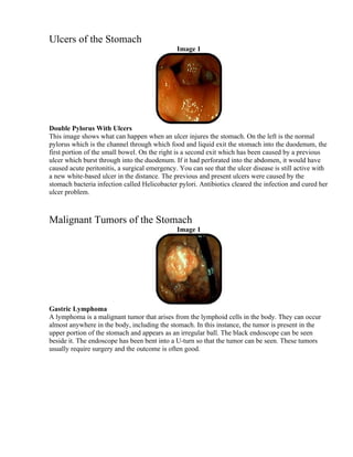
- 1. Ulcers of the Stomach Image 1 Double Pylorus With Ulcers This image shows what can happen when an ulcer injures the stomach. On the left is the normal pylorus which is the channel through which food and liquid exit the stomach into the duodenum, the first portion of the small bowel. On the right is a second exit which has been caused by a previous ulcer which burst through into the duodenum. If it had perforated into the abdomen, it would have caused acute peritonitis, a surgical emergency. You can see that the ulcer disease is still active with a new white-based ulcer in the distance. The previous and present ulcers were caused by the stomach bacteria infection called Helicobacter pylori. Antibiotics cleared the infection and cured her ulcer problem. Malignant Tumors of the Stomach Image 1 Gastric Lymphoma A lymphoma is a malignant tumor that arises from the lymphoid cells in the body. They can occur almost anywhere in the body, including the stomach. In this instance, the tumor is present in the upper portion of the stomach and appears as an irregular ball. The black endoscope can be seen beside it. The endoscope has been bent into a U-turn so that the tumor can be seen. These tumors usually require surgery and the outcome is often good.
- 2. Gastroesophageal Reflux Disease (GERD) Image 1 Image 2 Image 3 Image 4 Examples of GERD When acid from the stomach bathes the lower esophagus, a feeling of heartburn occurs. In some people, actual tissue damage occurs. This can cause some mild inflammation or reddening of the lower esophagus as seen in Image 1 or a slight erosion which has a whitish center surrounded by inflamed red tissue seen in Image 2. In the severe cases, extensive deep ulcerations occur. Images 3 and 4 show these ulcers. Benign Tumors of the Esophagus Image 1 Esophageal Polyp A polyp is a growth. Most polyps are benign but certain types can become malignant. They can occur anywhere in the intestinal tract, especially the colon. Infrequently, they develop in the esophagus. They are rather easy to remove through the endoscope. This 1 inch polyp in the lower esophagus was an incidental finding. The whitish material is coagulated saliva. On pathology exam, it was a benign adenoma type polyp.
- 3. Duodenal Ulcer Image 1 Image 2 Image 3 Examples of Duodenal Ulcer Ulcers often occur within the duodenum, usually in the first part just beyond the stomach which is called the duodenal cap. Ulcers can come in all shapes and sizes. Image 1 shows a single white- based ulcer. Image 2 shows numerous small ulcers scattered across the duodenal cap. Image 3 demonstrates a huge duodenal ulcer almost filling the cap. Note the brownish areas across the whitish base which is caused by some old blood. Some fresh bleeding is seen at the edge. Duodenal ulcers are caused by the bacteria, Helicobacter pylori, or by arthritis medications such as Advil, Motrin, Naprosyn, aspirin and many others. All of these ulcers healed under appropriate therapy.
- 4. Duodenal Tumors Image 1 Lymphoid Polyposis This is a benign but peculiar looking condition. The intestine is packed with lymphoid tissue which is necessary as part of the body's immune system which protects us from infection. Most of the intestinal lymph deposits are below the surface and not usually seen. In this instance, the duodenum is full of these benign lymph nodules. They cause no problem and do not need treated. They are just an endoscopic oddity Malignant Tumors Image 1 Image 2 Image 3 Image 4 Colon Cancer Most cancers in the colon originate in adenoma type polyps. These adenomas start out being benign. Then, as they get larger, they can become cancerous. That is why polyps are always removed from the colon when they are found. Cancers come in all sizes. Image 1 shows a small cancerous polyp about the size of an olive. The chance for a complete cure in this case is probably very good. Image 2 shows the same site after the polyp has been removed with cautery. The base of the polyp looks
- 5. nice and clean. Images 3 and 4 show more advanced cancers in the rectum and cecum or right colon respectively. At this point, surgery is needed to remove the diseased segment of colon. Diverticular Disease Image 1 Image 2 Diverticular Inflammatory Bowel Disease This is an unusual condition. Please review the information on diverticulosis first, so you know what diverticuli are. Inflammatory bowel disease is a chronic inflammation of the bowel lining. It may be called either ulcerative colitis or Crohn's disease. A third type of inflammation can occur with severe diverticulosis. The lining can become very reddened and then bleed. In these cases, the inflammation remains localized in the area with diverticulosis and never extends further. Treatment is usually the same as for ulcerative colitis or Crohn's disease. Here you see two images of the sigmoid colon. Image 1 a shows small black opening of a diverticuli on the left. Above it is the markedly reddened and inflamed fold. Image 2 shows scattered areas of inflammation.