Top Rated Bangalore Call Girls Ramamurthy Nagar ⟟ 9332606886 ⟟ Call Me For G...
Skull in newborn by DR.ARSHAD
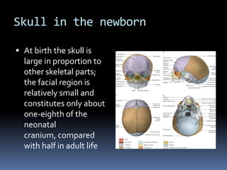
1. Skull in the newborn
At birth the skull is
large in proportion to
other skeletal parts;
the facial region is
relatively small and
constitutes only about
one-eighth of the
neonatal
cranium, compared
with half in adult life
2. Smallness of the face at birth is largely due to
the rudimentary stage of development of the
mandible and maxillae - the teeth are
unerupted.
The nose lies almost entirely between the
orbits, and the lower border of the nasal
aperture is only slightly lower in position than
the orbital floors
3. The large size of the calvaria, especially the
neurocranium, reflects early cerebral
maturation.
Bones of the cranial vault are unilaminar and
lack diploë.
4. Frontal and parietal
tuberosities are prominent;
in the frontal view, the
greatest width occurs
between the parietal
tuberosities.
The glabella, superciliary
arches and mastoid
processes are not
developed
Cranial base is relatively
short and narrow
5. Ossification is incomplete, and many bones
are still in several elements united by fibrous
tissue or cartilage.
Two halves of the frontal bone and
mandible, and the squamous, lateral and
basilar parts of the occipital bone are all
separate
6. Parts of the temporal bones are separate
except that fusion of the tympanic with the
petrous and squamous parts has started.
The fibrous membrane that forms the cranial
vault before ossification is unossified at the
angles of the parietal bones, producing six
fontanelles: two median (anterior and
posterior) and two lateral pairs
(sphenoidal/anterolateral and
mastoid/posterolateral).
7. The anterior fontanelle is the
largest and measures
approximately 4 cm in
anteroposterior and 2.5 cm in
transverse dimensions.
It occupies the junction between
the sagittal, coronal and frontal
sutures and is therefore
rhomboid in shape.
.The posterior fontanelle lies at
the junction between the
sagittal and lambdoid sutures
and is therefore triangular.
8. The sphenoidal
(anterolateral) and mastoid
(posterolateral) fontanelles
are small, irregular and
occur at the sphenoidal
and mastoid angles of the
parietal bones respectively.
9. At birth the orbits appear relatively large.
The developing tooth germs are generally
contained within the alveolar
crypts, although eruption of the upper central
incisor teeth can occur prior to, or shortly
after, birth
10. Temporal bones differ greatly from their
adult form.
The internal ear, tympanic cavity, auditory
ossicles and mastoid antrum are all almost
adult in size
The tympanic plate is an incomplete ring
which has usually started to fuse with the
squamous part, and the mastoid process is
absent.
11. The external aspect of the tympanic
membrane faces more inferiorly than
laterally
The stylomastoid foramen is exposed on the
lateral surface of the skull, the styloid process
has not fused with the temporal bone, the
mandibular fossa is flat and more lateral, and
its articular tubercle is undeveloped.
12. The mandibular fossa is flat and more
lateral, and its articular tubercle is
undeveloped.
The paranasal sinuses are rudimentary or
absent and only the maxillary sinuses are
usually identifiable
13. During birth the skull is moulded by slow
compression.
That part of the scalp which is more central in
the birth canal is often temporarily
oedematous as a result of interference with
venous return, and is called the caput
succedaneum
14. Fontanelles and the openness and width of
the sutures allow bones of the cranial vault
some overlap.
The skull is compressed in one plane with
compensatory orthogonal elongation. These
effects disappear within the first week after
birth.