development of diagnostic enzyme assay to detect leuser virus
The structure and folding behavior of a solenoid
1. The Structure And Folding Behavior Of A Solenoid Protein: A Molecular Dynamics Study
Sally Q. Fisher and Carol A. Parish
Department of Chemistry, University of Richmond, Richmond VA 23173
Force Fields
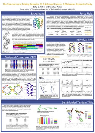
A number of standard force fields and explicit solvent models were tested with both the CTPR and
P46824 individual TPR systems. Of these, the amber03 and charmm27 force fields and the tip3p explicit
water model were found to be suitable for the CTPR systems, producing dynamics that were qualitatively
comparable to its experimental behavior in the literature. Invariably, all ensembles generated using the
charmm27 force field are more helical than those generated using the amber03 force field (Table 1). This
finding is in agreement with a wide body of literature showing that charmm27 tends to favor alpha helices.
Because our objective is to investigate if the P46824 domain does adopt these highly helical TPR structures,
we focus our analysis on the ensembles generated with the amber03 force field. It is our belief that this force
field will better allow us to observe any non alpha-helical structure if it is indeed present.
Repeat proteins are a class of proteins that contain a number of
highly homologous secondary structure elements arranged in tandem.
Unlike their globular counterparts, the folding pathway of repeat
proteins is thought to be fairly simple: short-range and regularized
interactions between the tandem secondary structure elements give
rise to a repetitive and often elongated structure. In many repeat
proteins, this elongated structure functions as a binding site. One such
family of repeat proteins is comprised of tetratricopeptide repeat (TPR)
proteins. A single TPR unit is made up of 34 amino acids in a
helix-loop-helix motif. Three or more TPRs in tandem gives rise to
super helical tertiary structure, with a groove suitable for binding
the alpha helix of target proteins and other small molecules.
Figure 1. Two TPRs in tandem.
Figure 2. Superhelix
formed by 13 TPRs.
Background
Figure 3. Kinesin cargo-binding P46824 primary sequence. Six regions suggested
to form TPR motifs are highlighted.
It is believed that one such TPR protein is the Kinesin molecular
motor protein. Kinesin’s motor functionality allows it to actively transport
intracellular cargos by walking along microtubules. The binding activity is
facilitated by the tail domain, which is believed to contain six TPRs. The
objective of this research is to determine if the tail domain contains TPRs
and if these TPRs give rise to a superhelical structure.
Table 1. Fraction Helicity Data for the Consensus Sequence Crystal Ensembles
amber03/tip3p charmm27/tip3p
Minimum Average Minimum Average
34 residues 0.588 0.793 0.048 0.735 0.822 0.008
30 residues 0.667 0.858 0.050 0.833 0.897 0.011
All subsequent
ensembles were
generated with:
amber03
and tip3p
In order to obtain a template structure for our homology modeling and to
validate our methods, we selected CTPR, a TPR protein that has a known crystal
structure and is highly homologous to the portion of the Kinesin sequence
thought to contain TPRs. CTPR is TPR protein whose primary sequence was
designed as a consensus among all known TPRs. It contains eight highly
conserved residues, termed scaffold residues: 4 (W/L/F), 7 (L/I/M), 8 (G/A/S),
11 (Y/L/F), 20 (A/S/E), 24 (F/Y/L), 27 (A/S/L), 32 (P/K/E). These positions have a
low tolerance for substitutions, as their pattern of large and small amino acids
governs the overall structure of the motif.
Designed Consensus: CTPR
Figure 6. Consensus sequence
(CTPR) with scaffold residues.
TRP 4
LEU 7
GLY 8
TYR 11
ALA 20
PRO 32
ALA 27
TYR 24
Figure 4. Alignment of consensus sequence with regions of the Kinesin sequence
thought to form TPRs. Scaffold residues that agree with the consensus residue
type are colored in blue and those that do not are colored in red.
Many repeat proteins, including TPR proteins have been shown
to follow a non two-state folding pathway. Because of the difficult
nature of constructing a homology model for large protein systems,
we used our homology models for the individual TPRs to construct a
model from the semi-folded ensemble.
Semi-Folded Tandem TPRs
Figure 5. Helix wheel maps for a typical TPR with helices
A and B, respectively. Consensus residues are labeled.
Individual TPRs
Models for the six Kinesin TPRs were
constructed by performing the appropriate
residue mutations on CTPR, according to the
sequence alignments. The systems were
prepared with various explicit water models
and standard ions. Following a standard
relaxation and equilibration protocol, 100 ns
simulations were performed for each system
using a variety of standard force fields. Data for
the amber03/tip3p ensemble are shown. Figure 7. Average structures for the CTPR and P4 TPRs ensembles
aligned by C atoms with STRIDE. (A) shows the alignment for CTPR
and P4 TPRs 1-6. (B) shows the alignment for CTPR and P4 TPRs 2-6.
CTPR
P46824
TPR1
TPR2
TPR3
TPR4
TPR5
TPR6
Table 2. Global Backbone Average and Maximum
RMSD Relative to Starting Structure
Maximum (Å) Average (Å)
TPR1 4.924 3.248 1.008
TPR2 3.391 0.831 0.368
TPR3 2.202 0.925 0.245
TPR4 1.973 0.761 0.169
TPR5 2.918 1.627 0.438
TPR6 3.238 0.949 0.335
Figure 9. Root mean square deviation per residue of the C backbone atoms for
TPR structures generated with amber03/tip3p taken at 10 ns intervals relative to
the starting structure.
Predicted Structural Perturbations:
1) TPR1: Gly8 Val8
2) TPR4: Ala20 Val20
3) TPR5: Ala27 Val27
TPR1
Local unfolding at N-terminus due to positively
charged Arg2 contributing to destabilizing
macrodipole of H-bonding pattern of helix.
Figure 10. Average structure for P4 TPR1
ensemble with macrodipoles for residues in the
region of the A helix displayed.
Figure 8. Fraction helicity given by STRIDE secondary structure assignments for the
ensembles generated with amber03/tip3p.
Table 3. C- Root Mean Square Deviations Between
Individual TPR Ensemble Average Structures and
Representative Multi-TPR Structures
amber03/tip3p charmm27/tip3p
1-5 1-5
TPR1 4.833 0.836
TPR2 2.230 0.459
TPR3 2.169 0.492
TPR4 2.867 0.654
TPR5 1.221 0.868
The above RMSD values (reported in Angstroms) are taken between
the ensemble average structures from the individual TPR simulations
and the corresponding TPR in the final structure from the tandem TPR
simulations. All CA atoms are used in the RMSD.
Tandem
P46824
TPR1
TPR2
TPR3
TPR4
TPR5
Figure 13. Average structures for the P46824 Individual TPR ensembles
aligned with representative multi-TPR structure by C atoms with STRIDE.
(A) shows the alignment for ensembles generated with amber03/tip3p.
(B) shows the alignment for ensembles generated with charmm27/tip3p.
Figure 11. Kinesin TPR1-TPR5 model structure
from the semi-folded ensemble.
Figure 12. Fraction helicity per residue given by STRIDE
secondary structure assignments for the ensembles generated
with amber03/tip3p. Positions comprising TPRs are colored as
indicated by the legend.