1. Maria R. Moore, MSPT, DPT'
Mary Ann Wilmarth, PT, DPT,
OCS, MTC, Cert. MDT, CEAS2
Marie B. Corkery, PT, DPT,
MHS, FAAOMPT3
Differentiating Hip Versus Back Pathology
with a Patient Status Post Lumbar
Laminectomy and Fusion: A Case Study
'Physical Therapist, Lighthouse Healthcare, Inc, Sprineeld VA
2Assistant Professor, DPT Program CPS/BCHS Northeastern University and ChitfofP7; Harvard University Health Sciences
3Associate Clinical Professor, Physical Therapy Departmen4 Bouve College of Health Sciences, Northeastern Universios Boston, MA
ABSTRACT
Background: In older adults with
normal degenerative changes, it can be dif-
ficult to differentiate low back pain from
hip pain. The purpose of this report is to
describe the role of differential diagnosis
of hip from back pathology in a patient
post lumbar laminectomy and fusion. Case
Description: The patient was a 56-year-old
female seen in physical therapy two months
postoperatively. A thorough examination led
to a differential diagnosis of intraarticular
hip pathology and the patient was referred
to an orthopaedic specialist. Outcomes: The
patient was diagnosed with severe right hip
osteoarthritis and scheduled for a right hip
arthroplasty. Three months after surgery
the patient had complete resolution of pain
and return of function. Discussion: Physi-
cal therapists should always complete dif-
ferential diagnosis and screen for additional
pathologies. Groin pain, decreased hip inter-
nal rotation, decreased hip strength, and gait
dysfunction are key findings to differentiate
between back and hip pathology.
Key Words: differential diagnosis, hip
osteoarthritis, hip pain, physical therapy
INTRODUCTION
Differentiating low back from hip
pathology can be difficult due to overlap-
ping pain referral patterns.' Intraarticular
hip pathology can commonly refer pain to
the groin, anterior thigh, buttock, anterior
knee, and lateral thigh regions.' Similarly,
with lumbar pathology, pain can be referred
into the proximal, middle, and distal ante-
rior thigh regions from the LI , L2, and L3
nerve roots respectively' and buttock pain
can originate from the L4 and L5 nerve
roots. A thorough history and physical exam
of the low back and hip areas can guide the
practitioner in establishing the correct diag-
nosis in a timely and efficient manner, while
helping to avoid the costly care and manage-
ment of misdiagnosis. In older adults with
normal lumbar degenerative changes, it can
be especially difficult to differentiate pain
originating from the low back from pain
originating in the hip. A common example
is differentiating degenerative back patholo-
gies from intraarticular hip pathologies,
such as hip osteoarthritis (OA).
Low back pain (LBP) is the second most
common cause of disability in adults in
the United States.' More than 80% of the
United States population will experience an
episode of LBP sometime during their lives,'
making LBP a diagnosis very commonly
seen by primary care clinicians and physical
therapists. Over the past few decades, there
has been an increase in the number of medi-
cal procedures for the low back, including
spinal injections and surgery. Among elderly
Americans, the increase in procedures is
closely related to an increase in diagnostic
imaging.' Although magnetic resonance
imaging (MRI) is highly sensitive, studies
have questioned the relevance of MRI find-
ings in terms of specificity and correlation
with clinical findings." In one such study,
MRI was performed on 67 individuals who
had never had an episode of LBP or sciatica.
In the group that was younger than 60 years
of age, 20% were found to have a herniated
nucleus pulposus. In the group that was 60
years of age or older, all but one person dem-
onstrated degeneration or bulging of a disc in
at least one lumbar level." In another study,
200 subjects without a history of LBP were
given an initial exam and MRI of the low
back and were followed over a 5-year period.
Fifty-one subjects developed a new onset
of LBP and had a new MRI taken within
6 to 12 weeks of the pain onset. Only 4%
of the subjects showed MRI changes with
probable clinical significance.' These studies
demonstrate the importance of correlating
normal lumbar degenerative changes, such
as joint space narrowing, disc degeneration,
stenosis, osteophyte formation, and lumbar
spondylolisthesis, with a thorough history
and clinical exam, especially in older adults
who would be expected to have age-related
degenerative changes.
Hip arthritis is a much less common
condition compared to LBP, with estimates
being 3.2% of the population older than
55 years of age.' Despite the fact that hip
OA is less common than LBP, the two con-
ditions often coexist. Hip OA can cause
abnormal gait and spinal sagittal plane
alignment and is associated with LBW° Hip
and spine arthritis are also part of the same
age-related degenerative process and there-
fore often coexist." In a prospective study
looking at 344 patients waiting to receive
hip arthroplasties, 49% reported LBP. Of
these patients, 66% had resolution of back
symptoms following their hip surgeries."
In a study by Hsieh et a1, 2 LBP was found
in 21% of patients who were scheduled to
receive a hip arthroplasty, and the presence
of LBP was statistically more common in
those with a longer duration of hip symp-
toms. In their study of patients with coex-
isting lumbar spine and hip pathologies
described as hip-spine syndrome, Ben Galim
et al'" followed 25 patients with severe hip
OA and at least moderate LBP. They found
that in the spine and hip regions, both pain
and function improved significantly follow-
ing hip arthroplasties. A key point recog-
nized by the authors after the study was that
in patients with both hip OA and LBP, the
hip should be treated first."
Whether a patient is seen by direct access
or is referred for any type of low back or hip
pain, the physical therapist should be cogni-
zant of the potential structures that could be
causing the patient's symptoms. As a practic-
ing autonomous provider, it is the therapist's
responsibility to perform a medical screen,
assess regions above and below the involved
area, and to rule in or rule out other poten-
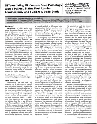
tial conditions. Figure 1 provides an algo-
rithm for evaluation of low back versus hip
pain with differential diagnosis based on the
location of pain.' There are other examples
in the literature of using an algorithmic
approach to help guide the practitioner in
developing a hip differential diagnosis based
on location of pain and information from
172 Orthopaedic Practice Vol 26;3:14
2. Groin or anterior
thigh pain
History and
Physical
Visceral
complaints
Lumbar pain
radiating into
the buttock or
leg
Differential diagnosis:
-diverticulitis/
diverticulosis
-inflammatory bowl
disease
-urintary tract infections
-nephrolithiasis
-appendicitis
-abdominal aortic
aneurysms
-prostatitis
-epididymitis
-hydroceles
-varicoceles
-testicular torsions
-testicular neoplasms
-ectopic pregnancy
-dysmenorrhea
-endometriosis
-pelvic inflammatory
disease
Internal
medicine
consultation
Differential diagnosis:
-sacroiliac joint
-zygapophyseal joint
-diskogenic
-radiculopathy
-lumbar stenosis
-piriformis syndrome
-spondylolisthesis
-lumbosacral
plexopathy
-peripheral nerve
lesion
-spine, pelvic or
femoral fracture
Diagnostics:
-X-ray
-MRI
-Bone Scan
-EMG
-Diagnostic injection
-Musculoskeletal
ultrasound
Differential diagnosis:
-osteoarthritis hip and
knee
-avascular necrosis
-adductor, iliopsoas
tendonitis
-septic arthritis
-osteomyelitis
-metastatic lesions
-pelvic or femoral
fractures
-hip dislocation
-labral tear
-peripheral nerve lesion
-spinal etiology
Diagnostics:
-X-ray
-MRI
-MRI arthrogram
-EMG
-Musculoskeletal
ultrasound
-Sed rate, ANA, RA titre
-Hip aspiration: cell count,
crystals, culture &
sensitivity
Figure 1. Algorithm for evaluation of low back and hip pain. Reprinted with
permission from Pract Neural.' Copyright 2008, BMJ Publishing Group.
the clinical exam, such as range of motion
(ROM) and special tests.'`
The purpose of this case report is to
describe the role of the physical therapist
in differentiating hip pathology from back
pathology in a patient post lumbar lami-
nectomy and fusion. After the patient's sur-
gery, the key signs and symptoms of groin
pain, decreased hip internal rotation ROM,
decreased hip strength, and gait deficits led
to a differential diagnosis of intraarticular
hip pathology with a referral to an orthopae-
dic hip specialist.
CASE DESCRIPTION
The patient was a 56-year-old female
who was referred to outpatient physical
therapy two months after a lumbar laminec-
tomy and fusion of L4-5 due to a lack of
any improvement in both pain and function
following the surgery. Her occupation as a
materials engineer primarily consisted of
desk work; however, she also needed to peri-
odically climb ladders in tight spaces. A pre-
surgery MRI indicated degenerative changes
throughout the lumbar spine with a 4 mm
anterolisthesis of L4 on L5. According to
Watters et al,'' a degenerative lumbar spon-
dylolisthesis can be defined as a forward or
backward slip by at least 3 mm. Presurgery,
the patient completed approximately one
month of physical therapy treatment with a
different physical therapist and also had cor-
ticosteroid injections with minimal relief.
The patient completed a 6-week course of
physical therapy following her back surgery.
During that postsurgery time, her main
complaints included overall right lower
extremity weakness, hip stiffness, difficulty
with stairs walking more than one-quarter
mile, and pain with sitting or standing for
longer than 30 minutes. The patient's pain
was reported mainly in the right groin area
as well as the right lateral thigh and the lat-
eral and anterior aspects of the right knee
region.
Tests and Measures
At the initial evaluation, the patient's
resting pain score was 3/10 on the visual
analog scale. Her Oswestry Disability Ques-
tionnaire score was 34% and her Roland
Morris Questionnaire score was 10 out of
24. Both of these questionnaires are widely
used to assess pain-related disability in
persons with LBP, and both have demon-
strated valid and reliable measures that are
responsive to change." The patient's lower
extremity neural screen was negative, dem-
onstrating normal deep tendon reflexes,
normal sensation, no abnormal strength
findings in the L2-S1 myotomes, and nega-
tive neural tension in the sciatic and femo-
ral nerves. Although her lumbar ROM was
globally limited, it was painfree. Her single
leg static balance was decreased for the right
lower extremity as she was able to maintain
balance for about 5 seconds. Her initial
strength measurements assessed by standard
manual muscle testing (MMT) are listed in
Table 1. Her hip goniometric passive ROM
measurements, which were taken in supine,
are listed in Table 2. Most notable were the
strength deficits in the musculature of the
right hip and right hip ROM deficits in
several planes. Significant flexibility deficits
were noted bilaterally in the piriformis, glu-
teus maximus, and hip adductor muscles,
with the right being more restricted than
the left. The patient's gait lacked hip exten-
sion on the right, with a short right antalgic
stride, and a notable Trendelenburg pattern.
After her initial evaluation and throughout
treatment, the patient complained of sig-
nificant right groin pain, especially with hip
internal rotation, and pain at both the ante-
rior and lateral aspects of the right knee with
passive hip flexion, internal rotation, and
external rotation.
Orthopardir Practirr Vol. 26;3:1-1 173
3. Table 1. Manual Musde Testing Measurements
Hip movement
Initial
MMT right
Initial
MMT left
Re-evaluation
MMT right
Re-evaluation
MMT left
Flexion 4-/5 5/5 4-/5 4+/5
Extension 4/5 5/5 515 5/5
Abduction 4-/5 4/5 4/5 4/5
Adduction 4-/5 4+15 4-15 4/5
Internal rotation 4/5 5/5 4/5 5/5
External rotation 4-/5 5/5 4-15 5/5
Abbreviation: MMT, manual muscle testing
Table 2. Goniometric Measurements
Hip movement
PROM
right
PROM
left
Re-evaluation
PROM right
Re-evaluation
PROM left
Flexion 100° 115° 110° 125°
Extension NT NT NT NT
Abduction NT NT 30° 55°
Adduction NT NT NT NT
Internal Rotation 25° 45° 20° 50°
External Rotation 25° 55° 40° 52°
Abbreviations: PROM, passive range of motion; NT, not tested
Re-evaluation measurements were taken
4 to 6 weeks after the initial evaluation. The
patient's resting pain level at re-evaluation
was 2/10, Oswestry Disability score was
20%, and Roland Morris score was 8 out of
24. She continued to have significant groin
pain and only mild improvements in func-
tional deficits. Table 2 lists changes in passive
ROM measurements after 4 to 6 weeks of
treatment. Hip passive ROM measurements
remained limited in several planes, with
an actual decrease in hip internal rotation.
Changes in hip strength, as seen in Table
1, were minimal, with no changes in hip
flexion, internal rotation, external rotation,
or adduction. Her gait deficits remained
unchanged.
Diagnosis and Prognosis
According to the Guide to Physical Thera-
pist Practice," the physical therapist diag-
nosis for this patient was within Pattern 4E
and included impaired joint mobility, motor
function, muscle performance, and range
of motion associated with localized inflam-
mation. At the time of initial evaluation,
the patient's prognosis was good for making
significant functional gains in a reasonable
length of time with skilled physical therapy
intervention. Her goals were to use stairs
without a handrail, ambulate community
distances, put on shoes without difficulty,
transfer from a low chair without diffi-
culty, and sit for at least 60 minutes with-
out discomfort. Over the 6-week treatment
period, as objective and subjective measures
improved minimally and the patient was
unable to progress with exercises, it was clear
that the prognosis with skilled physical ther-
apy was no longer favorable and the patient
required an additional work-up by a special-
ist before continuing care.
Intervention
Following the patient's lumbar laminec-
tomy and fusion, her treatment consisted
of lumbo-pelvic core stabilization exer-
cises, soft tissue mobilization of the pelvic
girdle musculature, lower extremity stretch-
ing, lower extremity strengthening with
focus on hip musculature, postural educa-
tion and awareness training, instruction in
body mechanics, gait training, and range of
motion exercises for the spine and hip. There
was minimal exercise progression over the
6-week period due to the patient's pain level
and tolerance.
Outcomes
Based on the patient's symptoms and
clinical examination, a differential diagno-
sis of hip intraarticular pathology was made
by the physical therapist. The patient was
referred to an orthopaedic hip specialist and
was diagnosed with severe osteoarthritis of
the right hip and scheduled to receive a hip
arthroplasty with a lateral approach. The
patient initiated physical therapy about 4
weeks after her hip replacement for reha-
bilitation according to a standard hip
replacement protocol. At the initial physical
therapy exam status post hip arthroplasty,
the patient reported almost complete resolu-
tion of groin, thigh, and knee pain. Her ini-
tial Hip Condition Questionnaire score was
19 out of 50. This is a questionnaire used
in some physical therapy settings to assess
pain-related disability due to hip pain, and
it has been found to have moderate to suffi-
cient psychometric properties. 16 The patient
was discharged from physical therapy after
two months of care. At that time, she had
returned to work full time without limita-
tions, was using stairs without compen-
sations, and demonstrated a normal gait
pattern without an assistive device. She con-
tinued to have some hip ROM and strength
limitations post-hip arthroplasty but overall
was making significant improvements. The
patient reported a return to 95% of her
prior function without pain and scored 3
out of 50 on the Hip Condition Question-
naire, indicating a significant improvement
in overall function. Her home exercise pro-
gram included continuation of her ROM
exercises, gait and functional training, and
strengthening exercises.
DISCUSSION
This case demonstrates how findings of
groin pain, decreased hip internal ROM,
decreased hip strength, and gait deficits,
which were present after a lumbar lami-
nectomy and fusion, led to a diagnosis of
intraarticular hip pathology and referral
to a hip orthopaedic specialist. The groin
region, a common pain referral area from
intraarticular hip pathology, has been shown
in various studies to be a key symptom of
hip pathology.2'17.18 A retrospective analysis
completed by Brown et air on 97 patients
with lower extremity pain revealed which
signs and symptoms were the best predic-
tors for primary sources of pain in the hip or
spine. They found that patients with groin
pain were 7 times more likely to have a hip
disorder, or hip and spine disorder, rather
than a spine disorder only.'' In another
study, 113 patients with end-stage hip dis-
ease were evaluated for pain patterns prior
to hip arthroplasty. The most common areas
of pain before surgery included the groin,
174 Orthopaedic Practice Vol 26:3:14
4. BETTER TOOLS FOR A QUICKER
COMEBACK
Upper Body & Back Kit - Stabilizes the back during rowing and other back muscle exercises.
Includes modular handles, combination mount and two 3-ft tubes with metal clips on both ends.
To order. , ,sa our
website or call
800-886-6621
f. ,
Gear to reduce pain, rehab injuries.
anterior thigh, buttock, anterior knee, and
greater trochanter! In the clinical guidelines
for hip pain and mobility deficits for hip
osteoarthritis, the Orthopaedic Section of
the American Physical Therapy Association
(APTA) recommended that key symptoms
be present for diagnosis and classification
of unilateral coxarthrosis or to identify the
impairment based category of hip pain and
mobility deficits. One key symptom that
should be present includes "moderate lateral
or anterior hip pain during weight bear-
ing."i9 The patient presented in this case
report fit this criterion as she experienced
anterior pain in the groin region throughout
her treatment.
Cibulka et al,'" in their review of guide-
lines for hip pain and mobility, stated that
limited hip internal rotation by more than
15° when compared to the nonpainful side
is a useful clinical finding that fits the uni-
lateral coxarthrosis criterion. In the criteria
for the classification of OA of the hip, the
American College of Rheumatology also
recognized decreased hip internal rotation
as a key sign.'" Many studies have demon-
strated that decreased hip internal rotation
and painful internal rotation are key signs or
clinical predictors in patients with hip OA
or other hip intraarticular pathologies.".23-25
Brown et al" reported that patients with
limited hip internal rotation were 14 times
more likely to have a hip, or hip and spine
disorder, rather than a spine disorder only.
In an additional study, 195 patients over the
age of 40 presenting with a new episode of
hip pain were tested for radiological evidence
of hip OA. Hip flexion, internal rotation,
and external rotation were tested to identify
which were most discriminatory of hip OA.
Internal rotation limitations were found
to be the most predictive of hip OA!' The
patient presented in this case study clearly
had a significant decrease in hip internal
rotation compared with the uninvolved side,
and hip internal rotation was painful, repro-
ducing her groin pain.
Another key finding that suggested
intraarticular hip pathology in this patient
was significant hip weakness in a non-
myotomal pattern that did not significantly
improve over the 6-week period of physi-
cal therapy. In addition, she had a painful
gait with significant deficits, including a
Trendelenburg pattern, which can indicate
hip weakness. In a study by Rasch et al,""
22 patients with known hip osteoarthritis
who were scheduled for hip arthroplasty
were tested for hip and knee strength. Hip
extension, flexion, adduction, abduction,
and knee extension strength of the involved
limbs were reduced by 11% to 29% when
compared to the uninvolved limbs. These
scores were significant and confirmed mus-
cular impairment, which likely led to func-
tional losses such as decreased ambulatory
capacity!" In the study by Brown et al,"
patients with a limp were 7 times more
likely to have hip, or hip or spine disorder,
rather than spine disorder only. In Cibulka
and Threlkeld's case study," key findings in a
patient with hip OA included significant hip
weakness and gait deficits. Pain, as well as
weakness, can lead to gait disturbances. The
Orthopaedic Section, APTA, recognized
pain with weight bearing in adults over the
age of 50 as an important clinical indicator
in the diagnosis and classification of unilat-
eral coxarthrosis and identification of the
impairment based category of hip pain and
mobility deficits.'" The patient presented
in this case study had significant strength
deficits of the hip, and gait abnormalities,
including a painful limp and Trendelenburg
pattern.
The patient's evaluation and treatment
notes, which were completed by a differ-
ent physical therapist prior to her lumbar
laminectomy and fusion, indicated that her
main complaints were LBP and groin pain
with standing and walking. Her hip ROM
was significantly limited in several direc-
tions, especially internal rotation. She dem-
onstrated significant right hip weakness in a
non-myotomal pattern and had gait abnor-
malities. She also had a positive Flexion
Abduction and External Rotation (FABER)
test and hip scour test, both of which have
been associated with hip pathology. 23,24 Her
MRI prior to back surgery demonstrated
lumbar degenerative changes throughout
with a 4 mm anterolisthesis of L4 on L5.
According to the Evidence-based Clinical
Guidelines for the Diagnosis and Treatment
of Degenerative Lumbar Spondylolisthesis
of the North American Spine Society, the
majority of symptomatic patients with an
absence of neurologic changes did well with
conservative care." If this patient had ini-
tially been thoroughly evaluated with a com-
prehensive medical screen and differential
diagnostic approach by a medical provider
prior to her back surgery, her outcome may
have been different, and she may have even
been spared a costly back surgery.
The following are examples in the litera-
ture of patients who have been treated, and
perhaps in some cases even over-treated,
for back pathologies while overlooking
coexisting hip pathologies. In a retrospec-
tive study that examined the prevalence of
coexisting spine and hip disease using initial
kidney, ureter, bladder (KUB) radiographs
in patients who underwent spinal surgery,
388 patients were evaluated for hip pathol-
ogy. Discernable hip pathology was found
in 32.5% of the patients and most had a
diagnosis of significant hip OA." In another
study, a retro analysis was performed on 43
patients with hip OA. Twenty-four of the
patients had been previously diagnosed with
hip OA; however, 19 of them were treated
solely for coexisting back pathologies with-
out treatment for the hip. 28 As previously
stated, several studies have indicated the
Orthopaedic Practice Vol. 26;3:14 175
5. kigit prevalence of LBP in patients with pri- 7.
?nary hip pathology.21213 Low back pain was
often found to resolve after treating the hip
first.'" Differentiating signs and symptoms
of low back pain from hip pathology can lead
to early diagnosis and in turn, the avoidance 8.
of costly, unwarranted medical care.
CONCLUSION
This case report demonstrated the
importance of screening and developing
differential diagnoses in a physical therapy 9.
setting. Despite the specific diagnosis for
referral, physical therapists should practice
as autonomous providers and critically eval-
uate and perform a medical screen so they
can inform physicians of additional issues
when they arise. Published research has 10.
demonstrated that the key signs and symp-
toms of groin pain, decreased hip internal
rotation, hip weakness, and gait dysfunction
can be indicative of hip pathology. Further
research is warranted to guide practitioners 11.
in developing additional criterion to distin-
guish between low back and hip pathologies.
ACKNOWLEDGEMENT
This paper was submitted in partial ful- 12.
fillment of the Transitional Doctor of Physi-
cal Therapy degree, CPS/Bouve College of
Health Sciences, Northeastern University,
Boston, MA. 13.
REFERENCES
1. Gehret JA, Freedman MK, Sher L. Low
back vs. hip pain: how to decide? Pract
Neurol. 2008:12-15.
2. Hsieh PH, Chang Y, Chen DW, Lee 14.
MS, Shih HN, Ueng SW. Pain distribu-
tion and response to total hip arthro-
plasty: a prospective observational study
in 113 patients with end-stage hip dis- 15.
ease.] Orthop Sci. 2012;17(3):213-218.
3. Centers for Disease Control and Pre-
vention. Prevalence of disabilities and
associated health conditions among 16.
adults-United States, 1999. MMWR
Morb Mortal Wkly Rep. 2001;50(8):149.
4. Rubin DI. Epidemiology and risk
factors for spine pain. Neurol Clin. 17.
2007;25(2):353-371.
5. Verrilli D, Welch HG. The impact ofdiag-
nostic testing on therapeutic interven-
tions. /AMA 1996;275(15):1189-1191. 18.
6. Boden SD, Davis DO, Dina TS, Patro-
nas NJ, Wiese! SW. Abnormal mag-
netic-resonance scans of the lumbar
spine in asymptomatic subjects. A pro- 19.
spective study. J Bone Joint Surg Am.
1990;72-A(3):403-408.
Carragee E, Alamin T, Cheng I, Frank-
lin T, van den Haak E, Hurwitz E. Are
first-time episodes of serious LBP asso-
ciated with new MRI findings? Spine J.
2006;6(6):624-635.
Lee JH, Lee SH. Physical exami-
nation, magnetic resonance imag-
ing, and electrodiagnostic study of
patients with lumbosacral disc hernia-
tion or spinal stenosis. J Rehabil Med.
2012;44(10):845-850.
Fear J, Hillman M, Chamberlain MA,
Tennant A. Prevalence of hip problems
in the population aged 55 years and
over: access to specialist care and future
demand for hip arthroplasty. Br J Rheu-
matol 1997;36(1):74-76.
Ben-Galim P, Ben-Galim T, Rand N,
et al. Hip-spine syndrome. The effect
of total hip replacement surgery on low
back pain in severe osteoarthritis of the
hip. Spine. 2007;32(19):2099-2102.
Parvizi J, Pour AE, Hillibrand A, Gold-
berg G, Sharkey PF, Rothman RH. Back
pain and total hip arthroplasty: a prospec-
tive natural history study. Clin Orthop
Relat Res. 2010;468(5):1325-1330.
Margo K, Drezner J, Motzkin D. Evalu-
ation and management of hip pain:
an algorithmic approach. J Fam Pract.
2003;52(8):607-617.
Watters WC, Bono C, Gilbert T, et al.
Degenerative lumbar spinal stenosis:
an evidence-based clinical guideline for
the diagnosis and treatment of degen-
erative lumbar spinal stenosis. Spine J.
2008;8(2):305-310.
Resnik L, Dobrzykowski E. Guide to
outcomes measurement for patients
with low back pain syndromes. J Orthop
Sports Phys Ther. 2003;33 (6): 307-318.
American Physical Therapy Association.
Guide to Physical Therapist Practice. 2nd
ed. Alexandria, VA: American Physical
Therapy Association; 2003.
Schunk C, Rutt R. TAOS Functional
Index: Orthopaedic rehabilitation out-
comes tool.] Rehabil Outcomes Measure.
1998;2(2):55-61.
Brown MD, Gomez-Marin 0, Brook-
field KR Li PS. Differential diagnosis
of hip disease versus spine disease. Clin
Orthop Rekt Res. 2004;419:280-284.
Lesher JM, Dreyfuss P, Hager N, Kaplan
M, Furman M. Hip joint pain referral
patterns: a descriptive study. Pain Med.
2008;9(1):22-25.
Cibulka MT, White DM, Woehrle J, et
al. Hip pain and mobility deficits--hip
osteoarthritis: clinical practice guide-
lines linked to the International Clas-
sification of Functioning, Disability,
and Health from the Orthopaedic Sec-
tion of the American Physical Therapy
Association. J Orthop Sports Phys Ther.
2009;39(4):A1 -A25
20. Altman R, Alarcon G, Appelrouth
D, et al. The American College of
Rheumatology criteria for the clas-
sification and reporting of osteoar-
thritis of the hip. Arthritis Rheumatol.
1991;34(5):505-514.
21. Birrell F, Croft P, Cooper C, et al. Pre-
dicting radiographic hip osteoarthritis
from range of movement. Rheumatology.
2001;40(5):506-512.
22. Cibulka MT, Threlkeld J. The early
clinical diagnosis of osteoarthritis of
the hip. J Orthop Sports Phys Ther.
2004;34(8):461-467.
23. Masiowski E, Sullivan W, Forster Har-
wood J, et al. The diagnostic validity of
hip provocation maneuvers to detect
intra-articular hip pathology. PM R.
2010;2(3):174-181.
24. Reijman M, Hazes JM, Koes BW,
Verhagen AE Bierma-Zeinstra SM.
Validity, reliability, and applicability
of seven definitions of hip osteoarthri-
tis used in epidemiological studies: a
systematic appraisal. Ann Rheum Dis.
2004;63(3):226-232.
25. Sutlive TG, Lopez HR Schnitker DE,
et al. Development of a clinical pre-
diction rule for diagnosing hip osteo-
arthritis in individuals with unilateral
hip pain. J Orthop Sports Phys Ther.
2008;38(9):542-550.
26. Rasch A, Bystrom AH, Dalen N, Berg
HE. Reduced muscle radiological den-
sity, cross-sectional area, and strength
of major hip and knee muscles in 22
patients with hip osteoarthritis. Acta
Orthopaedica. 2007;78 (4) :505-510.
27. Lee BH, Mood SH, Lee HM, Kim TH,
Lee SJ. Prevalence of hip pathology in
patients over age 50 with spinal condi-
tions requiring surgery. Indian J Orthop.
2012;46(3):291 -296.
28. Swezey RL. Overdiagnosed sciatica and
stenosis, underdiagnosed hip arthritis.
Orthopedics. 2003;26 (2):173-174.
176 Orthopaedic Practice VoL