Chapter 6: Human Nutrition - Digestion and Absorption
•
31 likes•10,771 views

This document summarizes human nutrition and digestion. It begins with the ingestion of food and outlines the processes of digestion, absorption, assimilation, and egestion. It then describes the organs and structures involved in digestion, including the mouth, esophagus, stomach, pancreas, small intestine, colon, rectum, anus, liver, and salivary glands. It also discusses peristalsis and the roles of enzymes and hormones in breaking down nutrients like carbohydrates, proteins, and fats at different stages of digestion.

Recommended
More Related Content
What's hot
What's hot (20)
Viewers also liked
Viewers also liked (20)
Similar to Chapter 6: Human Nutrition - Digestion and Absorption
Similar to Chapter 6: Human Nutrition - Digestion and Absorption (20)
More from Sofian Muhd
More from Sofian Muhd (13)
Recently uploaded
Recently uploaded (20)
Chapter 6: Human Nutrition - Digestion and Absorption
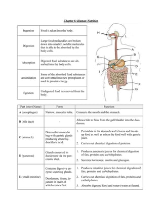
- 1. Chapter 6: Human Nutrition Ingestion Food is taken into the body. Large food molecukles are broken down into smaller, soluble molecules Digestion that is able to be absorbed by the body cells. Digested food substances are ab- Absorption sorbed into the body cells. Some of the absorbed food substances Assimilation are converted into new protoplasm or used to provide energy. Undigested food is removed from the Egestion body. Part letter (Name) Form Function A (oesophagus) Narrow, muscular tube. Connects the mouth and the stomach. Allows bile to flow from the gall bladder into the duo- B (bile duct) - denum. Distensible muscular 1. Peristalsis in the stomach wall churns and breaks bag with gastric glands up food as well as mixes the food well with gastric C (stomach) juice. producing dilute hy- drochloric acid. 2. Carries out chemical digestion of proteins. Gland connected to 1. Produces pancreatic juices for chemical digestion D (pancreas) duodenum via the pan- of fats, proteins and carbohydrates. creatic duct. 2. Secretes hormones: insulin and glucagon. Contains digestive en- 1. Produces intestinal juices for chemical digestion of zyme secreting glands. fats, proteins and carbohydrates. E (small intestine) 2. Carries out chemical digestion of fats, proteins and Duodenum, ileum, je- carbohydrates. junum in order of which comes first. 3. Absorbs digested food and water (water at ileum).
- 2. Absorbs water and mineral salts from the undigested F (colon) - food materials. Rectum is a short mus- Rectum: cular tube. Anus is an G (rectum, anus) Stores faeces temporarily, contracts to expel faeces opening where faeces through anus. leave the body. Mouth: Allows food to be ingested. Teeth: Masticate food to break them into smaller pieces of H (mouth, teeth, tongue) - food to increase surface area to volume ratio for chem- ical digestion. Tongue: Rolls food into bolus, mixes food with saliva. U-shaped part of small I (duodenum) Most of the chemical digestion occurs here. intestine J (gall bladder) Greenish-yellow bag Stores bile temporarily, contracts to release bile. Largest gland. Dark red. Connected to he- K (liver) patic vein, hepatic por- REFER TO LATER PART on assimilation. tal vein and hepatic artery. Produces saliva containing mucin, a substance that L (salivary glands) - makes food soft. Peristalsis are rhythmic wave-like contractions of the muscles to mix and propel the contents of the ali- mentary canal (especially in the oesophagus and the small intestine). To push food forward, … To allow food to enter, … Circular muscles contract, Circular muscles relax, longitudinal muscles re- longitudinal muscles con- lax. tract. Wall of gut will constrict Wall of gut will dilate (gut (gut becomes longer and becomes shorter and wid- narrower) and food is er) and the lumen is wi- squeezed forward. dened for food to enter.
- 3. Nutrient Carbohydrates Proteins Fats Chemical: starch maltose - - (via salivary amylase from salivary glands) Mouth Physical: Physical: Physical: Teeth grinds food into Teeth grinds food into smaller Teeth grinds food into smaller pieces to improve pieces to improve chemical di- smaller pieces to improve chemical digestion. gestion. chemical digestion. Oesophagus - - - Chemical: 1. caseinogen (s) casein (i) (via rennin from prorennin - from gastric glands) - 2. proteins polypeptides (via pepsin from pepsino- Stomach gen from gastric glands) Physical: Physical: Physical: Stomach muscles churn Stomach muscles churn food Stomach muscles churn food breaks up the food breaks up the food particles and food breaks up the food particles and mixes them mixes them with digestive en- particles and mixes them with digestive enzymes. zymes. with digestive enzymes. Chemical: Activation: Physical: 1. starch maltose trypsinogen trypsin big fat drops tiny fat (via pancreatic amylase (via enterokinase) droplets from pancreas) (via bile salts from liver) Chemical: 2. maltase glucose 1. proteins polypeptides Chemical: (via maltase from in- (via trypsin from trypsinogen fats fatty acids + glyce- testinal glands) from intestinal glands) rol Duodenum (via lipase from intestinal 3. sucrose Gl + Fr 2. polypeptides amino acids glands) (via sucrase from in- (via erepsin from intestinal testinal glands) glands) 4. lactose Gl + Ga (via lactase from in- testinal glands) Colon - - - Rectum - - -
- 4. Structural adaptation to in- How it works? crease absorption rates Less time is wasted diffusing through the cells and the digested food can travel One cell thick epithelium quickly from the intestines to the blood vessels. Capillaries transport away the blood which contains digested food to maintain the steep concentration gradient for dif- Extensive capillary network fusion. Lacteal transports fats while the capillaries transport sugars and amino acids. Folds are on the intestinal level, villi are finger-like projections on the folds and microvilli are finger-like projections on Folds, villi, microvilli the epithelial cells of villi. These in- crease the surface area to volume ratio, increasing the rate of diffusion of sub- stances. Length allows the digested food to be Intestine is long mostly absorbed. Active transport transports glucose from the intestines to the capillaries against Active transport the concentration gradient to increase the rate of glucose absorption. The hepatic portal vein transports most of the food absorbed from the small intestine. Functions of liver What is it? When there is too little glucose, islets of Langer- hans are stimulated to secrete glucagon. Glucagon stimulates the liver to convert glycogen to glucose. Carbohydrate metabolism When there is too much glucose, islets of Langer- hans are stimulated to secrete insulin. Insulin sti- mulates the liver to convert excess glucose to gly- cogen. The liver produces bile which is essential in the Bile production physical digestion of fat.
- 5. When the red blood cells wear out, they are de- stroyed in the spleen, a gland found near the liver. Breakdown of red blood cells The haemoglobin is brought to the liver and it stores the iron released. Bile pigments are also pro- duced in the breakdown. When there is excess amino acids, deamination occurs. The amino group is converted to ammonia Amino acids metabolism, formation of urea then urea which is removed in urine. The carbon residue is converted to glucose then glycogen which is stored in muscle cells and the liver. In the liver, the enzyme alcohol dehydrogenase breaks down alcohol into acetaldehyde which is further broken down to compounds usable in respi- Breakdown on alcohol ration. However, excessive alcohol consumption stimu- lates acid secretion in the stomach leading to the increased risk of gastric ulcers.
- 6. Chapter 7: Plant Nutrition Photosynthesis is a process whereby chlorophyll traps light energy and converts it into chemical energy for the formation of carbohydrates and their subsequent storage. What condition is needed for pho- How to test? tosynthesis? Carbon dioxide 1. Destarch two potted plants for 2 days by placing it in the dark. 2. Enclose the plants with polythene bags and secure the bags to the stem. 3. Leave the plants in bell jars: one with soda lime and potassium hydroxide and the other with pebbles and water. 4. Remove a leaf from each plants and test them for starch. Sunlight 1. Destarch a potted plant for 2 days by placing it in the dark. 2. Remove one leaf and test it for starch. 3. Sandwich a leaf which is still attached to the plant between two pieces of an opaque material (eg. black paper) and fasten with paper clips. 4. After a few hours, remove the leaf and test it for starch. Chlorophyll 1. Destarch a potted variegated plant for 2 days by placing it in the dark. 2. Expose the plant to strong sunlight for a few hours. 3. Remove one leaf and take note of the distribution of the green parts of the leaf which contain chlorophyll. 4. Test the leaf for starch. Testing for starch in leaves: 1. Remove the leaf and immediately place it in boiling water for 2 mi- nutes. 2. Put the boiled leaf into a boiling tube containing alcohol. Place the boiling tube in a beaker of hot water. The alcohol should turn green and the leaf should be brittle. 3. Carefully remove the leaf and place it on a white tile. Add a few drops of iodine solution. If starch is present, it should turn bluish black. If it is absent, it remains brown.
- 7. Equation for photosynthesis: Limiting factors are factors that directly affect a process if its quantity is changed. Effect of light intensity Effect of CO2 concentration Effect of temperature As light intensity increases, the As CO2 concentration increases, Since photosynthesis is an en- rate of photosynthesis also in- the rate of photosynthesis also zyme-catalysed process, increas- creases up till a maximum point. increases up till a maximum ing the temperature increases the From then on, increasing the point. From then on, increasing rate of photosynthesis up till a light intensity does not affect the the CO2 concentration does not maximum point at the optimum rate of photosynthesis and only affect the rate of photosynthesis temperature. From then on, in- the other factors affect it. and only the other factors affect creasing the temperature only it. lowers the rate of photosynthesis. What happens to these products? Outcome 1. Used immediately by plant cells during tissue respiration to pro- vide energy for cellular activities. 2. Converted to sucrose to be transported to be transported to sto- rage organs (eg. roots and tubers) or starch to be stored tempora- rily in the leaves. Glucose 3. Used to form amino acids and proteins when reacted with ni- trates. Amino acids are then transported to growing regions to form protoplasm. 4. Used to form fats as a storage mechanism in storage organs which might be used in cellular respiration. Oxygen diffuses out of the cells into the intercellular air spaces and Oxygen out into the surroundings via the stoma of the leaves.
- 8. Why is photosynthesis important? 1. Supports food chains: Photosynthesis stores energy within the carbohydrate molecules. With plants being the producers on the food chains, animals depend both directly and indirectly on plants to photosynthesise and make food for their consumption for energy for their activities. 2. Maintains the carbon dioxide-oxygen balance: Carbon dioxide is taken in during photosynthesis and oxygen is produced. The oxygen is needed by living organisms to carry out respiration to release energy for their activities. 3. Production of coal: When plants die, after thousands of years, coal is formed. It is a fuel that releases energy when burnt. Also it can be converted to liquid fuels to power vehicles. Single layer of closely packed cells covered on the top by a waxy cuticle that prevents excessive Upper epidermis evaporation of water. Transparent to allow sunlight to penetrate the leaf. One or two layers of closely packed, long and cylindrical cells. Palisade mesophyll Has most number of chloroplasts to absorb maximum sunlight. Cells are irregular in shape. Contains large intercellular air spaces to allow rapid diffusion of gases. Contains less chloroplasts Spongy mesophyll than the palisade tissue. Mesophyll cells are covered with a film of moisture so that CO2 can dissolve in it. Transport tissues (xylem and phloem) present here. Generally no chloroplasts found here as most light has been absorbed above. Covered on the Lower epidermis bottom by a waxy cuticle that prevents water loss through epidermal cells. Stoma Important in gaseous exchange.
- 9. How to they open? [IN THE LIGHT] How do they close? [IN THE DARK] 1. In sunlight, the concentration of K+ ions in- 1. The K+ ions diffuse out of the guard cells into creases in the guard cells. the epidermal cells. 2. Chloroplasts in the guard cells photosynthesise 2. This increase the water potential in the guard and the energy is used to pump K+ ions into the cells and water leaves the guard cells by osmo- guard cells from the neighbouring epidermal sis. cells. This lowers the water potential in the guard cells. 3. Hence, the guard cells become flaccid and the stomatal pore closes. 3. Water from neighbouring epidermal cells en- ters the guard cells by osmosis. The cells then turn turgid. 4. Since the guard cells have a thicker cellulose cell wall on the stomatal side, the guard cells become more curved and pull the stoma open. How to get carbon dioxide? In daylight when photosynthesis occurs, the carbon dioxide in the leaf is rapidly used up. The carbon dio- xide concentration in the intercellular air spaces is then lower than the carbon dioxide concentration in the surrounding air. When the guard cells open, carbon dioxide diffuses into the intercellular air spaces down a concentration gradient and dissolves into the thin film of moisture before diffusing into the cells for use. How to get water? REFER TO CHAPTER 9
- 10. Chapter 8: Human Transport Component of blood Function 1. Red blood cells contain haemoglobin which is an iron-containing protein. RBCs can transport oxygen from the lungs to all cells in the body be- cause haemoglobin combines reversely with oxygen in the equation: haemoglobin (purplish-red) + O2 ⇌ oxyhaemoglobin (bright red). As blood passes through the lungs, oxygen diffuses from the alveoli into the blood. Due to the affinity of haemoglobin to oxygen it combines and Red blood cells (RBCs) travels along the blood vessels until the tissues which have a low oxygen concentration, where it diffuses through the vessel wall into the tissues. 2. RBCs are circular, flattened biconcave discs. This shape increases the cell’s surface area to volume ratio so that it can absorb and release oxy- gen faster. 3. RBCs are elastic and can turn bell-shaped in order to squeeze through smaller BVs, usually smaller in diameter than itself. 1. Carries out phagocytosis or produces antibodies to protect our bodies against illnesses. Phagocytosis: Phagocytes engulf and ingests foreign particles, forming pus after it is digested. Antibody production: Antibodies are produced to destroy the bacteria by attaching to them to cause the bacterial surface membrane to rupture, to cause the bacteria to agglutinate for easier phagocytosis, to neutralise the toxins produced by bacteria and to attach to viruses, making them unable to bind to the host cell. White blood cells (WBCs) 2. Rejects foreign tissue Organ transplanted into body may be rejected by the recipient’s immune system. The organ may be treated as a foreign body and hence recipient’s lymphocytes may respond by producing antibodies to destroy the trans- planted organ. To prevent tissue rejection: i. Use of immunosuppresive drugs ii. Using tissues or organ from close relatives iii. Using tissue from other undamaged parts of the body Pale yellowish liquid that is 90% water. Transports: 1. Digested food substances (from small intestine to all parts of body) 2. Urea, uric acid and creatinine (from all parts of the body to the kidney) Plasma (PLM) 3. Carbon dioxide (from respiring tissues to the lungs) 4. Hormones (from the glands to target organs) 5. Heat (from respiring tissues to the entire body) 6. WBCs and RBCs and soluble proteins (eg. prothrombin and albumin)
- 11. Involved in clotting of blood. Blood clotting process: 1. When blood vessels are damaged, damaged tissues and blood platelets release thrombokinase, an enzyme. Platelets (PLT) 2. Prothrombin Thrombin (via thrombokinase and Ca2+ ions) 3. Soluble fibrinogen Insoluble fibrin threads (via thrombin) Fibrin threads entangle blood cells and the whole mass forms a clot. In normal undamaged blood vessels, heparin, an anti-clotting substance, pre- vents clotting. However, when damaged, the thrombokinase neutralises the heparin so that clotting takes place. Blood group A B AB O (recipient below, (antigen A) (antigen B) (antigen A & B) (no antigen) donor right) Not compatible. Not compatible. A Compatible. Agglutination will Agglutination will Compatible. (antibody B) occur. occur. Not compatible. Not compatible. B Agglutination will Compatible. Agglutination will Compatible (antibody A) occur. occur. AB Compatible. Compatible. Compatible. Compatible. (no antibody) Not compatible. Not compatible. Not compatible. O Agglutination will Agglutination will Agglutination will Compatible. (antibody A&B) occur. occur. occur. When common antigens and antibodies are present, the antibodies will cause the blood of the recipient to agglutinate (react with the antigens present on the RBCs). As such O is the universal donor and AB is the universal recipient. When checking for agglutination, look at ANTIGEN OF DONOR and ANTIBODY OF RECIPIENT.
- 12. Blood vessel Characteristic Function 1. Arterial blood pressure is high. Blood flow through A is fast, in spurts, reflecting rhythmic pumping of heart. 2. Thick and elastic muscular walls to withstand high pres- Carry oxygenated Arteries (A) sure. blood away from the heart. 3. Semi-lunar valves absent since blood has enough force to move forward on it own. 4. Ratio of lumen diameter to external diameter is small. 1. Venous blood pressure is low. Blood flow through V is slow and smooth. 2. Thin, slightly muscular walls since pressure is rather low. Carry deoxygenated Veins (V) 3. Semi-lunar valves are present to prevent backflow of blood to the heart. blood. Helped by contraction of skeletal muscles also to move blood. 4. Ratio of lumen diameter to external diameter is large. 1. Capillary blood pressure is lowest to allow time for diffu- sion of oxygen and carbon dioxide to and from tissue. Connects the A and V Capillaries (C) 2. One-cell thick walls to allow rapid diffusion of gases to via organs. and from tissue. 3. Semi-lunar valves absent.
- 13. Structure of heart The heart is a muscular pump that keeps blood flowing in one direction in blood vessels of the body. It is supplied with food and oxygen via the coronary arteries. The left side of the heart has thicker walls than the right side of the heart. The left ventricle has a thicker wall and more muscular walls than the right ventricle. It pumps blood by contracting its muscles. The median septum separates the two halves of the heart. Also, semi-lunar, bicuspid and tricuspid valves keep blood from backflowing into the ventricles or atria. The aorta (aka aortic arch) allows oxygenated blood to flow from the heart to the rest of the body. The vena cava (superior and inferior) allows deoxygenated blood to flow from rest of body to the heart. The pulmonary vein allows oxygenated blood to flow from the lungs to the heart. The pulmonary artery allows deoxygenated blood to flow from the heart to the lungs. *Tendon is known as chordae tendinae.
- 14. Cardiac cycle Phase of car- Left atrium Right atrium Left ventricle Right ventricle diac cycle Relaxes then blood Relaxes then blood I enters from pul- enters from vena Relaxes. Relaxes. monary veins. cava. Contracts. Results Contracts. Results in slightly high in slightly high pressure which pressure which Blood from left Blood from right II propels blood for- propels blood for- atrium rushes in. atrium rushes in. ward into the left ward into the right ventricle. ventricle. Contracts. Results in Contracts. Results in III high pressure which high pressure which (Ventricular Relaxes. Relaxes. propels blood for- propels blood for- systole) ward into the pul- ward into the aorta. monary vein. IV (effects on bi- Increase in blood pressure in ventricles forces bicuspid and tricuspid valves to close pre- and tricuspid venting backflow. valves) V (effects on Increase in blood pressure in ventricles forces semi-lunar valves to open releasing blood semi-lunar into the aorta and pulmonary vein. valves) VI (Ventricular - Relaxes. Relaxes. diastole) VII (effects on bi Decrease in blood pressure in ventricles forces bicuspid and tricuspid valves to open. and tricuspid valves) VIII (effects on Decrease in blood pressure in ventricles forces semi-lunar valves to close. Cycle then semi-lunar repeats itself. valves)
- 15. 4 Point What happens here? Phase… 1 Bicuspid and tricuspid valves close. IV 2 Blood pressure increases as ventricles contract. III 3 Semi-lunar valves open, releasing blood into aorta. V 4 Semi-lunar valves close, preventing backflow of blood into the ventricles. VIII 5 Blood pressure decreases as ventricles relax. VI 6 Bicuspid and tricuspid valves open. VII 7 Blood pressure in ventricles increases as it fills with blood. I Atherosclerosis causes fatty substances to be deposited on the inner surface of the coronary arteries. This narrows the lumen so less blood can flow through per unit time and the blood pressure increases. This increases the risk of a blood clot (thrombosis) in the arteries. If it occurs, oxygen supply is cut off the heart muscles which get damaged. A heart attack follows. Causes of atherosclerosis and coronary heart disease: 1. Diet rich in cholestrol and saturated animal fats 2. Emotional stress 3. Smoking Preventive measures against atherosclerosis and coronary heart disease: 1. Proper diet with polyunsatured plant fats and dietary fibre 2. Proper stress management 3. Avoiding smoking 4. Regular physical exercise
- 16. Chapter 9: Plant Transport Transport vessel Function Adaptations 1. A hard substance, lignin is deposited on the in- Conducts water and mineral ner walls of xylem vessels. Provides mechanical salts from the roots to the support and prevents the plants from collapse. Xylem stems and leaves. Provides mechanical support for the 2. Xylem have empty lumen without protoplasm plant. or cross-walls. Reduces resistance for water flowing through xylem vessels. 1. Companion cells have many mitochondria to provide energy for loading of sugars from the Conducts manufactured food mesophyll cells into the sieve tubes via active (sucrose + amino acids) from transport. Phloem the green parts of the plants to the rest of the plant. 2. Holes in sieve plate allow rapid flow of manu- factured food substances without it having to pass through cross-walls. Transport vessel Diagram Xylem (stem) Xylem (root) Phloem
- 17. How does WATER enter the plant? How does MINERAL SALTS enter the plant? 1. Root hair cells take in mineral salts via active The sap of the epidermal root hair cell is a relative- transport when the concentration of mineral ly concentrated solution of sugars and various min- salts in the sap of the root hair cells is higher eral salts as compared to the dilute solution of min- than the concentration of mineral salts in the eral salts in the thin film of liquid surrounding each thin film of liquid. soil particle. Thus, the sap has a lower water poten- 2. Root hair cells take in mineral salts via diffu- tial than the soil soultion. sion when the concentration of mineral salts in Since these two solutions is separated by a partially the sap of the root hair cells is lower than the permeable membrane, water enters from the thin concentration of mineral salts in the thin film film of liquid into the root hair cells by osmosis. of liquid. This increases the water potential in the epidermal root hair cells as compared to the inner root cells in How is the root hair cell adapted for its function? the cortex. Finger-like projection increases surface area to vo- As such water from the epidermal root hair cells lume ratio and hence increase rate of absorption of enters the inner root cells by osmosis and continues mineral salts and water. Cell surface membrane moving in until it reaches the xylem vessels and it prevents cell sap from leaking out into the soil. moves up the plant. Lots of mitochondria in root hair cells enables root hair cells to produce energy for active transport to move ions into the root hair cells. Root pressure Capillary action Transpiration pull Due to the living cells pumping Water tends to move up inside Transpiration = loss of water va- ions into the xylem vessels to very narrow tubes due to cohe- pour from the aerial parts of the reduce water potential in the ves- sion between the water molecules plants of a plant, especially sels, water passes from the living and the xylem’s inner walls and through the stomata of the leaves. cells into the xylem vessels by adhesion between the water mo- osmosis and flows upwards. lecules. This pulls water upwards When water evaporates from the in the narrow tubes. leaves, water is removed from the xylem vessels. This results in a suction force which pulls water upwards also known as transpira- tion pull. Transpiration: Step 1: Water continuously moves out of the mesophyll cells to form a thin film of moisture over their surfaces. Water evaporates from this thin film of moisture and moves into the intercellular air spaces. Water vapour accumulates in the large sub-stomatal air spaces. Step 2: Water vapour then diffuses through the stomata to the drier air outside the leaf. As water evapo rates from the mesophyll cells, the water potential of the cell sap decreases. The mesophyll cells begin to absorb water by osmosis from the cells deeper in the leaf. These cells, in turn, remove water from the veins, that it from the xylem vessels. This results in a suction force which pulls the whole column of water up the xylem vessels.
- 18. Factors affecting rate of transpiration How it affects? Wind blows away the water vapour that accumulates outside the stomata. This maintains the water vapour concentration gra- dient between the leaf and the atmosphere since the surrounding Wind air is less humid. As such when wind increases, rate of transpi- ration increases and when wind decreases, rate of transpiration decreases. A rise in temperature causes the rate of evaporation of water from the mesophyll cells and hence an increase in the rate of Temperature transpiration. As such when temperature increases, rate of transpiration increases and when temperature decreases, rate of transpiration decreases. When humidity increases, the rate of evaporation of water from the mesophyll cells decreases since water vapour is saturated in Humidity the air. As such when humidity increases, rate of transpiration decreases and when humidity decreases, rate of transpiration increases. When light intensity increases, rate of transpiration increases Light intensity and when light intensity decreases, rate of transpiration de- creases. The rate of photosynthesis is measured using a potometer in units g/h or cm3/h. When the rate of transpiration increases due to wind, temperature, humidity or light intensity, the rate of transpiration may exceed the rate of absorption of water in the roots. As such, the cells lose their water and then their turgor and they become flaccid. This causes them to lose support and hence wilt. Advantages of wilting: Reduces the rate of transpiration to prevent too much water from transpiring away. Disadvantages of wilting: Reduces the amount of carbon dioxide and hence lowers rate of photosynthesis. Translocation is the transport of manufactured food in plants in the phloem tissue. Test for path of water in stems Diagram Celery experiment: Add a celery stem into a beaker of water which is dyed red and after a few hours, cut thin transverse sections and observe for the dyed parts. Test of path of food in stems Diagram ‘Ringing’ experiment: Cut off a complete ring of bark including the phloem and cambium from the main stem of a woody twih. Place the twig in water. Prepare one with the ring above the water and one below the water and one without any cuts.
- 19. Using aphids: A feeding aphid is anaethetised with carbon dioxide. The body of the aphid is cut off, leaving the proboscis behind. An analysis of the liquid exuding out of the cut end of the proboscis (e) shows that it contains sucrose and amino acids. If the stem is sectioned and examined, it is seen that the proboscis is inserted into the phloem sieve tube. Using carbon-14 isotopes: A leaf is provided with carbon dioxide with radioactive carbon-14. When photosynthesis takes place, the sugars produced and transported in the phloem will have ra- dioactive carbon that is shown on the X-ray film after exposed onto an X-ray photographic film.