SlideShare une entreprise Scribd logo
1  sur  30
65V.S. Dogra, G.T. MacLennan (eds.), Genitourinary Radiology: Kidney, Bladder and Urethra,
DOI 10.1007/978-1-84800-245-6_2, © Springer-Verlag London 2013
Introduction
The diagnosis of renal inflammatory diseases is primarily•
based on a combination of clinical and laboratory findings
such as bacteriuria, pyuria, positive urine culture, and
leukocytosis.
In general, imaging is not required for diagnosis and treat-•
ment of uncomplicated infections in adult patients.
However, patients suspected of having an acute renal•
infection and those who are not responding to intravenous
antibiotic treatment within 72 h require imaging.
Imaging of urinary tract infection (UTI) in children is•
more important than in adults because of the much higher
potential for significant irreversible renal damage with
sequelae of hypertension and chronic renal failure.
Acute Pyelonephritis
Acute Bacterial Renal Infection in Children
General Information
Approximately 1–3 % of prepubertal children are diag-•
nosed with UTI as a bacterial infection of the renal paren-
chyma, bladder, or urethra.
Acute pyelonephritis is a bacterial or fungal infection of•
the renal parenchyma and collecting system.
Bacterial pyelonephritis may occur by hematogenous•
seeding, while an ascending route from the bladder is the
most common pathway in children.
Imaging
Intravenous Pyelography
Intravenous pyelography (IVP) can be used to assess the•
morphology of the urinary tract, if renal function is
adequate.
Approximately 20–25 % of patients with acute renal infec-•
tionhaveabnormalimagingfindings,includingdilatationof
the ureter, loss of visualization of a portion of renal outline,
diffuse renal enlargement, and decreased nephrogram.
Ultrasound
Ultrasound findings of acute bacterial renal infection in•
children are given in Table 2.1 (Fig. 2.1a, b).
Focal pyelonephritis or lobar nephronia is a localized•
infection without major suppuration. It usually appears as
a poorly defined mass with focal loss of the regional cor-
ticomedullary differentiation on ultrasound. The abnor-
mal focus can be relatively anechoic with low-level echoes
or can be more echogenic than the kidney.
Renal ultrasound is also useful in the diagnosis of compli-•
cations secondary to acute pyelonephritis such as abscess,
pyonephrosis, and perirenal fluid collections (Figs. 2.1
and 2.2a, b).
Combining gray-scale ultrasound and power Doppler•
ultrasound can provide a comprehensive evaluation of the
renal parenchyma in acute pyelonephritis with impaired
renal function.
Inflammatory Conditions
of the Kidney
Kemal Ödev, Ahmet Tuncay Turgut,
and Gregory T. MacLennan
2
K. Ödev, M.D.(*)
Department of Radiology,
Meram Medical School of Selcuk University,
Konya, Turkey
E-mail: kemalodev50@yahoo.com
A.T. Turgut, M.D.
Department of Radiology, Ankara Training and Research Hospital,
Ankara, Turkey
G.T. MacLennan, M.D.
Division of Anatomic Pathology, University Hospitals Case Medical
Center, Case Western Reserve University,
Cleveland, OH, USA
Table 2.1 Ultrasound findings in acute bacterial renal infection in
children
Renal enlargement
Decreased or increased parenchymal echogenicity
Minimal dilatation of renal pelvis
Loss of normal corticomedullary differentiation
Focal, poorly defined mass with variable echogenicity
66 K. Ödev et al.
a
b
c
d e
Fig. 2.1 Complicated acute bacterial pyelonephritis (ABP) in a
3-year-old girl with immunodeficiency. Ultrasound scans of right (a)
and left (b) kidneys reveal bilateral diffuse enlargement with loss of
the corticomedullary differentiation (arrow), multiple scattered areas
of decreased echogenicity, and dilated renal pelvis (arrow) represent-
ing diffuse spread of the inflammatory process, features consistent
with complicated acute pyelonephritis, and pyonephrosis. (c) Contrast-
enhanced CT scan demonstrates bilateral diffuse renal involvement
with scattered nonenhancing foci representing microabscesses (arrow-
heads) and a large cystic fluid collection in the left kidney (arrow)
suggestive of a renal abscess. The maximum intensity projection image
from excretory magnetic resonance urography (MRU) (d) and the max-
imum intensity projection image from T2-weighted half-Fourier acqui-
sition single-shot turbo spin-echo (HASTE) sequence (e) reveal the
true pathology that is consistent with bilateral multifocal renal
abscesses associated with complicated ABP (dashed arrows)
672 Inflammatory Conditions of the Kidney
Power Doppler ultrasound is superior to color-flow•
Doppler in defining extent of areas of hypoperfusion
because most pyelonephritic lesions are ischemic.
Computed Tomography
Computed tomography (CT) is a sensitive study in evalu-•
ation of the renal parenchyma in acute renal infection.
Inflammatory edema and microabscess may be demon-•
strated in the abnormal renal parenchyma.
Rounded or irregular parenchymal abnormalities•
appear as a patchy low-density areas on nonenhanced
CT scans.
After administration of radiographic contrast, these•
regions are seen as areas of decreased enhancement
(Fig. 2.1c).
Magnetic Resonance Imaging
Parenchymal alterations in complicated renal infections•
can be demonstrated on MRI.
AbscessformationsappearashypointenseonT1-weighted•
and hyperintense on T2-weighted images (Fig. 2.1d, e).
Nuclear Scintigraphy
In children, radionuclide imaging is strongly recom-•
mended both at early and late stages of the acute pyelone-
phritis because of the potential of significant functional
deficits within areas of inflammation and suppuration.
Radionuclide imaging with Tc-99m dimercaptosuccinic•
acid (DMSA) is considered the most sensitive modality
for the imaging of renal cortex in children.
It may show areas of peripheral decreased uptake related•
to acute pyelonephritis or scar formation.
Differential Diagnosis
The differential diagnosis of acute pyelonephritis is given in•
Table 2.2. Focal pyelonephritis causing a focal area of edema
should be differentiated from various mass lesions, includ-
ing a renal abscess, which usually appears more complex.
Pearls and Pitfalls
It is difficult to exclude the diagnosis of acute pyelone-•
phritis with imaging studies because IVP, CT, and ultra-
sound findings can be normal in up to 75 % of the cases.
On ultrasound, abnormal echogenicity areas can occa-•
sionally be misinterpreted as mass lesions.
Although renal infection can be localized by DMSA, it•
occasionally cannot be definitely distinguished from a
sterile inflammation by using any kind of radiotracer.
Acute Bacterial Renal Infection in Adults
General Information
The incidence of acute bacterial pyelonephritis (ABP) is•
high, with an increasing rate of hospitalization.
In the United States, approximately 250,000 cases of ABP•
with more than 100,000 hospitalizations occur each year.
Imaging
Intravenous Pyelography
The main role of IVP in the imaging of infection involves•
screening for obstruction, although this is now better
demonstrated by other modalities.
Only 25 % of cases of ABP will have positive IVP findings,•
including enlargement of the affected kidney, delayed con-
trast excretion, and compression of the collecting system.
Table 2.2 Differential diagnosis of acute pyelonephritis in children
Ureteric obstruction
Contusion
Tubular obstruction
Hypotension
Scarring
Autosomal recessive polycystic kidney disease
Renal abscess
a
b
Fig. 2.2 Acute bacterial pyelonephritis. Transverse (a) and longitudi-
nal (b) ultrasound scans of a 13 months old boy performed after an
episode of UTI reveal echogenic caliceal debris associated with ABP
(arrows) (Courtesy of Alparslan Ünsal, MD, Aydın, Turkey)
68 K. Ödev et al.
Ultrasound
Sonographically, variable echotexture changes of the•
renal parenchyma have been described for ABP.
They are in the form of hypoechoic lesions or to a lesser•
degree, hyperechoic foci.
The former are attributed to edema, whereas the latter•
may be due to hemorrhage.
Ultrasound can also be used to detect and monitor the•
progress of focal inflammatory masses.
Thus, ultrasound is widely used either as a complement to•
the IVP or as a screening modality, which may or may not
be followed by CT scan, depending on the findings.
Computed Tomography
CT is considered the imaging modality of choice in the•
evaluation of patients with ABP.
It is superior to both ultrasound and IVP in detecting mor-•
phologic and functional abnormalities, defining the extent
of disease, and detecting the formation of renal subcapsu-
lar or perinephric abscess or hematoma (Fig. 2.3a–c).
In hemorrhagic bacterial pyelonephritis, nonenhanced CT•
may demonstrate, on occasion, wedge-shaped or rounded
areas with increased attenuation due to parenchymal
bleeding.
Nonenhanced CT is an excellent study for identifying cal-•
culi, gas formation, hemorrhage, parenchymal
calcification, urinary obstruction, renal enlargement, and
inflammatory masses.
Typical findings of ABP after intravenous contrast admin-•
istration include ill-defined, wedge-shaped lesions of
decreased attenuation radiating from the papilla in the
medulla to the cortical surface.
These wedge-shaped lesions probably represent areas of•
poorly or nonfunctioning parenchyma due to vasospasm,
tubular obstruction, and/or interstitial edema.
The nonspecific appearance of a striated nephrogram in•
the excretory phase on CT comprises linear bands of
alternating hyper- and hypoattenuation parallel to the axes
of tubules and collecting ducts, corresponding to
a b
c
Fig. 2.3 Hemorrhagic acute pyelonephritis in a 45-year-old woman
presented with left flank pain. (a) Nonenhanced CT scan demonstrates
a low-attenuation collection in the left perirenal space (arrow). (b)
Contrast-enhanced CT scan reveals a perirenal fluid collection with
mixed attenuation (arrow) and thickening of Gerota’s fascia.
Preoperative diagnosis was perirenal abscess. (c) Left nephrectomy
revealed an acute pyelonephritis with perirenal hematoma
692 Inflammatory Conditions of the Kidney
obstructed tubules with intervening normal tubules, which
is associated with stasis of contrast material in dilated
ducts due to edematous renal parenchyma (Table 2.3).
Pathology
Acute bacterial pyelonephritis results from an ascending•
infection that originates in the lower urinary tract.
Grossly, the collecting system is thickened, with yellow-•
white abscesses located throughout the renal paren-
chyma. An abscess is composed of central necrosis,
surrounded by a zone of preserved neutrophils, all of
which is surrounded by dilated vessels and fibroblasts.
Fibroblastic proliferations serve to organize and confine
processes and cause adjacent structures to contract.
Cortical abscesses may result in hematogenous spread,
further seeding the kidney with microorganisms
(Fig. 2.4).
Histologically, the predominant finding is neutrophils;•
neutrophils are present in the interstitium and in the renal
tubules (eventually forming white blood cell casts)
(Fig. 2.5). In early infection, neutrophils and bacteria may
be seen in the collecting ducts.
Differential Diagnosis
On CT, differential diagnosis for focal areas of hypoat-•
tenuation includes renal infarction, tumors (renal cell car-
cinoma), and scarring.
The hypoattenuating areas associated with infarcts and•
tumors persist following antibiotic treatment, in contrast
to those caused by ABP.
Furthermore, scarring rarely occurs in adult patients.•
Pearls and Pitfalls
On ultrasound, the combination of a hypoechoic area and•
a focal inflammatory bulge to the perinephric space may
be misinterpreted as a neoplasm.
On CT, streak artifacts during the excretory phase may•
give a false impression of abnormal nephrogram.
The appearance of a “striated nephrogram” is not•
pathognomonic for ABP and can be seen in kidneys fol-
lowing renal trauma, in medullary sponge kidney and in
autosomal recessive polycystic kidney disease
(Table 2.3).
Chronic Pyelonephritis
General Information
Chronic pyelonephritis is a chronic interstitial nephritis•
that may arise secondary to a host of etiologic causes,
including recurrent infection, autoimmune diseases, cal-
culous disease, and chronic obstruction.
Imaging
The imaging findings of chronic pyelonephritis are summa-
rized in Table 2.4.
Intravenous Pyelography
Renal size is usually decreased, unless chronic obstruc-•
tion is present, in which case the renal size may be
increased.
Renal contour blurring.•
Cortical thinning.•
Table 2.3 Causes of striated nephrogram on CT
Acute pyelonephritis
Acute urinary obstruction
Renal vein thrombosis
Renal contusion
Hypotension
Medullary sponge kidney
Autosomal recessive polycystic kidney disease
Tubular obstruction (myoglobinuria)
Fig. 2.4 Acute pyelonephritis. Patient died of sepsis after inadvertent
ligation of one ureter. The kidney is swollen and pale. Microabscesses
are visible on the cortical surface (From MacLennan GT, Cheng L.
Atlas of Genitourinary Pathology. New York: Springer; 2011. Reprinted
with permission)
70 K. Ödev et al.
Focal nonfunctional area in nephrogram phase.•
Ultrasound
Focal or diffuse cortical thinning•
Renal scar formation•
Distortion of renal contours•
Computed Tomography
The kidney is usually shrunken and has an irregular outer•
margin (Fig. 2.6).
Renal scar formation presents as defect in the margin of•
the kidney between two calyces.
Decreased contrast excretion on enhanced CT.•
Fibrotic distortion of the collecting system.•
Pathology
Chronic pyelonephritis is a general term implying chronic•
interstitial renal infection due to long-standing obstruc-
tion or vesicoureteric reflux, resulting in loss of renal
parenchyma, with scarring. Chronic pyelonephritis
accounts for 5–15 % of cases of end-stage renal failure.
Most patients are asymptomatic until hypertension or ure-
mia develops.
The gross findings in chronic pyelonephritis are variable.•
When the condition is obstruction related, the kidney may
be large due to dilatation of the collecting system, and the
Fig. 2.5 Acute pyelonephritis.
Abundant neutrophils are present
in the interstitium and within
renal tubules
Fig.2.6 Forty-year-oldwomanwithrecurrentpyelonephritis.Contrast-
enhanced CT scan reveals severe parenchymal scarring of and decreased
contrast excretion from the right kidney (arrow). The left kidney is
normal
Table 2.4 Imaging findings in chronic pyelonephritis
Renal scarring
Renal atrophy
Cortical thinning
Hypertrophy of residual normal tissue
Caliceal clubbing
Dilatation of the calyceal system
Renal asymmetry
712 Inflammatory Conditions of the Kidney
cortical surface is distorted by irregular scars (Fig. 2.7).
Beneath the scars, the renal cortex is thin, and the papillae
are scalloped or obliterated, causing the affected calyces
to appear dilated.
Microscopically, chronic pyelonephritis consists of intersti-•
tial fibrosis, tubular atrophy, and sclerosis of glomeruli, often
patchyindistribution(Fig.2.8).Theinterstitialinflammation
is mixed but is composed mainly of lymphocytes. Tubules
usually contain eosinophilic material, a feature known as
“thyroidization.” Arteries and arterioles commonly show
fibrosis, mural hyalinization, and occlusion.
Differential Diagnosis
Several conditions involving the loss of renal parenchy-•
mal tissue such as infarction or papillary necrosis and
fetal lobulation should be considered in the differential
diagnosis of renal scarring.
Pearls and Pitfalls
On ultrasound, the limited depiction of calyces may•
preclude the distinction of a renal scarring in cases with
chronic pyelonephritis.
Inchronicpyelonephritis,tubulesusuallycontaineosino-•
philic material, a feature known as “thyroidization.”
Vesicoureteral Reflux and Reflux Nephropathy
General Information
Vesicoureteral reflux (VUR) is described as the existence•
of retrograde urine flow from the bladder to the ureter and
the upper urinary tract.
This condition usually develops secondary to the primary•
abnormality of the vesicoureteral junction and may cause
focal or diffuse acute and/or chronic pyelonephritis.
The prevalence of VUR in asymptomatic children is less than•
0.5 %, but VUR is seen in up to 50 % of children with UTIs.
Renal damage, also termed “reflux nephropathy,” develops in
only 10 % of the children with symptomatic UTI.
Imaging
Intravenous Pyelography
IVP can demonstrate cortical contour deformity associ-•
ated with a clubbed calix.
In the past, IVP was the preferred diagnostic method in•
the demonstration of renal scarring and parenchymal
function but was replaced by ultrasound and DMSA
renal scintigraphy because of its low sensitivity in iden-
tifying these abnormalities, the nephrotoxic potential of
contrast materials used, and the necessity for exposure to
radiation.
Voiding Cystoureterography
The gold standard method for the evaluation of VUR is•
the conventional voiding cystoureterography (VCUG)
(Fig. 2.9a).
The international system of radiologic grading of vesi-•
coureteral reflux has assigned five grades of reflux on
VCUG:
Grade 0: Indicates no VUR
Grade 1: VUR that does not reach the renal pelvis
Grade 2: VUR extending to the renal pelvis without dilatation
Grade 3: VUR extending to the kidney with mild ureteral
dilatation and mild to moderate pelvicalyceal dilatation
Grade 4: VUR extending to the kidney with moderate
ureteral dilatation and complete obliteration of the
sharp angles of all fornices
Fig. 2.7 Chronic obstructive pyelonephritis. The renal damage in a
setting of obstructed renal drainage results from pressure-related atro-
phy and chronic infection. This patient had long-standing distal ureteral
obstruction. The renal pelvis and calyces are dilated, with flattening or
obliteration of papillae and global thinning of renal cortex
72 K. Ödev et al.
Grade 5: VUR extending to the kidney with a tortuous
ureter and moderate dilatation of the renal pelvis to
extreme dilatation of the entire upper urinary tract
The sensitivity of VCUG depends on the severity of the•
VUR. It has been demonstrated that VCUG has 100 %
sensitivity in the diagnosis of grade 4 and grade 5
VUR.
Ultrasound
Ultrasound may be helpful for the detection of parenchy-•
mal damage and dilatation of the collecting system.
Features of VUR that may be evident by ultrasound•
include diminished renal length and dilatation of the ure-
ter, the renal pelvis, and the renal calyces.
Ureteral dilatation was found to be the most helpful diag-•
nostic feature in detection of the higher grades of VUR by
ultrasound.
Recently, it has been advocated that echo contrast-•
enhanced cystosonography may be useful in diagnosing,
grading, and following up on patients with VUR.
Magnetic Resonance Imaging
Multiplanar cross-sectional T1- and T2-weighted mag-•
netic resonance imaging (MRI) combined with unen-
hanced 3D magnetic resonance urography (MRU) is
potentially useful for morphological evaluation of the uri-
nary system, without the hazards related to the use of
intravenous iodinated contrast agents.
In addition to morphological evaluation, contrast-•
enhanced MRU provides functional information about
renal perfusion, concentration, and excretion of contrast
agent (Fig. 2.9b, c).
Gadolinium-enhanced T1-weighted excretory MRU may•
demonstrate the parenchymal scars and perfusion defects
associated with reflux nephropathy.
Probably, the greatest advantage of the use of this•
T1-weighted gadolinium-enhanced 3D-MRU technique
is the visualization of the urinary tract of patients with
impaired renal function.
Unenhanced T2-weighted MRU technique allows excel-•
lent visualization of the urinary tract and is useful in
patients with dilated collecting systems. However, a non-
dilated urinary tract is either invisible or incompletely
visualized with this technique, whereas excellent visual-
ization is achieved with contrast-enhanced T1-weighted
excretory MRU.
Nuclear Scintigraphy
Direct radionuclide cystography with a• 99m
Tc-labeled
agent (sulfur colloid, diethylenetriamine penta-acetate
[DTPA], or pertechnetate) is a well-accepted alternative
to fluoroscopic VCUG for screening asymptomatic chil-
dren, for follow-up examination of children with vesi-
coureteral reflux (VUR), for postoperative evaluation
after ureteral reimplantation, and for excluding VUR.
• 99m
Tc-labeled DTPA scintigraphy is more sensitive than
VCUG in detection of VUR.
DMSA renal scintigraphy has been found to be more sen-•
sitive than IVP in the evaluation of focal renal scars.
Fig. 2.8 Chronic obstructive
pyelonephritis. The glomeruli are
sclerotic, the interstitium shows
fibrosis and is infiltrated by
lymphocytes, the tubules are
atrophic and contain eosinophilic
casts (thyroidization), and the
blood vessels show intimal
sclerosis and mural thickening
732 Inflammatory Conditions of the Kidney
a b
c
Fig. 2.9 VUR and reflux nephropathy in 5-year-old boy with recurrent
bladder infection. (a) Retrograde voiding cystourethrogram reveals
marked dilatation of the left ureter and left renal pelvicaliceal system
(arrows) representing reflux to the left kidney and trabeculations and
multiple diverticula in the bladder (open arrow). (b) Coronal contrast-
enhanced 3D T1-weighted gradient echo MRI demonstrates minimal
dilatation of the right collecting system (arrow) and severe hydroureter-
onephrosis on the left collecting system (open arrow). (c) On sagittal
contrast-enhanced T1-weighted gradient echo MRI, bladder diverticula
(arrow) and diffuse bladder wall thickening secondary to cystitis are
visualized excellently
74 K. Ödev et al.
Pathology
When chronic pyelonephritis is primarily reflux related,•
the affected kidney is usually small. Reflux nephropathy
usually results from vesicoureteral reflux (a congenital
disorder) combined with intrarenal reflux, thus allowing
infection access to the renal parenchyma.
The kidneys are small and contracted, with broad•
depressed sometimes saddle-shaped scars in the capsule
(Fig. 2.10). There is loss of renal pyramids with dilated
and thickened calyces, and the renal cortex overlying
these areas is thinned. Renal parenchyma between scarred
areas may be normal or hypertrophic.
When these findings are present in a hypertensive patient•
known to have had long-standing VUR, the renal lesion is
sometimes described as “Ask-Upmark kidney”
(Fig. 2.11).
The predominant histologic finding is an interstitial lym-•
phoid infiltrate with extensive renal tubular atrophy.
Residual tubules contain eosinophilic casts (thyroidiza-
tion) (Fig. 2.12). Additional findings include glomerular
and vascular sclerosis.
Differential Diagnosis
On VCUG, scarring from reflux nephropathy should be•
differentiated from scarring due to other etiologies such
as parenchymal infarction.
The deformed appearance of the calyx underlying the scar•
in reflux nephropathy may be a clue for the differential
diagnosis.
Pearls and Pitfalls
Fetal lobation and other normal variants of renal develop-•
ment may be mistaken for renal scarring on ultrasound.
With VCUG, misinterpretation may be possible, for•
example, the soft tissue of the bowel wall may infre-
quently simulate contrast in a ureter, or overlying stool
and intestinal gas may obscure the kidney, precluding the
diagnosis of lesser degrees of VUR.
HIV-Associated Nephropathy
General Information
The kidneys are affected in HIV-infected patients by a•
wide spectrum of entities including HIV-associated neph-
ropathy, atypical infections, malignancy, and drug-related
renal disease.
The nephropathy may be the first manifestation of HIV•
infection and often precedes opportunistic infections.
It is the leading cause of renal failure in HIV-positive•
patients.
Fig. 2.10 Reflux nephropathy. Some portions of renal cortex are
relatively preserved; in the scarred areas, there is virtually no resid-
ual cortex. This is an example of an Ask-Upmark kidney, part of a
clinical scenario including chronic urinary tract infections associated
with vesicoureteric reflux, hypertension, and a small kidney with
saddle-shaped scars
Fig. 2.11 Ask-Upmark kidney. Gray-scale ultrasound demonstrates
saddle shape of the kidney (arrow) secondary to scar formation from
VUR
752 Inflammatory Conditions of the Kidney
Imaging
Ultrasound
On ultrasound, cardinal imaging findings for HIV-•
associated nephropathy include normal-sized or enlarged
kidneys, increased echogenicity of the cortex, renal pelvi-
caliceal thickening, and loss of the fat appearance of the
renal sinus.
Renal biopsy provides the definitive diagnosis of the dis-•
ease; the aforementioned imaging findings are only
suggestive.
Decreased corticomedullary differentiation and paren-•
chymal heterogeneity are late findings of HIV-associated
nephropathy.
Computed Tomography
CT findings include enlarged kidneys, a hyperattenuating•
medulla on the unenhanced scan, and a striated nephro-
graphic appearance.
Various opportunistic renal infectious agents such as•
Pneumocystis jirovecii, fungal organisms, and mycobac-
teria may affect the kidneys of patients with AIDS, giving
rise to a wide spectrum of imaging findings with low
specificity, such as renal cortical calcification, focal areas
of increased echogenicity in the cortex and medulla, and
focal microabscesses and hydronephrosis.
The presence of renal calcifications and nephrocalcinosis•
may suggest HIV-associated infection despite not being
specific for any specific pathogen.
Non-Hodgkin lymphoma and Kaposi sarcoma are the•
most commonly encountered malignancies in patients
with HIV infection.
CT is the imaging modality of choice for the evaluation of•
all patients with lymphoma. Among the common patterns
of renal involvement by the disease are multiple paren-
chymal masses, solitary lesions, direct extension from
retroperitoneal adenopathy, perinephric disease, and
nephromegaly.
Likewise, renal involvement by Kaposi sarcoma associ-•
ated with AIDS usually gives rise to nonspecific clinical
or radiologic manifestations.
Finally, certain antiretroviral agents such as indinavir and•
nelfinavir may cause the development of renal calculi.
Magnetic Resonance Imaging
On MRI, loss of corticomedullary differentiation and•
renal enlargement may be seen.
Differential Diagnosis
The combination of hyperechoic kidneys and a thickened•
pelvicaliceal system by ultrasound can imply a possible
diagnosis of HIV-associated nephropathy. However,
infection and acute tubular necrosis should be considered
in the differential diagnosis.
The presence of calcification and nephrocalcinosis may•
suggest HIV-associated infection despite not being
specific for any specific pathogen.
Fig. 2.12 Reflux nephropathy.
In this Ask-Upmark kidney, the
renal tissue on the left is
essentially normal, whereas on
the right, in the area of scarring,
glomeruli are absent, and the
interstitium is fibrotic and
infiltrated by lymphocytes. The
tubules are atrophic and contain
eosinophilic casts
(thyroidization), and the blood
vessels show intimal sclerosis
76 K. Ödev et al.
Pearls and Pitfalls
An unusual pattern of nephrocalcinosis defined as “partial•
nephrocalcinosis” which involves the renal cortex and
medulla asymmetrically has been described in patients
with AIDS-related M. avium-intracellulare infection.
Renal and Perirenal Abscess
General Information
Pyelonephritis leading to renal abscess is quite rare,•
with an incidence of 0.01 %. Although the vast majority
of renal infections undergo resolution with effective
antibiotic treatment, one of the complication of inade-
quately or untreated acute pyelonephritis is renal
abscess.
Renal abscess is more common in patients with condi-•
tions predisposing to infection, such as diabetes or other
diseases associated with an immunocompromised status
(Figs. 2.1, 2.13, and 2.14), as well as with urinary tract
obstruction and/or renal stones.
Renal abscess and pyonephrosis may result from•
hematogenous seeding of infection in immunosup-
pressed patients without any urinary obstruction.
Furthermore, diabetes mellitus and immunosuppression
may predispose patients to the development of an abscess
(Fig. 2.15).
Abscess may also result from interventional procedures,•
such as diagnostic cyst aspiration (Fig. 2.16a, b), or alco-
hol embolization of the kidney.
Imaging
Plain Film Radiography
Abdominal radiography typically demonstrates enlarge-•
ment of the affected kidney.
Intravenous Pyelography
IVP may reveal nonfunctional or faint opacification of the•
involved kidney, with focal pelvicalyceal dilatation or
focal excretory defects.
Ultrasound
Ultrasound demonstrates a round, thickened, or smooth-•
walled complex mass.
At ultrasound, abscess is rarely anechoic (Figs.• 2.1a and
2.13a), and it may have internal septation and loculations
(Fig. 2.14a). These atypical findings are indistinguishable
from those of infected or hemorrhagic cysts (Fig. 2.16a).
Computed Tomography
CT is the best imaging method of determining extension•
of a renal abscess into adjacent structures.
On precontrast CT scans, abscess may be a solitary mass•
or may appear as multiple, round, well-defined, and low-
attenuation masses.
After the administration of intravenous contrast agent, an•
abscess becomes more conspicuous due to enhancement
(Figs. 2.1c, 2.13b, c, and 2.16b). Renal abscess may have
an enhancing rim but does not enhance centrally.
In some cases, Gerota’s fascia may be thickened, or peri-•
renal extension of the abscess may be seen.
Rupture of a renal abscess or an infected cyst through the•
renal capsule results in involvement of the perirenal space.
Perirenal abscess may result from direct extension of•
peritoneal and/or retroperitoneal infection.
Magnetic Resonance Imaging
On MRI, abscess presents a low or inhomogeneous signal•
intensity on T1- and T2-weighted images depending on
the amount of protein, fluid, and cellular debris.
Pathology
Perinephric abscesses occur in the perinephric fat.•
They most commonly result from extension or rupture of•
a renal abscess or pyonephrosis; however, they can also
occur secondary to surgery or transplantation (Figs. 2.17
and 2.18).
The offending organisms may be demonstrable in the•
fluid aspirated from abscess cavity (Fig. 2.19).
Differential Diagnosis
Renal abscesses should mainly be differentiated from a•
cystic renal cell carcinoma. The lack of vascularity on
Doppler imaging may be helpful for the relevant
differentiation.
The differential diagnosis of perinephric abscesses•
includes perinephric fluid collections like urinoma, sub-
capsular and perirenal hematoma, renal lymphangiomato-
sis, pancreatic pararenal fluid collections, and transudate
fluid associated with nephropathies.
For perinephric urinomas, the history of trauma or the•
finding of an obstructing stone usually suggests the cor-
rect diagnosis. Extravasation of excreted contrast material
can be depicted on delayed CT scans in patients with
trauma to ureters.
Pearls and Pitfalls
Several conditions may cause diagnostic pitfalls because•
each of them may be misinterpreted as perinephric fluid
collection. These conditions are:
772 Inflammatory Conditions of the Kidney
The hypoechoic rim at the periphery of infantile poly-–
cystic kidneys
The peripheral hypoechoic rim surrounding a kidney–
Acute cortical necrosis–
Perirenal non-Hodgkin’s lymphoma–
Retroperitoneal fibrosis–
Prominency of perirenal fat or thickened renal fascia–
causing a perirenal halo
Breathing artifact on CT–
Pyonephrosis
General Information
Chronic suppurative infection in an obstructed kidney is•
called “pyonephrosis.”
In pediatric patients, ureteropelvic junction obstruction,•
ureterocele, and ureteral ectopia are common causes of
obstruction (Fig. 2.20).
a
b c
Fig. 2.13 Renal abscess and pyohydronephrosis in 18-year-old immu-
nocompromised male with low-grade fever. (a) Ultrasound reveals an
anechoic fluid collection with thin walls (arrow) and echogenic mate-
rial within the collecting system. Early and late phase contrast-enhanced
CT scans (b, c) demonstrate a nonenhancing renal mass with regular
contour (arrow), corresponding to fluid collection defined in (a) and left
hydronephrosis. Note contrast layering over the dense contrast material
(arrow). All ultrasound and CT changes resolved after appropriate anti-
biotic therapy
78 K. Ödev et al.
In adults, pyonephrosis is usually due to undiagnosed•
obstructive nephropathy, such as calculi, tumor, com-
plicated pyelonephritis, strictures, or a congenital
anomaly.
Imaging
Intravenous Pyelography
IVP fails to demonstrate the collecting system in pyo-•
nephrosis, and it is seldom used for this purpose, except
for surgical planning or postoperative assessment.
Ultrasound
Ultrasound is useful in early and accurate diagnosis of•
pyonephrosis.
The classic ultrasound finding in pyonephrosis is the pres-•
ence of echogenic material in a dilated collecting system
(Figs. 2.1a, b and 2.20). There may be fine echoes, fluid–
fluid levels, and debris levels within the collecting
system.
Echogenic debris is the most reliable sign of pyonephrosis.•
Computed Tomography
CT may be useful in evaluating adult pyonephrosis and in•
detecting the cause of obstruction, which may not be evi-
dent on ultrasound.
CT scan can be performed without the use of intravenous•
contrast material, with an accuracy of 97 % in the detec-
tion of calculi in the urinary tract.
Contrast-enhanced CT may be useful in demonstrat-•
ing parenchymal and functional changes. It clearly
a b
c
Fig. 2.14 Renal abscess in 5-year-old immunocompromised boy.
(a) Ultrasound demonstrates a complex cystic mass with compression
of the renal parenchyma (arrow). (b) Coronal contrast-enhanced 3D
T1-weighted gradient echo image during early phase reveals the lack of
enhancement confirming the cystic nature of the lesion (arrow).
(c) Axial T2-weighted MRI reveals a hyperintense mass compressing
the parenchyma (dashed arrow). Renal outlines have become obscured
secondary to the perirenal inflammatory process. Note the hypointense
rim around the abscess cavity (arrow). The pathological diagnosis was
renal abscess
792 Inflammatory Conditions of the Kidney
demonstrates thickening of the collecting system wall
(>2 mm), parenchymal or perinephric inflammatory
changes, and dilatation or obstruction of the collect-
ing system.
Magnetic Resonance Imaging
MRU may show the presence of a fluid/fluid level within•
the dilated collecting system and is useful in establishing
the severity of dilatation, the site, and the most likely
cause of obstruction in pyonephrosis.
Fig. 2.17 Perirenal abscess, bacterial. Perirenal abscesses are com-
posed of necrotic renal or perirenal tissue and loculated inflammatory
exudate
Fig. 2.18 Perirenal abscess, fungal. This perirenal fungal abscess
developed in a 64-year-old man who was immunosuppressed following
liver transplant. Despite efforts at conservative management, nephrec-
tomy was required. The abscess involves both renal parenchyma and
perirenal fat (arrowheads) (Image courtesy of Stacy Kim, M.D.)
Fig. 2.15 Perirenal abscess in 40-year-old diabetic woman with
Escherichia coli in blood and urine cultures. Contrast-enhanced CT
scan demonstrates a large perirenal abscess which displaces the left kid-
ney (arrow)
a
b
Fig. 2.16 Infected renal cyst in a 65-year-old man with left flank pain
following several months of percutaneous cyst drainage. (a) Ultrasound
reveals a large multilocated cystic lesion with thick wall and internal
septations (arrow). (b) Contrast-enhanced CT scan shows that the
attenuation of cystic lesion is slightly higher than water and that the
lesion had an enhancing wall (arrow). Infected cyst was confirmed by
nephrectomy
80 K. Ödev et al.
Pathology
In pyonephrosis, the kidney is largely replaced by a purulent•
collection of necrotic renal tissue and inflammatory exudate
composedofacuteandchronicinflammatorycellsandedema
fluid, forming, in effect, a contained abscess (Fig. 2.21).
Differential Diagnosis
The main differential diagnosis of pyonephrosis includes•
hydronephrosis.
Ultrasound findings such as dilatation of the pelvicaliceal•
system, echogenic collecting system debris, and fluid–fluid
levels within the collecting system may be helpful in dis-
tinguishing pyonephrosis from simple hydronephrosis.
CT findings including renal pelvic wall thickness exceed-•
ing 2 mm, dilatation of the pelvicaliceal system, paren-
chymal or perinephric inflammatory changes, and layering
of contrast material with the purulent fluid on excretory
studies may also provide a correct diagnosis.
MRI findings suggestive for pyonephrosis are similar to•
those seen at CT.
Pearls and Pitfalls
Rarely, a primary tumor of the upper collecting system,•
such as transitional cell carcinoma of the renal pelvis,
may exhibit low-level echoes on ultrasound and may
mimic pyonephrosis.
Other clinical material, such as urine cytologic findings,•
may suggest the diagnosis of transitional cell carcinoma.
Settlement or movement of debris within the lesion•
with changes in patient position, although observed
infrequently, may enable the diagnosis of
pyonephrosis.
MRI may be helpful for the diagnosis in equivocal cases.•
Emphysematous Pyelonephritis
General Information
Emphysematous pyelonephritis (EPN) is a life-threaten-•
ing necrotizing infection of the kidneys characterized by
gas formation within the renal parenchyma or the peri-
nephric space.
Up to 97 % of patients with EPN are diabetics; in contrast,•
EPN is quite rare in nondiabetics.
The commonest infecting organisms include Escherichia•
coli, Klebsiella pneumoniae, and Proteus mirabilis.
Women are affected twice as often as men.
Imaging
Plain Film Radiography
Conventional radiography may demonstrate gas bubbles•
or abnormal gas collection within the renal fossa
(Fig. 2.22a, b).
Fig. 2.19 Perirenal abscess,
fungal. Fungal organisms
consistent with Aspergillus sp.
were identified within the
abscess cavity shown in Fig. 2.18
812 Inflammatory Conditions of the Kidney
Intravenous Pyelography
IVPmaydemonstrateapersistentnephrogramontheaffected•
side secondary to delayed excretion of contrast agent.
Ultrasound
Ultrasound demonstrates an enlarged kidney with high-•
amplitude echoes within the renal parenchyma and often
with low-level posterior dirty acoustic shadowing, known
as reverberation artifacts (Fig. 2.23a–c).
Gas in the collecting system can be seen after certain•
interventional procedures, and it should not be confused
with EPN.
Computed Tomography
A CT classification scheme divides EPN into two types:•
– Type I is characterized by renal parenchymal destruc-
tion that manifests with either streaky or mottled areas
of gas (Fig. 2.24). This is seen in 33 % of patients, and
the associated mortality is approximately 68 %. Intra-
or extrarenal fluid collections are notably absent.
– Type II is characterized by bubbly or loculated gas
within fluid collections in the renal parenchyma, col-
lecting system, or perirenal space (Figs. 2.22b and
2.23c). This type is seen in 66 % of patients, and the
mortality rate is approximately 18 %.
Pathology
Emphysematous pyelonephritis is a rare life-threatening•
complication of acute pyelonephritis, usually of bacterial
origin. It is associated clinically with diabetes mellitus
and urinary tract obstruction.
Abscesses are accompanied by renal papillary necrosis•
and cortical infarcts. Renal papillary necrosis refers to
necrosis of portions of the medulla. There is a cystic
appearance to the kidney secondary to gas formation
(Fig. 2.25).
Micro: Empty spaces lacking epithelial cell linings and•
distorting the parenchyma with areas of vascular throm-
bosis, ischemic necrosis, suppurative inflammation, and
abscess formation.
Differential Diagnosis
Retroperitoneal perforation of abdominal viscera•
Psoas abscess due to gas-forming organisms•
Reflux of air from the bladder•
Bronchorenal, enterorenal, or cutaneorenal fistulae•
Air in a focal renal abscess•
Fig. 2.21 Pyonephrosis. The term “pyonephrosis” implies obstructed
renal drainage, resulting in a contained abscess with varying degrees of
renal parenchymal destruction. The collecting system contains abun-
dant purulent material, and only a thin rim of residual renal tissue
remains. This condition resulted from an obstruction at mid-ureter
level; the etiology of the ureteral obstruction was indeterminate (From
MacLennan GT, Cheng L. Atlas of Genitourinary Pathology. New
York: Springer; 2011. Reprinted with permission)
Fig. 2.20 Pyonephrosis in 8-month-old boy with suspected upper UTI.
On ultrasound, the dilated pelvicalyceal system is filled with echogenic
material attributable to debris and organized pus in the renal pelvis and
ureter due to ureterovesical junction obstruction (p renal pelvis,
u ureter)
82 K. Ödev et al.
A history of urologic intervention, such as nephrostomy•
insertion, or retrograde pyelogram
Pearls and Pitfalls
On ultrasound, gas within the kidney or renal pelvis may•
mimic renal calculi.
The distal shadowing with reverberations and “dirty shad-•
owing” of low-level echoes may be helpful for the differ-
entiation of EPN from renal calculus.
Xanthogranulomatous Pyelonephritis
General Information
Xanthogranulomatous pyelonephritis (XGP) is a rare•
form of chronic pyelonephritis, occurring most commonly
in women, with a peak incidence in the fifth and sixth
decades.
Clinical symptoms are often vague, and laboratory•
findings are nonspecific.
The classic urographic triad in XGP consists of:•
Unilaterally decreased/absent renal excretion–
Staghorn calculus (70 %)–
Poorly defined mass or diffuse renal enlargement–
Imaging
Intravenous Pyelography
May demonstrate an enlarged nonfunctional kidney with•
staghorn calculi.
Ultrasound
On ultrasound, the kidney is usually enlarged, with mul-•
tiple hypoechoic or anechoic areas corresponding to
dilated calyces and areas of parenchymal destruction.
Central echogenic foci representing staghorn calculus•
may be seen. In focal XGP, ultrasound findings are
nonspecific, and it is not possible to differentiate this from
a renal abscess or necrotic and/or cystic renal cell
neoplasm.
Computed Tomography
CT is more informative than ultrasound in the evaluation•
of XGP.
CT findings of diffuse XGP are listed in Table• 2.5.
In diffuse XGP, the inflammatory process may involve the•
perinephric space, the adrenal gland, the ipsilateral psoas
muscle, and/or the subdiaphragmatic area (Fig. 2.26a–d).
In focal XGP, a focal mass of low attenuation with rim•
enhancement is seen on CT, often associated with calcu-
lus (Fig. 2.27).
Magnetic Resonance Imaging
MRI is valuable for diagnosing XGP and is extremely•
sensitive for identifying the lipid-laden xanthogranuloma-
tous tissues (Fig. 2.26b–d).
MRI is less accurate in diagnosing focal XGP with a lim-•
ited component of lipid-laden macrophages.
Pseudocystic masses with thick septa of XGP have low•
signal intensity in T1-weighted images and high signal
a
b
Fig. 2.22 Bilateral type 2 EPN with extensive parenchymal destruc-
tion in a 60-year-old diabetic woman. (a) IVP demonstrates lucent air
which outlines the right pelvicalyceal system and the right ureter
(arrows). (b) Contrast-enhanced CT scan reveals abscess with gas bub-
bles and fluid contents in the right collecting system and in bilateral
renal parenchyma (arrows)
832 Inflammatory Conditions of the Kidney
intensity on T2-weighted images, compared with the renal
parenchyma.
Diffuse XGP may show a wide range of signal intensities•
on MRI, due to the heterogeneous composition of the
chronic suppurative renal inflammation.
Pathology
The process may be diffuse, segmental, or focal. In the•
diffuse form of this entity, the kidney is completely
obstructed, most commonly by a staghorn calculus.
Grossly, the collecting system is thickened, with yellow-•
white nodules present in the renal pyramids (Fig. 2.28).
The process begins with suppurative inflammation in the
pelvis and adjacent sinus fat, with the cortex, perinephric
fat, and retroperitoneal tissue potentially becoming
involved.
a b
c
Fig. 2.23 Type 1 EPN in a 50-year-old diabetic woman. Ultrasound (a,
b) reveals echogenic foci with “dirty” shadowing in the left kidney
(arrow in a). Acoustic shadowing represents stone in the left pelvis
renalis (arrow in b). (c) Unenhanced CT scan demonstrates multiple
parenchymal gas collections in the left kidney (arrow)
Fig. 2.24 EP in a 45-year-old diabetic woman. Unenhanced CT scan
demonstrates air bubbles within the renal collecting system and ureter
and moderate hydroureteronephrosis bilaterally (arrows)
84 K. Ödev et al.
Histologically, there is a zonal pattern within the xan-•
thogranulomatous nodules and thickened areas. The cen-
tral zone is composed of necrosis and neutrophils. This
area is admixed with/surrounded by foamy histiocytes
(Fig. 2.29). The outermost zone is composed of
fibroblasts.
Differential Diagnosis
The differential diagnosis may include infiltrative renal
masses, such as:
Transitional cell carcinoma•
Renal cell carcinoma•
Lymphoma•
Nonneoplastic pseudotumoral conditions such as renal or•
perirenal abscess
Pyonephrosis•
Renal tuberculosis (TB)•
Focal or diffuse nephritis•
Fungal infection•
The definitive diagnosis must be confirmed by surgery.
Pearls and Pitfalls
Gas is rarely seen and may be misinterpreted as pyoneph-•
rosis or EPN.
Renal Hydatid Cyst Disease
General Information
Hydatid disease, mainly caused by• Echinococcus granu-
losus, frequently involves the liver (60 %) and lungs
(25 %); renal involvement occurs only in 3 % of cases.
The kidney is involved primarily by hydatid disease via•
the systemic circulation, but secondary involvement by
spread from a hydatid cyst in an adjacent organ is
possible.
Imaging
Imaging findings of renal hydatid cyst are summarized in
Table 2.6.
Plain Film Radiography
Radiography may show a soft tissue mass in the kidney•
area with amorphous and/or curvilinear wall calcification,
although similar calcifications may be seen in other renal
lesions, including simple cyst, hematoma, XGP, and renal
cell carcinoma.
Intravenous Pyelography
IVP may show a rounded soft tissue shadow with curvi-•
linear calcifications, signs of compression of the pelvica-
lyceal system, and ureter or nonfunctioning kidney.
If the cyst ruptures into the renal collecting system, IVP•
may show filling defects, due to daughter cyts in the pel-
vicalyceal system, or it may disclose the presence of an
irregular mass with or without urinary obstruction and a
nonfunctioning kidney.
Ultrasound
Various ultrasound appearances of hydatid cysts have•
been described by Gharbi.
In the Gharbi classification, a Type I echinococcal cyst•
appears as a well-defined anechoic lesion on ultrasound
and is indistinguishable from a simple cystic lesion
(Fig. 2.30a). Multiple echogenic foci due to “hydatid
sand” may be seen in the cyst by repositioning of the
patient.
Fig. 2.25 Emphysematous pyelonephritis. This term denotes gas bub-
ble formation in the renal parenchyma or perirenal tissues; it is often
accompanied by abscess and cortical infarcts. This kidney is from a
teenaged diabetic female who died of urosepsis. Tiny gas bubbles are
visible within the purulent infiltrates in the renal cortex (From
MacLennan GT, Resnick MI, Bostwick DG. Pathology for urologists.
Philadelphia: Saunders; 2003, with permission)
Table 2.5 Computed tomography findings of diffuse xanthogranu-
lomatous pyelonephritis
Enlarged nonfunctioning kidney
Staghorn or solitary calculus filling the pelvis renalis
Caliceal and/or pelvic dilatation
Multiple nonenhancing cystic masses filled with pus and debris
Rim enhancement with contrast medium administration
Calcifications within the masses
Low-attenuation areas of lipid-rich xanthogranulomatous tissue
within the masses
Absence of renal stone
Small, contracted kidney
Abundant perirenal fat tissue
852 Inflammatory Conditions of the Kidney
The findings in cases of Type II echinococcal cyst are•
generally specific and characteristic for echinococcal dis-
ease. In these lesions, floating and detached membranes
or daughter cysts that are pathognomonic for hydatid dis-
ease may be seen in the cyst (Fig. 2.31a).
Complete detachment of the membranes inside the cyst•
has been referred to as sonographic water-lily sign because
of its similarity to the radiographic water-lily sign in pul-
monary cysts.
Both Type II and Type III lesions (multivesicular) are•
almost pathognomonic for hydatid disease (Fig. 2.31a).
Type IV and Type V lesions demonstrate heterogenous•
solid appearances with a combination of liquid and solid
cystic contents.
a
cc
b
d
Fig. 2.26 Diffuse XGP in a 40-year-old diabetic woman. (a) Contrast-
enhanced CT scan demonstrates diffuse enlargement of the nonfunc-
tioning left kidney with calculus (dashed arrow) and subcapsular fluid
collection (arrow) and diffuse inflammatory process extending from the
left kidney into the left perirenal and left posterior pararenal space
(open arrow). Coronal contrast-enhanced 3D T1-weighted gradient
echo sequence image (b) and coronal true fast imaging with steady-
state precession (FISP) sequence (c) reveal the nonfunctioning left kid-
ney and the fluid-filled areas in the left perirenal and subdiaphragmatic
regions (arrows). (d) Macroscopic specimen revealed a stone in the left
renal pelvis. The renal pelvis and calyces are surrounded by inflamed
tissue and infected fluid collections corresponding to xanthogranuloma-
tous inflammation
86 K. Ödev et al.
Type V lesions demonstrate calcifications in the cyst wall•
and germinative membranes.
Computed Tomography
On CT, the contents of a closed simple (intact) unilocular•
cyst are homogenous, with a density close to that of water
(10 HU) (Fig. 2.30b).
Calcification of the cyst wall or internal septa is easily•
demonstrated on CT (Fig. 2.31b).
CT may reveal detachment of the germinative membranes•
from the ectocyst, which gives the pathognomonic appear-
ance of a ruptured cyst.
On CT, the presence of daughter cysts is pathognomonic•
of renal cystic hydatid cyst (Figs. 2.31b and 2.32a).
Magnetic Resonance Imaging
MRI diagnosis of intact unilocular renal hydatid cysts is•
based on demonstration of a relatively thick cyst wall
(Fig. 2.30c).
Although cyst wall calcification is more clearly demon-•
strated on CT, MRI is superior in demonstrating hydatid
cyst morphology and the exact extent of the disease
(Figs. 2.30c, 2.32b, c, 2.33, and 2.34a–c).
On T1-weighted images, the parasitic cyst wall is isoin-•
tense relative to the fluid in the cyst and appears on
T2-weighted images as a low-signal-intensity rim sur-
rounding the homogeneous high signal intensity cyst con-
tents (Figs. 2.30c and 2.32b).
On MRI, intact renal unilocular hydatid cyst may show•
homogeneous hypointense signal on T1-weighted images
and homogeneous hyperintense signal on T2-weighted
images.
Perforated hydatid cysts exhibit detached germinative•
membranes.
The membranes are seen as floating structures within the•
cyst and appear dark on both T1- and T2-weighted images
(Figs. 2.33 and 2.34a–c).
Pathology
Gross: Large multiloculated cyst containing numerous•
hydatid cysts (daughter cysts).
Micro: The cysts are enveloped by a fibrous pseudocap-•
sule and may be empty or contain scolices with evident
hooklets. The daughter cysts have an outer layer of lami-
nated chitin.
Differential Diagnosis
Sonographically, a Type I hydatid cyst may be indistin-•
guishable from a simple renal cyst, but a double contour
thick wall, involvement of other organs, or a history of
living in endemic regions strongly favors the diagnosis of
hydatid cyst.
On CT, enhancement of the wall of the cyst with contrast•
helps in differentiating hydatid cyst from simple renal
cyst or abscess.
Fig. 2.27 Contrast-enhanced CT scan of a 51-year-old female patient
showing a focal mass of low attenuation with rim enhancement in the
left kidney (arrow) which was histopathologically proven to be a focal
XGP. Note that the lesion is not associated with any calculus (Courtesy
of Ugur Toprak, MD, Ankara, Turkey)
Fig. 2.28 Xanthogranulomatous pyelonephritis. This is typically a
renal calculus-associated inflammatory process that may be difficult to
distinguish radiologically from a renal neoplasm. The cut surface exhib-
its multiple gray-white to yellow nodules. The papillae are blunted, and
a staghorn calculus (black arrow) is present. The inflammatory process
often extends into the perirenal and sinus fat (yellow arrow) (Image
courtesy of Douglas Hartman, M.D.)
872 Inflammatory Conditions of the Kidney
The “rim sign” on MRI corresponding to low-signal-•
intensity cyst wall is also helpful for differentiating uni-
locular hydatid cysts from simple renal cysts, although
this sign has also been described in hepatocarcinoma,
amoebic liver abscess, adenoma, and hematoma.
The diffuse and bilateral nature of polycystic kidney dis-•
ease is helpful in distinguishing this entity from Type III
hydatid cysts.
On CT, the presence of daughter cysts is a helpful in dis-•
tinguishing Type 3 hydatid cysts from simple renal cysts,
renal abscess, and cystic renal cell carcinoma.
The heterogenous appearance of Type IV lesions may be•
difficult to distinguish from infected renal cysts, abscesses,
pyonephrosis, hydronephrosis, and neoplasms.
CT may be useful in the diagnosis of Type IV hydatid•
cysts with a “pseudotumor” appearance.
Pearls and Pitfalls
The Gharbi classification is used in classifying hydatid•
cyst disease.
Postoperative imaging findings should not be misinter-•
preted as a recurrence.
Renal Malakoplakia
General Information
Malakoplakia is a rare granulomatous inflammatory dis-•
ease associated with chronic coliform infections caused
by abnormal macrophage function.
Fewer than 200 cases of renal malakoplakia have been•
reported in the literature so far.
Clinically, it is most often accompanied by fever, flank•
pain, or a palpable flank mass.
Imaging
The imaging findings of malakoplakia are nonspecific and•
can often mimic other lesions, such as renal neoplasms;
therefore, the diagnosis is typically made only after surgi-
cal intervention.
Plain Film Radiography
Usually reveals the presence of an enlarged kidney.•
Fig.2.29 Xanthogranulomatous
pyelonephritis. There is a
polymorphous assortment of
inflammatory cells, including
neutrophils centrally, with a
broad zone of large pale
lipid-laden macrophages, plasma
cells, eosinophils, and
lymphocytes and sometimes
multinucleated giant cells, as in
this case. There is usually a
fibrous reparative reaction at the
periphery of the inflammatory
nodules
Table 2.6 Sonographic appearance of hydatid cysts in Gharbi
classification
Type I – Well-defined, purely anechoic lesions that may be
indistinguishable from simple renal cysts. Multiple echogenic foci
due to hydatid sands may be seen in the cyst
Type II – A focal or diffuse detachment of the inner germinal layer
results in a floating membrane inside the cyst
Type III – Multiseptated cysts with multiple daughter cysts
Type IV – Heterogeneous solid appearance with infolded mem-
branes and internal echoes
Type V – Solid appearance calcifications in the cyst wall and
germinative membranes
88 K. Ödev et al.
Intravenous Pyelography
On IVP, one or both kidneys are large, with smooth con-•
tours and with diminished or absent function.
Filling defects in the collecting system with calyceal•
distortion due to foci of malakoplakia have been
described.
Ultrasound
Ultrasound demonstrates distortion and compression of•
the central echo complex by well-defined masses with
variable echogenicity.
Ultrasound findings may be observed unilaterally or•
bilaterally.
Computed Tomography
CT may show nonspecific masses or an enlarged kidney•
with poor contrast uptake.
Nonenhancing areas in the kidney may be seen in con-•
trast-enhanced CT.
Magnetic Resonance Imaging
Irregularly contoured kidneys with multiple 1- to 2-cm•
poorly defined nodules.
Nodules have low signal intensity on T1- and T2-weighted•
MRI sequences.
Differential Diagnosis
XGP (unilaterality and the presence of upper tract calculi•
are more in keeping with XGP than with malakoplakia)
Localized abscesses•
Lymphoma•
Multifocal primary or metastatic tumors•
a b
c
Fig. 2.30 Thirty-year-old woman with renal hydatid cyst. (a)
Ultrasound demonstrates a large, unilocular cyst with a well-defined
contour and thick wall (arrow) with inflammatory reaction adjacent to
the cyst. (b) A sagittal oblique maximum intensity projection CT recon-
struction image of the left kidney shows an intense enhancement of the
cyst wall (arrow). (c) T2-weighted MRI reveals a large unilocular cyst
containing high-intensity fluid and having a low-intensity rim “peri-
cyst” (arrow)
892 Inflammatory Conditions of the Kidney
Pathology
Malakoplakia is a chronic granulomatous disease usually•
resulting from recurrent urinary tract infections in mid-
dle-aged women. It is an uncommon disorder that most
often involves the urinary bladder but very rarely occurs
in the upper tracts.
In this entity, the renal parenchyma and pelvis are coated•
with multiple yellow-brown soft plaques.
The plaques are composed of large eosinophilic his-•
tiocytes (von Hansemann histiocytes), many of which
contain basophilic cytoplasmic inclusions (Michaelis–
Gutmann bodies) (Fig. 2.35). The inclusions represent
a
b
c
Fig. 2.32 Forty-year-old man with renal hydatid cyst. (a) Contrast-
enhanced CT scan demonstrates a lobulated cystic mass with enhance-
ment located on the right kidney (arrow). The cyst wall is not apparent.
(b) On coronal T2-weighted MRI, a low-signal-intensity peripheral rim
of capsule (arrow) and daughter cysts are well demonstrated. In this
image, hydatid cyst is demonstrated much better than on CT. (c)
Macroscopic specimen revealed the hydatid cyst containing numerous
daughter cysts
a
b
Fig. 2.31 Thirty-five-year-old woman with renal hydatid cyst. (a)
Ultrasound demonstrates a multilocular cyst in the right kidney (arrow).
(b) Contrast-enhanced CT scan reveals multilocular appearance due to
numerous daughter cysts. Note curvilinear calcification of the cyst wall
(arrow)
90 K. Ödev et al.
incompletely digested bacilli and can be highlighted with
the PAS stain and special stains for calcium (von Kossa)
and iron.
Pearls and Pitfalls
The radiological appearance of renal malakoplakia,•
depending on the pattern of involvement, is usually
nonspecific.
The malakoplakia nodules have low signal intensity on•
T1- and T2-weighted MRI sequences.
The plaques are composed of large eosinophilic his-•
tiocytes (von Hansemann histiocytes), many of which
contain basophilic cytoplasmic inclusions (Michaelis–
Gutmann bodies).
Renal Tuberculosis
General Information
Genitourinary involvement by tuberculosis (TB) develops•
in approximately 8 % of patients with pulmonary disease.
Less than 50 % of patients with renal TB have a known•
history of TB.
Abnormal radiographic findings typical of pulmonary•
TB are present in only 50 % of patients with renal TB,
and only one-half of these patients show signs of active
TB.
Fig. 2.33 Thirteen-year-old girl with ruptured right renal hydatid cyst.
On T2-weighted MRI, detached germinative membranes in hydatid
fluid (arrow) are seen
a
b
c
Fig. 2.34 Thirty-year-old woman with a ruptured renal hydatid cyst.
(a) T1-weighted MRI reveals a low-signal-intensity mass in the left kid-
ney (arrow). Cyst wall is isointense relative to cyst contents. On
T2-weighted MRI (b, c), the detached germinative membranes (arrow
in b) are shown much better than on T1-weighted MRI. Daughter cysts
(arrow in c) are identified within the mother cyst. Note both detached
membranes and pericyst are of low intensity
912 Inflammatory Conditions of the Kidney
Imaging
Imaging findings of renal TBC are summarized in
Table 2.7.
Intravenous Pyelography
IVP may show calcified parenchymal masses and irregu-•
larity of the caliceal contour due to necrotizing
papillitis.
Development of infundibular, pelvic, or ureteral strictures•
is a common finding and is nearly pathognomonic of TB.
Ultrasound
Tuberculous granulomas in the renal parenchyma may•
appear as masses of variable echogenicity, with or with-
out areas of caseous necrosis and calcifications.
Computed Tomography
CT is more sensitive than other methods in detecting renal•
calcifications, which are present in 50 % of the patients.
Renal calcifications show various patterns: punctate, cur-•
vilinear foci, lobar disruption, and calcifications of the
entire renal parenchyma and renal pelvis.
The most common findings on CT are caliectasis, strictures•
of the infundibulum, renal pelvis and ureter, generalized
hydronephrosis, and parenchymal scarring (Fig. 2.36a, b).
The end result may be a “putty kidney,” composed of•
residual calcified nonfunctional renal tissue.
Pathology
In tuberculosis, the kidneys become infected by the•
hematogenous spread of mycobacteria, most often M.
tuberculosis, resulting in the formation of multiple caseat-
ing granulomas (Fig. 2.37).
Most patients are middle aged with no clinically apparent•
pulmonary disease.
Fig. 2.35 Malakoplakia.
Microscopic picture
demonstrating eosinophilic
histiocytes and Michaelis–
Gutmann bodies (arrows)
Table 2.7 Imaging findings of renal tuberculosis
Intravenous pyelography
Calcified parenchymal masses
Irregularity of the caliceal contour
Stricture formations in infundibula and renal pelvis
Ultrasound
Mass lesions with variable echogenicity
Necrotic areas of caseation
Calcifications
Computed tomography
Punctate, curvilinear, lobar, or diffuse calcifications in renal
parenchyma and pelvis renalis
Caliectasis
Strictures of infundibulum and renal pelvis
Hydronephrosis
Parenchymal scarring
92 K. Ödev et al.
Caseating granulomas are composed of central necrosis•
and epitheloid histiocytes, surrounded by lymphocytes,
giant cells, and fibroblasts (Fig. 2.38). The mycobacte-
ria can be highlighted by special stains for acid-fast
bacilli.
Differential Diagnosis
Chronic pyelonephritis.•
Papillary necrosis.•
XGP.•
Medullary sponge kidney.•
Renal cell carcinoma.•
Transitional cell carcinoma.•
Multiplicity of abnormal findings favors for the diagnosis•
of renal TB.
The disease should be considered in the differential diag-•
nosis, when findings consistent with chronic renal
inflammatory disease are detected in the presence of peri-
ureteric or peripelvic fibrosis.
Pearls and Pitfalls
Normal urographic findings are detected in approximately•
up to 15 % of patients presenting with active renal TB.
Development of infundibular, pelvic, or ureteral strictures is a•
common finding and is nearly pathognomonic of renal TB.
Acknowledgements The authors thank Alaaddin Nayman, MD, for
his help in the preparation of the illustrations and to Mehmet Kılınç,
MD, Recai Gürbüz, MD, and Giray Karalezli, MD, for their help in
clinical materials.
The authors also thank to Alparslan Ünsal, MD, Ugur Toprak, MD,
and Nevzat Karabulut, MD, for providing Figs. 2.2, 2.27, and 2.36,
respectively.
a b
Fig. 2.36 Renal tuberculosis. Contrast-enhanced axial CT scans (a, b)
in nephrographic phase in a 61-year-old man with left renal tuberculo-
sis. (a) Enlarged left kidney has asymmetrically delayed parenchymal
enhancement with amorphous parenchymal calcifications (arrow).
(b) Hydronephrosis and thickening of renal pelvic wall (dashed arrow)
are evident in the left kidney (Courtesy of Nevzat Karabulut, MD,
Denizli, Turkey)
Fig. 2.37 Renal tuberculosis (M. chelonae). This explanted transplant
kidney had become infected with M. chelonae, a rapidly growing myco-
bacterium most often found in skin, soft tissue, or postoperative wound
infections. The pale gray-tan structure is a tuberculous perirenal abscess,
which necessitated nephrectomy
932 Inflammatory Conditions of the Kidney
Suggested Reading
Barbaric ZL. Principles of genitourinary radiology. 2nd ed. New York:
Georg Thieme Verlag; 1994.
Bellah RD, Epelman MS, Darge K. Sonography in the evaluation of
pediatric urinary tract infection. Ultrasound Clin. 2010;5:1–13.
Craig WD, Wagner BJ, Travis M, et al. Pyelonephritis: radiologic-
pathologic review. Radiographics. 2008;28:255–76.
Dharmadhikari R, Crisp A. Sequential changes in sonographic appear-
ances of childhood renal malakoplakia progressing to end-stage
renal failure. J Ultrasound Med. 2006;25:1219–22.
Kawashima A, Sandler CM, Goldman SM, et al. CT of renal
inflammatory disease. Radiographics. 1997;17:851–68.
Lebowitz RL, Olbing H, Parkkulainen KV, Smellie JM, Tamminen-
Möbius TE. International system of radiographic grading of vesi-
coureteric reflux: International Reflux Study in Children. Pediatr
Radiol. 1985;15(2):105–9.
Leroy S, Vantalon S, Larakeb A, Ducou-Le-Pointe H, Bensman A.
Vesicoureteral reflux in children with urinary tract infection: com-
parison of diagnostic accuracy of renal US criteria. Radiology.
2010;255:890–8.
Odev K, Kilinc M, Arslan A, et al. Renal hydatid cysts and the evalua-
tion of their radiologic images. Eur Urol. 1996;30:40–9.
Vourganti S, Agarwal PK, Bodner DR, et al. Ultrasonographic evalu-
ation of renal infections. Radiol Clin North Am. 2006;44:
763–75.
Fig. 2.38 Renal tuberculosis
(M. tuberculosis). This patient
had a previous well-established
history of pulmonary
tuberculosis. Patient developed a
renal mass which proved to be a
tuberculous granuloma, with
central caseating necrosis and
peripheral scarring. Central
necrosis is at right, and
granulomatous inflammation and
fibrosis are evident at left. A
multinucleated giant cell is noted
(arrow)
http://www.springer.com/978-1-84800-244-9

Contenu connexe

Tendances

Doppler ultrasound of lower limb arteries
Doppler ultrasound of lower limb arteriesDoppler ultrasound of lower limb arteries
Doppler ultrasound of lower limb arteriesSamir Haffar
 
Renal masses imaging
Renal masses imagingRenal masses imaging
Renal masses imagingSatish Naga
 
abdominal x ray radiology
abdominal x ray radiologyabdominal x ray radiology
abdominal x ray radiologysarfraj Ahmad
 
Radiology Spots PPT- 2 by Dr Chandni Wadhwani
Radiology Spots PPT- 2 by Dr Chandni WadhwaniRadiology Spots PPT- 2 by Dr Chandni Wadhwani
Radiology Spots PPT- 2 by Dr Chandni WadhwaniChandni Wadhwani
 
Ultrasound in abdominal emergencies
Ultrasound in abdominal emergenciesUltrasound in abdominal emergencies
Ultrasound in abdominal emergenciesAhmed Bahnassy
 
radiological imaging of pancreatic malignancy - solid neoplasms radiological ...
radiological imaging of pancreatic malignancy - solid neoplasms radiological ...radiological imaging of pancreatic malignancy - solid neoplasms radiological ...
radiological imaging of pancreatic malignancy - solid neoplasms radiological ...RagubharathiRavi
 
Doppler ultrasound of the kidneys
Doppler ultrasound of the kidneysDoppler ultrasound of the kidneys
Doppler ultrasound of the kidneysSamir Haffar
 
Imaging of portal hypertension
Imaging of portal hypertensionImaging of portal hypertension
Imaging of portal hypertensionDev Lakhera
 
Imaging of kidny i htn by dr.abd alla shady md
Imaging of kidny i htn by dr.abd alla shady mdImaging of kidny i htn by dr.abd alla shady md
Imaging of kidny i htn by dr.abd alla shady mdFarragBahbah
 
Ultrasound renal stone differential diagnosis .
Ultrasound renal stone differential diagnosis .Ultrasound renal stone differential diagnosis .
Ultrasound renal stone differential diagnosis .AHMED ESAWY
 
IMAGING OF CYSTIC PANCREATIC TUMOURS
IMAGING OF CYSTIC PANCREATIC TUMOURSIMAGING OF CYSTIC PANCREATIC TUMOURS
IMAGING OF CYSTIC PANCREATIC TUMOURSDr I Gurubharath .
 
Ultrasound of the urinary tract - Renal cysts
Ultrasound of the urinary tract - Renal cystsUltrasound of the urinary tract - Renal cysts
Ultrasound of the urinary tract - Renal cystsSamir Haffar
 
Radiological imaging of pleural diseases
Radiological imaging of pleural diseases Radiological imaging of pleural diseases
Radiological imaging of pleural diseases Pankaj Kaira
 
Presentation1.pptx radiological imaging of mediastinal masses .
Presentation1.pptx radiological imaging of mediastinal masses .Presentation1.pptx radiological imaging of mediastinal masses .
Presentation1.pptx radiological imaging of mediastinal masses .Abdellah Nazeer
 

Tendances (20)

Radiology Spotters
Radiology Spotters Radiology Spotters
Radiology Spotters
 
Doppler ultrasound of lower limb arteries
Doppler ultrasound of lower limb arteriesDoppler ultrasound of lower limb arteries
Doppler ultrasound of lower limb arteries
 
Renal masses imaging
Renal masses imagingRenal masses imaging
Renal masses imaging
 
abdominal x ray radiology
abdominal x ray radiologyabdominal x ray radiology
abdominal x ray radiology
 
Radiology Spots PPT- 2 by Dr Chandni Wadhwani
Radiology Spots PPT- 2 by Dr Chandni WadhwaniRadiology Spots PPT- 2 by Dr Chandni Wadhwani
Radiology Spots PPT- 2 by Dr Chandni Wadhwani
 
Spots with keys (2)
Spots with keys (2)Spots with keys (2)
Spots with keys (2)
 
Ultrasound in abdominal emergencies
Ultrasound in abdominal emergenciesUltrasound in abdominal emergencies
Ultrasound in abdominal emergencies
 
radiological imaging of pancreatic malignancy - solid neoplasms radiological ...
radiological imaging of pancreatic malignancy - solid neoplasms radiological ...radiological imaging of pancreatic malignancy - solid neoplasms radiological ...
radiological imaging of pancreatic malignancy - solid neoplasms radiological ...
 
Doppler ultrasound of the kidneys
Doppler ultrasound of the kidneysDoppler ultrasound of the kidneys
Doppler ultrasound of the kidneys
 
Imaging of portal hypertension
Imaging of portal hypertensionImaging of portal hypertension
Imaging of portal hypertension
 
Imaging of kidny i htn by dr.abd alla shady md
Imaging of kidny i htn by dr.abd alla shady mdImaging of kidny i htn by dr.abd alla shady md
Imaging of kidny i htn by dr.abd alla shady md
 
Radiology spotters
Radiology spottersRadiology spotters
Radiology spotters
 
Ultrasound renal stone differential diagnosis .
Ultrasound renal stone differential diagnosis .Ultrasound renal stone differential diagnosis .
Ultrasound renal stone differential diagnosis .
 
Doppler of the portal system
Doppler of the portal systemDoppler of the portal system
Doppler of the portal system
 
IMAGING OF CYSTIC PANCREATIC TUMOURS
IMAGING OF CYSTIC PANCREATIC TUMOURSIMAGING OF CYSTIC PANCREATIC TUMOURS
IMAGING OF CYSTIC PANCREATIC TUMOURS
 
Ultrasound of the urinary tract - Renal cysts
Ultrasound of the urinary tract - Renal cystsUltrasound of the urinary tract - Renal cysts
Ultrasound of the urinary tract - Renal cysts
 
Radiological imaging of pleural diseases
Radiological imaging of pleural diseases Radiological imaging of pleural diseases
Radiological imaging of pleural diseases
 
Presentation1.pptx radiological imaging of mediastinal masses .
Presentation1.pptx radiological imaging of mediastinal masses .Presentation1.pptx radiological imaging of mediastinal masses .
Presentation1.pptx radiological imaging of mediastinal masses .
 
Liver ultrasound
Liver ultrasoundLiver ultrasound
Liver ultrasound
 
Imaging of Renal Tumors
Imaging of Renal TumorsImaging of Renal Tumors
Imaging of Renal Tumors
 

En vedette

Presentation1.pptx, radiological imaging of male infertility.
Presentation1.pptx, radiological imaging of male infertility.Presentation1.pptx, radiological imaging of male infertility.
Presentation1.pptx, radiological imaging of male infertility.Abdellah Nazeer
 
radiology.Urinary tract lecture 2.(dr.nasr)
radiology.Urinary tract lecture 2.(dr.nasr)radiology.Urinary tract lecture 2.(dr.nasr)
radiology.Urinary tract lecture 2.(dr.nasr)student
 
Imaging of urethral pathologies
Imaging of urethral pathologiesImaging of urethral pathologies
Imaging of urethral pathologiesSunil Kumar
 
Obstructive uropathy
Obstructive uropathyObstructive uropathy
Obstructive uropathyPius Musau
 
RGU MCU and its interpretation in pathology of Urinary Bladder & Urethra
RGU MCU and its interpretation in pathology of Urinary Bladder & UrethraRGU MCU and its interpretation in pathology of Urinary Bladder & Urethra
RGU MCU and its interpretation in pathology of Urinary Bladder & Urethradbc9427
 
Methods of imaging of the urinary tract using contrast
Methods of imaging of the urinary tract using contrastMethods of imaging of the urinary tract using contrast
Methods of imaging of the urinary tract using contrastDr Abdalla M. Gamal
 
MRI SECTIONAL ANATOMY OF BRAIN
MRI SECTIONAL ANATOMY OF BRAIN MRI SECTIONAL ANATOMY OF BRAIN
MRI SECTIONAL ANATOMY OF BRAIN Vipin Kumar
 
Urinary tract imaging and pathology
Urinary tract imaging and pathologyUrinary tract imaging and pathology
Urinary tract imaging and pathologySultan Al-Abbadi
 
Carcinoma urinary bladder
Carcinoma urinary bladderCarcinoma urinary bladder
Carcinoma urinary bladderMusfirah Tahir
 
congenital anomalies of renal system
congenital anomalies of renal systemcongenital anomalies of renal system
congenital anomalies of renal systemRia Saira
 

En vedette (14)

Imaging of Urinary tract TB
Imaging of Urinary tract TBImaging of Urinary tract TB
Imaging of Urinary tract TB
 
Presentation1.pptx, radiological imaging of male infertility.
Presentation1.pptx, radiological imaging of male infertility.Presentation1.pptx, radiological imaging of male infertility.
Presentation1.pptx, radiological imaging of male infertility.
 
radiology.Urinary tract lecture 2.(dr.nasr)
radiology.Urinary tract lecture 2.(dr.nasr)radiology.Urinary tract lecture 2.(dr.nasr)
radiology.Urinary tract lecture 2.(dr.nasr)
 
Imaging of urethral pathologies
Imaging of urethral pathologiesImaging of urethral pathologies
Imaging of urethral pathologies
 
Obstructive uropathy
Obstructive uropathyObstructive uropathy
Obstructive uropathy
 
RGU MCU and its interpretation in pathology of Urinary Bladder & Urethra
RGU MCU and its interpretation in pathology of Urinary Bladder & UrethraRGU MCU and its interpretation in pathology of Urinary Bladder & Urethra
RGU MCU and its interpretation in pathology of Urinary Bladder & Urethra
 
Methods of imaging of the urinary tract using contrast
Methods of imaging of the urinary tract using contrastMethods of imaging of the urinary tract using contrast
Methods of imaging of the urinary tract using contrast
 
MRI SECTIONAL ANATOMY OF BRAIN
MRI SECTIONAL ANATOMY OF BRAIN MRI SECTIONAL ANATOMY OF BRAIN
MRI SECTIONAL ANATOMY OF BRAIN
 
Imaging in urology: part 1 kub & ivp
Imaging in urology: part 1  kub & ivpImaging in urology: part 1  kub & ivp
Imaging in urology: part 1 kub & ivp
 
Urinary tract infections
Urinary tract infectionsUrinary tract infections
Urinary tract infections
 
Urinary tract imaging and pathology
Urinary tract imaging and pathologyUrinary tract imaging and pathology
Urinary tract imaging and pathology
 
Carcinoma urinary bladder
Carcinoma urinary bladderCarcinoma urinary bladder
Carcinoma urinary bladder
 
MRI sequences
MRI sequencesMRI sequences
MRI sequences
 
congenital anomalies of renal system
congenital anomalies of renal systemcongenital anomalies of renal system
congenital anomalies of renal system
 

Similaire à Genitourinary radiology kidney, bladder and urethra

Role of imaging in urosepsis
Role of  imaging in urosepsisRole of  imaging in urosepsis
Role of imaging in urosepsisAhmed Bahnassy
 
Imaging ofsplenic diseases [Autosaved].pptx
Imaging ofsplenic diseases  [Autosaved].pptxImaging ofsplenic diseases  [Autosaved].pptx
Imaging ofsplenic diseases [Autosaved].pptxabelllll
 
Pediatric urinary tract infection..the role of imaging
Pediatric urinary tract infection..the role of imagingPediatric urinary tract infection..the role of imaging
Pediatric urinary tract infection..the role of imagingAhmed Bahnassy
 
Pediatric urinary tract infection..the role of imaging
Pediatric urinary tract infection..the role of imagingPediatric urinary tract infection..the role of imaging
Pediatric urinary tract infection..the role of imagingAlexandria Urology Hospital
 
23204946
2320494623204946
23204946radgirl
 
Urinary tract infections
Urinary tract infectionsUrinary tract infections
Urinary tract infectionsMilan Silwal
 
obstructivejaundice-190919140942.pdf
obstructivejaundice-190919140942.pdfobstructivejaundice-190919140942.pdf
obstructivejaundice-190919140942.pdfmonicaaneesha
 
genitourinary infection radiology.pptx
genitourinary infection radiology.pptxgenitourinary infection radiology.pptx
genitourinary infection radiology.pptxdrparamesh93
 
dr. jihad ajlan TB.pptx
dr. jihad ajlan TB.pptxdr. jihad ajlan TB.pptx
dr. jihad ajlan TB.pptxssuser0c1992
 
Presentation1.pptx, radiological imaging of obstructive jaundice.
Presentation1.pptx, radiological imaging of obstructive jaundice.Presentation1.pptx, radiological imaging of obstructive jaundice.
Presentation1.pptx, radiological imaging of obstructive jaundice.Abdellah Nazeer
 
Imaging of Non tubercular infections of the urinary tract
Imaging of Non tubercular infections of the urinary tractImaging of Non tubercular infections of the urinary tract
Imaging of Non tubercular infections of the urinary tractvinothmezoss
 
medicine.Kidney 2.(dr.ala)
medicine.Kidney 2.(dr.ala)medicine.Kidney 2.(dr.ala)
medicine.Kidney 2.(dr.ala)student
 

Similaire à Genitourinary radiology kidney, bladder and urethra (20)

Role of imaging in urosepsis
Role of  imaging in urosepsisRole of  imaging in urosepsis
Role of imaging in urosepsis
 
Painless haematuria for P.G Students
Painless haematuria for P.G Students Painless haematuria for P.G Students
Painless haematuria for P.G Students
 
Imaging ofsplenic diseases [Autosaved].pptx
Imaging ofsplenic diseases  [Autosaved].pptxImaging ofsplenic diseases  [Autosaved].pptx
Imaging ofsplenic diseases [Autosaved].pptx
 
Pediatric urinary tract infection..the role of imaging
Pediatric urinary tract infection..the role of imagingPediatric urinary tract infection..the role of imaging
Pediatric urinary tract infection..the role of imaging
 
Pediatric urinary tract infection..the role of imaging
Pediatric urinary tract infection..the role of imagingPediatric urinary tract infection..the role of imaging
Pediatric urinary tract infection..the role of imaging
 
23204946
2320494623204946
23204946
 
Urinary tract infections
Urinary tract infectionsUrinary tract infections
Urinary tract infections
 
obstructivejaundice-190919140942.pdf
obstructivejaundice-190919140942.pdfobstructivejaundice-190919140942.pdf
obstructivejaundice-190919140942.pdf
 
Imaging of Obstructive jaundice
Imaging of Obstructive jaundiceImaging of Obstructive jaundice
Imaging of Obstructive jaundice
 
genitourinary infection radiology.pptx
genitourinary infection radiology.pptxgenitourinary infection radiology.pptx
genitourinary infection radiology.pptx
 
dr. jihad ajlan TB.pptx
dr. jihad ajlan TB.pptxdr. jihad ajlan TB.pptx
dr. jihad ajlan TB.pptx
 
Presentation1.pptx, radiological imaging of obstructive jaundice.
Presentation1.pptx, radiological imaging of obstructive jaundice.Presentation1.pptx, radiological imaging of obstructive jaundice.
Presentation1.pptx, radiological imaging of obstructive jaundice.
 
Cystic hepatic lesions
Cystic hepatic lesionsCystic hepatic lesions
Cystic hepatic lesions
 
Ar 04 16-walshe
Ar 04 16-walsheAr 04 16-walshe
Ar 04 16-walshe
 
UTI 2.pptx
UTI 2.pptxUTI 2.pptx
UTI 2.pptx
 
Imaging of Non tubercular infections of the urinary tract
Imaging of Non tubercular infections of the urinary tractImaging of Non tubercular infections of the urinary tract
Imaging of Non tubercular infections of the urinary tract
 
Radiology 5th year, 4th lecture (Dr. Nasrin Alatrushi)
Radiology 5th year, 4th lecture (Dr. Nasrin Alatrushi)Radiology 5th year, 4th lecture (Dr. Nasrin Alatrushi)
Radiology 5th year, 4th lecture (Dr. Nasrin Alatrushi)
 
SOLID RENAL MASSES imaging
SOLID RENAL MASSES imagingSOLID RENAL MASSES imaging
SOLID RENAL MASSES imaging
 
medicine.Kidney 2.(dr.ala)
medicine.Kidney 2.(dr.ala)medicine.Kidney 2.(dr.ala)
medicine.Kidney 2.(dr.ala)
 
ARRS5.pptx
ARRS5.pptxARRS5.pptx
ARRS5.pptx
 

Plus de Springer

The chemistry of the actinide and transactinide elements (set vol.1 6)
The chemistry of the actinide and transactinide elements (set vol.1 6)The chemistry of the actinide and transactinide elements (set vol.1 6)
The chemistry of the actinide and transactinide elements (set vol.1 6)Springer
 
Transition metal catalyzed enantioselective allylic substitution in organic s...
Transition metal catalyzed enantioselective allylic substitution in organic s...Transition metal catalyzed enantioselective allylic substitution in organic s...
Transition metal catalyzed enantioselective allylic substitution in organic s...Springer
 
Total synthesis of natural products
Total synthesis of natural productsTotal synthesis of natural products
Total synthesis of natural productsSpringer
 
Solid state nmr
Solid state nmrSolid state nmr
Solid state nmrSpringer
 
Mass spectrometry
Mass spectrometryMass spectrometry
Mass spectrometrySpringer
 
Higher oxidation state organopalladium and platinum
Higher oxidation state organopalladium and platinumHigher oxidation state organopalladium and platinum
Higher oxidation state organopalladium and platinumSpringer
 
Principles and applications of esr spectroscopy
Principles and applications of esr spectroscopyPrinciples and applications of esr spectroscopy
Principles and applications of esr spectroscopySpringer
 
Inorganic 3 d structures
Inorganic 3 d structuresInorganic 3 d structures
Inorganic 3 d structuresSpringer
 
Field flow fractionation in biopolymer analysis
Field flow fractionation in biopolymer analysisField flow fractionation in biopolymer analysis
Field flow fractionation in biopolymer analysisSpringer
 
Thermodynamics of crystalline states
Thermodynamics of crystalline statesThermodynamics of crystalline states
Thermodynamics of crystalline statesSpringer
 
Theory of electroelasticity
Theory of electroelasticityTheory of electroelasticity
Theory of electroelasticitySpringer
 
Tensor algebra and tensor analysis for engineers
Tensor algebra and tensor analysis for engineersTensor algebra and tensor analysis for engineers
Tensor algebra and tensor analysis for engineersSpringer
 
Springer handbook of nanomaterials
Springer handbook of nanomaterialsSpringer handbook of nanomaterials
Springer handbook of nanomaterialsSpringer
 
Shock wave compression of condensed matter
Shock wave compression of condensed matterShock wave compression of condensed matter
Shock wave compression of condensed matterSpringer
 
Polarization bremsstrahlung on atoms, plasmas, nanostructures and solids
Polarization bremsstrahlung on atoms, plasmas, nanostructures and solidsPolarization bremsstrahlung on atoms, plasmas, nanostructures and solids
Polarization bremsstrahlung on atoms, plasmas, nanostructures and solidsSpringer
 
Nanostructured materials for magnetoelectronics
Nanostructured materials for magnetoelectronicsNanostructured materials for magnetoelectronics
Nanostructured materials for magnetoelectronicsSpringer
 
Nanobioelectrochemistry
NanobioelectrochemistryNanobioelectrochemistry
NanobioelectrochemistrySpringer
 
Modern theory of magnetism in metals and alloys
Modern theory of magnetism in metals and alloysModern theory of magnetism in metals and alloys
Modern theory of magnetism in metals and alloysSpringer
 
Mechanical behaviour of materials
Mechanical behaviour of materialsMechanical behaviour of materials
Mechanical behaviour of materialsSpringer
 

Plus de Springer (20)

The chemistry of the actinide and transactinide elements (set vol.1 6)
The chemistry of the actinide and transactinide elements (set vol.1 6)The chemistry of the actinide and transactinide elements (set vol.1 6)
The chemistry of the actinide and transactinide elements (set vol.1 6)
 
Transition metal catalyzed enantioselective allylic substitution in organic s...
Transition metal catalyzed enantioselective allylic substitution in organic s...Transition metal catalyzed enantioselective allylic substitution in organic s...
Transition metal catalyzed enantioselective allylic substitution in organic s...
 
Total synthesis of natural products
Total synthesis of natural productsTotal synthesis of natural products
Total synthesis of natural products
 
Solid state nmr
Solid state nmrSolid state nmr
Solid state nmr
 
Mass spectrometry
Mass spectrometryMass spectrometry
Mass spectrometry
 
Higher oxidation state organopalladium and platinum
Higher oxidation state organopalladium and platinumHigher oxidation state organopalladium and platinum
Higher oxidation state organopalladium and platinum
 
Principles and applications of esr spectroscopy
Principles and applications of esr spectroscopyPrinciples and applications of esr spectroscopy
Principles and applications of esr spectroscopy
 
Inorganic 3 d structures
Inorganic 3 d structuresInorganic 3 d structures
Inorganic 3 d structures
 
Field flow fractionation in biopolymer analysis
Field flow fractionation in biopolymer analysisField flow fractionation in biopolymer analysis
Field flow fractionation in biopolymer analysis
 
Thermodynamics of crystalline states
Thermodynamics of crystalline statesThermodynamics of crystalline states
Thermodynamics of crystalline states
 
Theory of electroelasticity
Theory of electroelasticityTheory of electroelasticity
Theory of electroelasticity
 
Tensor algebra and tensor analysis for engineers
Tensor algebra and tensor analysis for engineersTensor algebra and tensor analysis for engineers
Tensor algebra and tensor analysis for engineers
 
Springer handbook of nanomaterials
Springer handbook of nanomaterialsSpringer handbook of nanomaterials
Springer handbook of nanomaterials
 
Shock wave compression of condensed matter
Shock wave compression of condensed matterShock wave compression of condensed matter
Shock wave compression of condensed matter
 
Polarization bremsstrahlung on atoms, plasmas, nanostructures and solids
Polarization bremsstrahlung on atoms, plasmas, nanostructures and solidsPolarization bremsstrahlung on atoms, plasmas, nanostructures and solids
Polarization bremsstrahlung on atoms, plasmas, nanostructures and solids
 
Nanostructured materials for magnetoelectronics
Nanostructured materials for magnetoelectronicsNanostructured materials for magnetoelectronics
Nanostructured materials for magnetoelectronics
 
Nanobioelectrochemistry
NanobioelectrochemistryNanobioelectrochemistry
Nanobioelectrochemistry
 
Modern theory of magnetism in metals and alloys
Modern theory of magnetism in metals and alloysModern theory of magnetism in metals and alloys
Modern theory of magnetism in metals and alloys
 
Mechanical behaviour of materials
Mechanical behaviour of materialsMechanical behaviour of materials
Mechanical behaviour of materials
 
Magnonics
MagnonicsMagnonics
Magnonics
 

Genitourinary radiology kidney, bladder and urethra

  • 1. 65V.S. Dogra, G.T. MacLennan (eds.), Genitourinary Radiology: Kidney, Bladder and Urethra, DOI 10.1007/978-1-84800-245-6_2, © Springer-Verlag London 2013 Introduction The diagnosis of renal inflammatory diseases is primarily• based on a combination of clinical and laboratory findings such as bacteriuria, pyuria, positive urine culture, and leukocytosis. In general, imaging is not required for diagnosis and treat-• ment of uncomplicated infections in adult patients. However, patients suspected of having an acute renal• infection and those who are not responding to intravenous antibiotic treatment within 72 h require imaging. Imaging of urinary tract infection (UTI) in children is• more important than in adults because of the much higher potential for significant irreversible renal damage with sequelae of hypertension and chronic renal failure. Acute Pyelonephritis Acute Bacterial Renal Infection in Children General Information Approximately 1–3 % of prepubertal children are diag-• nosed with UTI as a bacterial infection of the renal paren- chyma, bladder, or urethra. Acute pyelonephritis is a bacterial or fungal infection of• the renal parenchyma and collecting system. Bacterial pyelonephritis may occur by hematogenous• seeding, while an ascending route from the bladder is the most common pathway in children. Imaging Intravenous Pyelography Intravenous pyelography (IVP) can be used to assess the• morphology of the urinary tract, if renal function is adequate. Approximately 20–25 % of patients with acute renal infec-• tionhaveabnormalimagingfindings,includingdilatationof the ureter, loss of visualization of a portion of renal outline, diffuse renal enlargement, and decreased nephrogram. Ultrasound Ultrasound findings of acute bacterial renal infection in• children are given in Table 2.1 (Fig. 2.1a, b). Focal pyelonephritis or lobar nephronia is a localized• infection without major suppuration. It usually appears as a poorly defined mass with focal loss of the regional cor- ticomedullary differentiation on ultrasound. The abnor- mal focus can be relatively anechoic with low-level echoes or can be more echogenic than the kidney. Renal ultrasound is also useful in the diagnosis of compli-• cations secondary to acute pyelonephritis such as abscess, pyonephrosis, and perirenal fluid collections (Figs. 2.1 and 2.2a, b). Combining gray-scale ultrasound and power Doppler• ultrasound can provide a comprehensive evaluation of the renal parenchyma in acute pyelonephritis with impaired renal function. Inflammatory Conditions of the Kidney Kemal Ödev, Ahmet Tuncay Turgut, and Gregory T. MacLennan 2 K. Ödev, M.D.(*) Department of Radiology, Meram Medical School of Selcuk University, Konya, Turkey E-mail: kemalodev50@yahoo.com A.T. Turgut, M.D. Department of Radiology, Ankara Training and Research Hospital, Ankara, Turkey G.T. MacLennan, M.D. Division of Anatomic Pathology, University Hospitals Case Medical Center, Case Western Reserve University, Cleveland, OH, USA Table 2.1 Ultrasound findings in acute bacterial renal infection in children Renal enlargement Decreased or increased parenchymal echogenicity Minimal dilatation of renal pelvis Loss of normal corticomedullary differentiation Focal, poorly defined mass with variable echogenicity
  • 2. 66 K. Ödev et al.
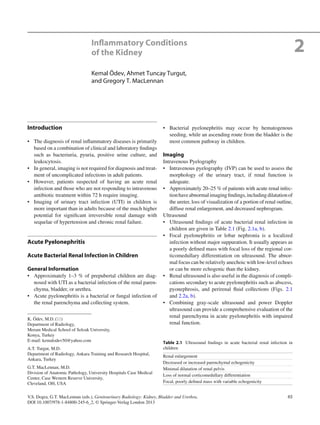
 a b c d e Fig. 2.1 Complicated acute bacterial pyelonephritis (ABP) in a 3-year-old girl with immunodeficiency. Ultrasound scans of right (a) and left (b) kidneys reveal bilateral diffuse enlargement with loss of the corticomedullary differentiation (arrow), multiple scattered areas of decreased echogenicity, and dilated renal pelvis (arrow) represent- ing diffuse spread of the inflammatory process, features consistent with complicated acute pyelonephritis, and pyonephrosis. (c) Contrast- enhanced CT scan demonstrates bilateral diffuse renal involvement with scattered nonenhancing foci representing microabscesses (arrow- heads) and a large cystic fluid collection in the left kidney (arrow) suggestive of a renal abscess. The maximum intensity projection image from excretory magnetic resonance urography (MRU) (d) and the max- imum intensity projection image from T2-weighted half-Fourier acqui- sition single-shot turbo spin-echo (HASTE) sequence (e) reveal the true pathology that is consistent with bilateral multifocal renal abscesses associated with complicated ABP (dashed arrows)
  • 3. 672 Inflammatory Conditions of the Kidney Power Doppler ultrasound is superior to color-flow• Doppler in defining extent of areas of hypoperfusion because most pyelonephritic lesions are ischemic. Computed Tomography Computed tomography (CT) is a sensitive study in evalu-• ation of the renal parenchyma in acute renal infection. Inflammatory edema and microabscess may be demon-• strated in the abnormal renal parenchyma. Rounded or irregular parenchymal abnormalities• appear as a patchy low-density areas on nonenhanced CT scans. After administration of radiographic contrast, these• regions are seen as areas of decreased enhancement (Fig. 2.1c). Magnetic Resonance Imaging Parenchymal alterations in complicated renal infections• can be demonstrated on MRI. AbscessformationsappearashypointenseonT1-weighted• and hyperintense on T2-weighted images (Fig. 2.1d, e). Nuclear Scintigraphy In children, radionuclide imaging is strongly recom-• mended both at early and late stages of the acute pyelone- phritis because of the potential of significant functional deficits within areas of inflammation and suppuration. Radionuclide imaging with Tc-99m dimercaptosuccinic• acid (DMSA) is considered the most sensitive modality for the imaging of renal cortex in children. It may show areas of peripheral decreased uptake related• to acute pyelonephritis or scar formation. Differential Diagnosis The differential diagnosis of acute pyelonephritis is given in• Table 2.2. Focal pyelonephritis causing a focal area of edema should be differentiated from various mass lesions, includ- ing a renal abscess, which usually appears more complex. Pearls and Pitfalls It is difficult to exclude the diagnosis of acute pyelone-• phritis with imaging studies because IVP, CT, and ultra- sound findings can be normal in up to 75 % of the cases. On ultrasound, abnormal echogenicity areas can occa-• sionally be misinterpreted as mass lesions. Although renal infection can be localized by DMSA, it• occasionally cannot be definitely distinguished from a sterile inflammation by using any kind of radiotracer. Acute Bacterial Renal Infection in Adults General Information The incidence of acute bacterial pyelonephritis (ABP) is• high, with an increasing rate of hospitalization. In the United States, approximately 250,000 cases of ABP• with more than 100,000 hospitalizations occur each year. Imaging Intravenous Pyelography The main role of IVP in the imaging of infection involves• screening for obstruction, although this is now better demonstrated by other modalities. Only 25 % of cases of ABP will have positive IVP findings,• including enlargement of the affected kidney, delayed con- trast excretion, and compression of the collecting system. Table 2.2 Differential diagnosis of acute pyelonephritis in children Ureteric obstruction Contusion Tubular obstruction Hypotension Scarring Autosomal recessive polycystic kidney disease Renal abscess a b Fig. 2.2 Acute bacterial pyelonephritis. Transverse (a) and longitudi- nal (b) ultrasound scans of a 13 months old boy performed after an episode of UTI reveal echogenic caliceal debris associated with ABP (arrows) (Courtesy of Alparslan Ünsal, MD, Aydın, Turkey)
  • 4. 68 K. Ödev et al. Ultrasound Sonographically, variable echotexture changes of the• renal parenchyma have been described for ABP. They are in the form of hypoechoic lesions or to a lesser• degree, hyperechoic foci. The former are attributed to edema, whereas the latter• may be due to hemorrhage. Ultrasound can also be used to detect and monitor the• progress of focal inflammatory masses. Thus, ultrasound is widely used either as a complement to• the IVP or as a screening modality, which may or may not be followed by CT scan, depending on the findings. Computed Tomography CT is considered the imaging modality of choice in the• evaluation of patients with ABP. It is superior to both ultrasound and IVP in detecting mor-• phologic and functional abnormalities, defining the extent of disease, and detecting the formation of renal subcapsu- lar or perinephric abscess or hematoma (Fig. 2.3a–c). In hemorrhagic bacterial pyelonephritis, nonenhanced CT• may demonstrate, on occasion, wedge-shaped or rounded areas with increased attenuation due to parenchymal bleeding. Nonenhanced CT is an excellent study for identifying cal-• culi, gas formation, hemorrhage, parenchymal calcification, urinary obstruction, renal enlargement, and inflammatory masses. Typical findings of ABP after intravenous contrast admin-• istration include ill-defined, wedge-shaped lesions of decreased attenuation radiating from the papilla in the medulla to the cortical surface. These wedge-shaped lesions probably represent areas of• poorly or nonfunctioning parenchyma due to vasospasm, tubular obstruction, and/or interstitial edema. The nonspecific appearance of a striated nephrogram in• the excretory phase on CT comprises linear bands of alternating hyper- and hypoattenuation parallel to the axes of tubules and collecting ducts, corresponding to a b c Fig. 2.3 Hemorrhagic acute pyelonephritis in a 45-year-old woman presented with left flank pain. (a) Nonenhanced CT scan demonstrates a low-attenuation collection in the left perirenal space (arrow). (b) Contrast-enhanced CT scan reveals a perirenal fluid collection with mixed attenuation (arrow) and thickening of Gerota’s fascia. Preoperative diagnosis was perirenal abscess. (c) Left nephrectomy revealed an acute pyelonephritis with perirenal hematoma
  • 5. 692 Inflammatory Conditions of the Kidney obstructed tubules with intervening normal tubules, which is associated with stasis of contrast material in dilated ducts due to edematous renal parenchyma (Table 2.3). Pathology Acute bacterial pyelonephritis results from an ascending• infection that originates in the lower urinary tract. Grossly, the collecting system is thickened, with yellow-• white abscesses located throughout the renal paren- chyma. An abscess is composed of central necrosis, surrounded by a zone of preserved neutrophils, all of which is surrounded by dilated vessels and fibroblasts. Fibroblastic proliferations serve to organize and confine processes and cause adjacent structures to contract. Cortical abscesses may result in hematogenous spread, further seeding the kidney with microorganisms (Fig. 2.4). Histologically, the predominant finding is neutrophils;• neutrophils are present in the interstitium and in the renal tubules (eventually forming white blood cell casts) (Fig. 2.5). In early infection, neutrophils and bacteria may be seen in the collecting ducts. Differential Diagnosis On CT, differential diagnosis for focal areas of hypoat-• tenuation includes renal infarction, tumors (renal cell car- cinoma), and scarring. The hypoattenuating areas associated with infarcts and• tumors persist following antibiotic treatment, in contrast to those caused by ABP. Furthermore, scarring rarely occurs in adult patients.• Pearls and Pitfalls On ultrasound, the combination of a hypoechoic area and• a focal inflammatory bulge to the perinephric space may be misinterpreted as a neoplasm. On CT, streak artifacts during the excretory phase may• give a false impression of abnormal nephrogram. The appearance of a “striated nephrogram” is not• pathognomonic for ABP and can be seen in kidneys fol- lowing renal trauma, in medullary sponge kidney and in autosomal recessive polycystic kidney disease (Table 2.3). Chronic Pyelonephritis General Information Chronic pyelonephritis is a chronic interstitial nephritis• that may arise secondary to a host of etiologic causes, including recurrent infection, autoimmune diseases, cal- culous disease, and chronic obstruction. Imaging The imaging findings of chronic pyelonephritis are summa- rized in Table 2.4. Intravenous Pyelography Renal size is usually decreased, unless chronic obstruc-• tion is present, in which case the renal size may be increased. Renal contour blurring.• Cortical thinning.• Table 2.3 Causes of striated nephrogram on CT Acute pyelonephritis Acute urinary obstruction Renal vein thrombosis Renal contusion Hypotension Medullary sponge kidney Autosomal recessive polycystic kidney disease Tubular obstruction (myoglobinuria) Fig. 2.4 Acute pyelonephritis. Patient died of sepsis after inadvertent ligation of one ureter. The kidney is swollen and pale. Microabscesses are visible on the cortical surface (From MacLennan GT, Cheng L. Atlas of Genitourinary Pathology. New York: Springer; 2011. Reprinted with permission)
  • 6. 70 K. Ödev et al. Focal nonfunctional area in nephrogram phase.• Ultrasound Focal or diffuse cortical thinning• Renal scar formation• Distortion of renal contours• Computed Tomography The kidney is usually shrunken and has an irregular outer• margin (Fig. 2.6). Renal scar formation presents as defect in the margin of• the kidney between two calyces. Decreased contrast excretion on enhanced CT.• Fibrotic distortion of the collecting system.• Pathology Chronic pyelonephritis is a general term implying chronic• interstitial renal infection due to long-standing obstruc- tion or vesicoureteric reflux, resulting in loss of renal parenchyma, with scarring. Chronic pyelonephritis accounts for 5–15 % of cases of end-stage renal failure. Most patients are asymptomatic until hypertension or ure- mia develops. The gross findings in chronic pyelonephritis are variable.• When the condition is obstruction related, the kidney may be large due to dilatation of the collecting system, and the Fig. 2.5 Acute pyelonephritis. Abundant neutrophils are present in the interstitium and within renal tubules Fig.2.6 Forty-year-oldwomanwithrecurrentpyelonephritis.Contrast- enhanced CT scan reveals severe parenchymal scarring of and decreased contrast excretion from the right kidney (arrow). The left kidney is normal Table 2.4 Imaging findings in chronic pyelonephritis Renal scarring Renal atrophy Cortical thinning Hypertrophy of residual normal tissue Caliceal clubbing Dilatation of the calyceal system Renal asymmetry
  • 7. 712 Inflammatory Conditions of the Kidney cortical surface is distorted by irregular scars (Fig. 2.7). Beneath the scars, the renal cortex is thin, and the papillae are scalloped or obliterated, causing the affected calyces to appear dilated. Microscopically, chronic pyelonephritis consists of intersti-• tial fibrosis, tubular atrophy, and sclerosis of glomeruli, often patchyindistribution(Fig.2.8).Theinterstitialinflammation is mixed but is composed mainly of lymphocytes. Tubules usually contain eosinophilic material, a feature known as “thyroidization.” Arteries and arterioles commonly show fibrosis, mural hyalinization, and occlusion. Differential Diagnosis Several conditions involving the loss of renal parenchy-• mal tissue such as infarction or papillary necrosis and fetal lobulation should be considered in the differential diagnosis of renal scarring. Pearls and Pitfalls On ultrasound, the limited depiction of calyces may• preclude the distinction of a renal scarring in cases with chronic pyelonephritis. Inchronicpyelonephritis,tubulesusuallycontaineosino-• philic material, a feature known as “thyroidization.” Vesicoureteral Reflux and Reflux Nephropathy General Information Vesicoureteral reflux (VUR) is described as the existence• of retrograde urine flow from the bladder to the ureter and the upper urinary tract. This condition usually develops secondary to the primary• abnormality of the vesicoureteral junction and may cause focal or diffuse acute and/or chronic pyelonephritis. The prevalence of VUR in asymptomatic children is less than• 0.5 %, but VUR is seen in up to 50 % of children with UTIs. Renal damage, also termed “reflux nephropathy,” develops in only 10 % of the children with symptomatic UTI. Imaging Intravenous Pyelography IVP can demonstrate cortical contour deformity associ-• ated with a clubbed calix. In the past, IVP was the preferred diagnostic method in• the demonstration of renal scarring and parenchymal function but was replaced by ultrasound and DMSA renal scintigraphy because of its low sensitivity in iden- tifying these abnormalities, the nephrotoxic potential of contrast materials used, and the necessity for exposure to radiation. Voiding Cystoureterography The gold standard method for the evaluation of VUR is• the conventional voiding cystoureterography (VCUG) (Fig. 2.9a). The international system of radiologic grading of vesi-• coureteral reflux has assigned five grades of reflux on VCUG: Grade 0: Indicates no VUR Grade 1: VUR that does not reach the renal pelvis Grade 2: VUR extending to the renal pelvis without dilatation Grade 3: VUR extending to the kidney with mild ureteral dilatation and mild to moderate pelvicalyceal dilatation Grade 4: VUR extending to the kidney with moderate ureteral dilatation and complete obliteration of the sharp angles of all fornices Fig. 2.7 Chronic obstructive pyelonephritis. The renal damage in a setting of obstructed renal drainage results from pressure-related atro- phy and chronic infection. This patient had long-standing distal ureteral obstruction. The renal pelvis and calyces are dilated, with flattening or obliteration of papillae and global thinning of renal cortex
  • 8. 72 K. Ödev et al. Grade 5: VUR extending to the kidney with a tortuous ureter and moderate dilatation of the renal pelvis to extreme dilatation of the entire upper urinary tract The sensitivity of VCUG depends on the severity of the• VUR. It has been demonstrated that VCUG has 100 % sensitivity in the diagnosis of grade 4 and grade 5 VUR. Ultrasound Ultrasound may be helpful for the detection of parenchy-• mal damage and dilatation of the collecting system. Features of VUR that may be evident by ultrasound• include diminished renal length and dilatation of the ure- ter, the renal pelvis, and the renal calyces. Ureteral dilatation was found to be the most helpful diag-• nostic feature in detection of the higher grades of VUR by ultrasound. Recently, it has been advocated that echo contrast-• enhanced cystosonography may be useful in diagnosing, grading, and following up on patients with VUR. Magnetic Resonance Imaging Multiplanar cross-sectional T1- and T2-weighted mag-• netic resonance imaging (MRI) combined with unen- hanced 3D magnetic resonance urography (MRU) is potentially useful for morphological evaluation of the uri- nary system, without the hazards related to the use of intravenous iodinated contrast agents. In addition to morphological evaluation, contrast-• enhanced MRU provides functional information about renal perfusion, concentration, and excretion of contrast agent (Fig. 2.9b, c). Gadolinium-enhanced T1-weighted excretory MRU may• demonstrate the parenchymal scars and perfusion defects associated with reflux nephropathy. Probably, the greatest advantage of the use of this• T1-weighted gadolinium-enhanced 3D-MRU technique is the visualization of the urinary tract of patients with impaired renal function. Unenhanced T2-weighted MRU technique allows excel-• lent visualization of the urinary tract and is useful in patients with dilated collecting systems. However, a non- dilated urinary tract is either invisible or incompletely visualized with this technique, whereas excellent visual- ization is achieved with contrast-enhanced T1-weighted excretory MRU. Nuclear Scintigraphy Direct radionuclide cystography with a• 99m Tc-labeled agent (sulfur colloid, diethylenetriamine penta-acetate [DTPA], or pertechnetate) is a well-accepted alternative to fluoroscopic VCUG for screening asymptomatic chil- dren, for follow-up examination of children with vesi- coureteral reflux (VUR), for postoperative evaluation after ureteral reimplantation, and for excluding VUR. • 99m Tc-labeled DTPA scintigraphy is more sensitive than VCUG in detection of VUR. DMSA renal scintigraphy has been found to be more sen-• sitive than IVP in the evaluation of focal renal scars. Fig. 2.8 Chronic obstructive pyelonephritis. The glomeruli are sclerotic, the interstitium shows fibrosis and is infiltrated by lymphocytes, the tubules are atrophic and contain eosinophilic casts (thyroidization), and the blood vessels show intimal sclerosis and mural thickening
  • 9. 732 Inflammatory Conditions of the Kidney a b c Fig. 2.9 VUR and reflux nephropathy in 5-year-old boy with recurrent bladder infection. (a) Retrograde voiding cystourethrogram reveals marked dilatation of the left ureter and left renal pelvicaliceal system (arrows) representing reflux to the left kidney and trabeculations and multiple diverticula in the bladder (open arrow). (b) Coronal contrast- enhanced 3D T1-weighted gradient echo MRI demonstrates minimal dilatation of the right collecting system (arrow) and severe hydroureter- onephrosis on the left collecting system (open arrow). (c) On sagittal contrast-enhanced T1-weighted gradient echo MRI, bladder diverticula (arrow) and diffuse bladder wall thickening secondary to cystitis are visualized excellently
  • 10. 74 K. Ödev et al. Pathology When chronic pyelonephritis is primarily reflux related,• the affected kidney is usually small. Reflux nephropathy usually results from vesicoureteral reflux (a congenital disorder) combined with intrarenal reflux, thus allowing infection access to the renal parenchyma. The kidneys are small and contracted, with broad• depressed sometimes saddle-shaped scars in the capsule (Fig. 2.10). There is loss of renal pyramids with dilated and thickened calyces, and the renal cortex overlying these areas is thinned. Renal parenchyma between scarred areas may be normal or hypertrophic. When these findings are present in a hypertensive patient• known to have had long-standing VUR, the renal lesion is sometimes described as “Ask-Upmark kidney” (Fig. 2.11). The predominant histologic finding is an interstitial lym-• phoid infiltrate with extensive renal tubular atrophy. Residual tubules contain eosinophilic casts (thyroidiza- tion) (Fig. 2.12). Additional findings include glomerular and vascular sclerosis. Differential Diagnosis On VCUG, scarring from reflux nephropathy should be• differentiated from scarring due to other etiologies such as parenchymal infarction. The deformed appearance of the calyx underlying the scar• in reflux nephropathy may be a clue for the differential diagnosis. Pearls and Pitfalls Fetal lobation and other normal variants of renal develop-• ment may be mistaken for renal scarring on ultrasound. With VCUG, misinterpretation may be possible, for• example, the soft tissue of the bowel wall may infre- quently simulate contrast in a ureter, or overlying stool and intestinal gas may obscure the kidney, precluding the diagnosis of lesser degrees of VUR. HIV-Associated Nephropathy General Information The kidneys are affected in HIV-infected patients by a• wide spectrum of entities including HIV-associated neph- ropathy, atypical infections, malignancy, and drug-related renal disease. The nephropathy may be the first manifestation of HIV• infection and often precedes opportunistic infections. It is the leading cause of renal failure in HIV-positive• patients. Fig. 2.10 Reflux nephropathy. Some portions of renal cortex are relatively preserved; in the scarred areas, there is virtually no resid- ual cortex. This is an example of an Ask-Upmark kidney, part of a clinical scenario including chronic urinary tract infections associated with vesicoureteric reflux, hypertension, and a small kidney with saddle-shaped scars Fig. 2.11 Ask-Upmark kidney. Gray-scale ultrasound demonstrates saddle shape of the kidney (arrow) secondary to scar formation from VUR
  • 11. 752 Inflammatory Conditions of the Kidney Imaging Ultrasound On ultrasound, cardinal imaging findings for HIV-• associated nephropathy include normal-sized or enlarged kidneys, increased echogenicity of the cortex, renal pelvi- caliceal thickening, and loss of the fat appearance of the renal sinus. Renal biopsy provides the definitive diagnosis of the dis-• ease; the aforementioned imaging findings are only suggestive. Decreased corticomedullary differentiation and paren-• chymal heterogeneity are late findings of HIV-associated nephropathy. Computed Tomography CT findings include enlarged kidneys, a hyperattenuating• medulla on the unenhanced scan, and a striated nephro- graphic appearance. Various opportunistic renal infectious agents such as• Pneumocystis jirovecii, fungal organisms, and mycobac- teria may affect the kidneys of patients with AIDS, giving rise to a wide spectrum of imaging findings with low specificity, such as renal cortical calcification, focal areas of increased echogenicity in the cortex and medulla, and focal microabscesses and hydronephrosis. The presence of renal calcifications and nephrocalcinosis• may suggest HIV-associated infection despite not being specific for any specific pathogen. Non-Hodgkin lymphoma and Kaposi sarcoma are the• most commonly encountered malignancies in patients with HIV infection. CT is the imaging modality of choice for the evaluation of• all patients with lymphoma. Among the common patterns of renal involvement by the disease are multiple paren- chymal masses, solitary lesions, direct extension from retroperitoneal adenopathy, perinephric disease, and nephromegaly. Likewise, renal involvement by Kaposi sarcoma associ-• ated with AIDS usually gives rise to nonspecific clinical or radiologic manifestations. Finally, certain antiretroviral agents such as indinavir and• nelfinavir may cause the development of renal calculi. Magnetic Resonance Imaging On MRI, loss of corticomedullary differentiation and• renal enlargement may be seen. Differential Diagnosis The combination of hyperechoic kidneys and a thickened• pelvicaliceal system by ultrasound can imply a possible diagnosis of HIV-associated nephropathy. However, infection and acute tubular necrosis should be considered in the differential diagnosis. The presence of calcification and nephrocalcinosis may• suggest HIV-associated infection despite not being specific for any specific pathogen. Fig. 2.12 Reflux nephropathy. In this Ask-Upmark kidney, the renal tissue on the left is essentially normal, whereas on the right, in the area of scarring, glomeruli are absent, and the interstitium is fibrotic and infiltrated by lymphocytes. The tubules are atrophic and contain eosinophilic casts (thyroidization), and the blood vessels show intimal sclerosis
  • 12. 76 K. Ödev et al. Pearls and Pitfalls An unusual pattern of nephrocalcinosis defined as “partial• nephrocalcinosis” which involves the renal cortex and medulla asymmetrically has been described in patients with AIDS-related M. avium-intracellulare infection. Renal and Perirenal Abscess General Information Pyelonephritis leading to renal abscess is quite rare,• with an incidence of 0.01 %. Although the vast majority of renal infections undergo resolution with effective antibiotic treatment, one of the complication of inade- quately or untreated acute pyelonephritis is renal abscess. Renal abscess is more common in patients with condi-• tions predisposing to infection, such as diabetes or other diseases associated with an immunocompromised status (Figs. 2.1, 2.13, and 2.14), as well as with urinary tract obstruction and/or renal stones. Renal abscess and pyonephrosis may result from• hematogenous seeding of infection in immunosup- pressed patients without any urinary obstruction. Furthermore, diabetes mellitus and immunosuppression may predispose patients to the development of an abscess (Fig. 2.15). Abscess may also result from interventional procedures,• such as diagnostic cyst aspiration (Fig. 2.16a, b), or alco- hol embolization of the kidney. Imaging Plain Film Radiography Abdominal radiography typically demonstrates enlarge-• ment of the affected kidney. Intravenous Pyelography IVP may reveal nonfunctional or faint opacification of the• involved kidney, with focal pelvicalyceal dilatation or focal excretory defects. Ultrasound Ultrasound demonstrates a round, thickened, or smooth-• walled complex mass. At ultrasound, abscess is rarely anechoic (Figs.• 2.1a and 2.13a), and it may have internal septation and loculations (Fig. 2.14a). These atypical findings are indistinguishable from those of infected or hemorrhagic cysts (Fig. 2.16a). Computed Tomography CT is the best imaging method of determining extension• of a renal abscess into adjacent structures. On precontrast CT scans, abscess may be a solitary mass• or may appear as multiple, round, well-defined, and low- attenuation masses. After the administration of intravenous contrast agent, an• abscess becomes more conspicuous due to enhancement (Figs. 2.1c, 2.13b, c, and 2.16b). Renal abscess may have an enhancing rim but does not enhance centrally. In some cases, Gerota’s fascia may be thickened, or peri-• renal extension of the abscess may be seen. Rupture of a renal abscess or an infected cyst through the• renal capsule results in involvement of the perirenal space. Perirenal abscess may result from direct extension of• peritoneal and/or retroperitoneal infection. Magnetic Resonance Imaging On MRI, abscess presents a low or inhomogeneous signal• intensity on T1- and T2-weighted images depending on the amount of protein, fluid, and cellular debris. Pathology Perinephric abscesses occur in the perinephric fat.• They most commonly result from extension or rupture of• a renal abscess or pyonephrosis; however, they can also occur secondary to surgery or transplantation (Figs. 2.17 and 2.18). The offending organisms may be demonstrable in the• fluid aspirated from abscess cavity (Fig. 2.19). Differential Diagnosis Renal abscesses should mainly be differentiated from a• cystic renal cell carcinoma. The lack of vascularity on Doppler imaging may be helpful for the relevant differentiation. The differential diagnosis of perinephric abscesses• includes perinephric fluid collections like urinoma, sub- capsular and perirenal hematoma, renal lymphangiomato- sis, pancreatic pararenal fluid collections, and transudate fluid associated with nephropathies. For perinephric urinomas, the history of trauma or the• finding of an obstructing stone usually suggests the cor- rect diagnosis. Extravasation of excreted contrast material can be depicted on delayed CT scans in patients with trauma to ureters. Pearls and Pitfalls Several conditions may cause diagnostic pitfalls because• each of them may be misinterpreted as perinephric fluid collection. These conditions are:
  • 13. 772 Inflammatory Conditions of the Kidney The hypoechoic rim at the periphery of infantile poly-– cystic kidneys The peripheral hypoechoic rim surrounding a kidney– Acute cortical necrosis– Perirenal non-Hodgkin’s lymphoma– Retroperitoneal fibrosis– Prominency of perirenal fat or thickened renal fascia– causing a perirenal halo Breathing artifact on CT– Pyonephrosis General Information Chronic suppurative infection in an obstructed kidney is• called “pyonephrosis.” In pediatric patients, ureteropelvic junction obstruction,• ureterocele, and ureteral ectopia are common causes of obstruction (Fig. 2.20). a b c Fig. 2.13 Renal abscess and pyohydronephrosis in 18-year-old immu- nocompromised male with low-grade fever. (a) Ultrasound reveals an anechoic fluid collection with thin walls (arrow) and echogenic mate- rial within the collecting system. Early and late phase contrast-enhanced CT scans (b, c) demonstrate a nonenhancing renal mass with regular contour (arrow), corresponding to fluid collection defined in (a) and left hydronephrosis. Note contrast layering over the dense contrast material (arrow). All ultrasound and CT changes resolved after appropriate anti- biotic therapy
  • 14. 78 K. Ödev et al. In adults, pyonephrosis is usually due to undiagnosed• obstructive nephropathy, such as calculi, tumor, com- plicated pyelonephritis, strictures, or a congenital anomaly. Imaging Intravenous Pyelography IVP fails to demonstrate the collecting system in pyo-• nephrosis, and it is seldom used for this purpose, except for surgical planning or postoperative assessment. Ultrasound Ultrasound is useful in early and accurate diagnosis of• pyonephrosis. The classic ultrasound finding in pyonephrosis is the pres-• ence of echogenic material in a dilated collecting system (Figs. 2.1a, b and 2.20). There may be fine echoes, fluid– fluid levels, and debris levels within the collecting system. Echogenic debris is the most reliable sign of pyonephrosis.• Computed Tomography CT may be useful in evaluating adult pyonephrosis and in• detecting the cause of obstruction, which may not be evi- dent on ultrasound. CT scan can be performed without the use of intravenous• contrast material, with an accuracy of 97 % in the detec- tion of calculi in the urinary tract. Contrast-enhanced CT may be useful in demonstrat-• ing parenchymal and functional changes. It clearly a b c Fig. 2.14 Renal abscess in 5-year-old immunocompromised boy. (a) Ultrasound demonstrates a complex cystic mass with compression of the renal parenchyma (arrow). (b) Coronal contrast-enhanced 3D T1-weighted gradient echo image during early phase reveals the lack of enhancement confirming the cystic nature of the lesion (arrow). (c) Axial T2-weighted MRI reveals a hyperintense mass compressing the parenchyma (dashed arrow). Renal outlines have become obscured secondary to the perirenal inflammatory process. Note the hypointense rim around the abscess cavity (arrow). The pathological diagnosis was renal abscess
  • 15. 792 Inflammatory Conditions of the Kidney demonstrates thickening of the collecting system wall (>2 mm), parenchymal or perinephric inflammatory changes, and dilatation or obstruction of the collect- ing system. Magnetic Resonance Imaging MRU may show the presence of a fluid/fluid level within• the dilated collecting system and is useful in establishing the severity of dilatation, the site, and the most likely cause of obstruction in pyonephrosis. Fig. 2.17 Perirenal abscess, bacterial. Perirenal abscesses are com- posed of necrotic renal or perirenal tissue and loculated inflammatory exudate Fig. 2.18 Perirenal abscess, fungal. This perirenal fungal abscess developed in a 64-year-old man who was immunosuppressed following liver transplant. Despite efforts at conservative management, nephrec- tomy was required. The abscess involves both renal parenchyma and perirenal fat (arrowheads) (Image courtesy of Stacy Kim, M.D.) Fig. 2.15 Perirenal abscess in 40-year-old diabetic woman with Escherichia coli in blood and urine cultures. Contrast-enhanced CT scan demonstrates a large perirenal abscess which displaces the left kid- ney (arrow) a b Fig. 2.16 Infected renal cyst in a 65-year-old man with left flank pain following several months of percutaneous cyst drainage. (a) Ultrasound reveals a large multilocated cystic lesion with thick wall and internal septations (arrow). (b) Contrast-enhanced CT scan shows that the attenuation of cystic lesion is slightly higher than water and that the lesion had an enhancing wall (arrow). Infected cyst was confirmed by nephrectomy
  • 16. 80 K. Ödev et al. Pathology In pyonephrosis, the kidney is largely replaced by a purulent• collection of necrotic renal tissue and inflammatory exudate composedofacuteandchronicinflammatorycellsandedema fluid, forming, in effect, a contained abscess (Fig. 2.21). Differential Diagnosis The main differential diagnosis of pyonephrosis includes• hydronephrosis. Ultrasound findings such as dilatation of the pelvicaliceal• system, echogenic collecting system debris, and fluid–fluid levels within the collecting system may be helpful in dis- tinguishing pyonephrosis from simple hydronephrosis. CT findings including renal pelvic wall thickness exceed-• ing 2 mm, dilatation of the pelvicaliceal system, paren- chymal or perinephric inflammatory changes, and layering of contrast material with the purulent fluid on excretory studies may also provide a correct diagnosis. MRI findings suggestive for pyonephrosis are similar to• those seen at CT. Pearls and Pitfalls Rarely, a primary tumor of the upper collecting system,• such as transitional cell carcinoma of the renal pelvis, may exhibit low-level echoes on ultrasound and may mimic pyonephrosis. Other clinical material, such as urine cytologic findings,• may suggest the diagnosis of transitional cell carcinoma. Settlement or movement of debris within the lesion• with changes in patient position, although observed infrequently, may enable the diagnosis of pyonephrosis. MRI may be helpful for the diagnosis in equivocal cases.• Emphysematous Pyelonephritis General Information Emphysematous pyelonephritis (EPN) is a life-threaten-• ing necrotizing infection of the kidneys characterized by gas formation within the renal parenchyma or the peri- nephric space. Up to 97 % of patients with EPN are diabetics; in contrast,• EPN is quite rare in nondiabetics. The commonest infecting organisms include Escherichia• coli, Klebsiella pneumoniae, and Proteus mirabilis. Women are affected twice as often as men. Imaging Plain Film Radiography Conventional radiography may demonstrate gas bubbles• or abnormal gas collection within the renal fossa (Fig. 2.22a, b). Fig. 2.19 Perirenal abscess, fungal. Fungal organisms consistent with Aspergillus sp. were identified within the abscess cavity shown in Fig. 2.18
  • 17. 812 Inflammatory Conditions of the Kidney Intravenous Pyelography IVPmaydemonstrateapersistentnephrogramontheaffected• side secondary to delayed excretion of contrast agent. Ultrasound Ultrasound demonstrates an enlarged kidney with high-• amplitude echoes within the renal parenchyma and often with low-level posterior dirty acoustic shadowing, known as reverberation artifacts (Fig. 2.23a–c). Gas in the collecting system can be seen after certain• interventional procedures, and it should not be confused with EPN. Computed Tomography A CT classification scheme divides EPN into two types:• – Type I is characterized by renal parenchymal destruc- tion that manifests with either streaky or mottled areas of gas (Fig. 2.24). This is seen in 33 % of patients, and the associated mortality is approximately 68 %. Intra- or extrarenal fluid collections are notably absent. – Type II is characterized by bubbly or loculated gas within fluid collections in the renal parenchyma, col- lecting system, or perirenal space (Figs. 2.22b and 2.23c). This type is seen in 66 % of patients, and the mortality rate is approximately 18 %. Pathology Emphysematous pyelonephritis is a rare life-threatening• complication of acute pyelonephritis, usually of bacterial origin. It is associated clinically with diabetes mellitus and urinary tract obstruction. Abscesses are accompanied by renal papillary necrosis• and cortical infarcts. Renal papillary necrosis refers to necrosis of portions of the medulla. There is a cystic appearance to the kidney secondary to gas formation (Fig. 2.25). Micro: Empty spaces lacking epithelial cell linings and• distorting the parenchyma with areas of vascular throm- bosis, ischemic necrosis, suppurative inflammation, and abscess formation. Differential Diagnosis Retroperitoneal perforation of abdominal viscera• Psoas abscess due to gas-forming organisms• Reflux of air from the bladder• Bronchorenal, enterorenal, or cutaneorenal fistulae• Air in a focal renal abscess• Fig. 2.21 Pyonephrosis. The term “pyonephrosis” implies obstructed renal drainage, resulting in a contained abscess with varying degrees of renal parenchymal destruction. The collecting system contains abun- dant purulent material, and only a thin rim of residual renal tissue remains. This condition resulted from an obstruction at mid-ureter level; the etiology of the ureteral obstruction was indeterminate (From MacLennan GT, Cheng L. Atlas of Genitourinary Pathology. New York: Springer; 2011. Reprinted with permission) Fig. 2.20 Pyonephrosis in 8-month-old boy with suspected upper UTI. On ultrasound, the dilated pelvicalyceal system is filled with echogenic material attributable to debris and organized pus in the renal pelvis and ureter due to ureterovesical junction obstruction (p renal pelvis, u ureter)
  • 18. 82 K. Ödev et al. A history of urologic intervention, such as nephrostomy• insertion, or retrograde pyelogram Pearls and Pitfalls On ultrasound, gas within the kidney or renal pelvis may• mimic renal calculi. The distal shadowing with reverberations and “dirty shad-• owing” of low-level echoes may be helpful for the differ- entiation of EPN from renal calculus. Xanthogranulomatous Pyelonephritis General Information Xanthogranulomatous pyelonephritis (XGP) is a rare• form of chronic pyelonephritis, occurring most commonly in women, with a peak incidence in the fifth and sixth decades. Clinical symptoms are often vague, and laboratory• findings are nonspecific. The classic urographic triad in XGP consists of:• Unilaterally decreased/absent renal excretion– Staghorn calculus (70 %)– Poorly defined mass or diffuse renal enlargement– Imaging Intravenous Pyelography May demonstrate an enlarged nonfunctional kidney with• staghorn calculi. Ultrasound On ultrasound, the kidney is usually enlarged, with mul-• tiple hypoechoic or anechoic areas corresponding to dilated calyces and areas of parenchymal destruction. Central echogenic foci representing staghorn calculus• may be seen. In focal XGP, ultrasound findings are nonspecific, and it is not possible to differentiate this from a renal abscess or necrotic and/or cystic renal cell neoplasm. Computed Tomography CT is more informative than ultrasound in the evaluation• of XGP. CT findings of diffuse XGP are listed in Table• 2.5. In diffuse XGP, the inflammatory process may involve the• perinephric space, the adrenal gland, the ipsilateral psoas muscle, and/or the subdiaphragmatic area (Fig. 2.26a–d). In focal XGP, a focal mass of low attenuation with rim• enhancement is seen on CT, often associated with calcu- lus (Fig. 2.27). Magnetic Resonance Imaging MRI is valuable for diagnosing XGP and is extremely• sensitive for identifying the lipid-laden xanthogranuloma- tous tissues (Fig. 2.26b–d). MRI is less accurate in diagnosing focal XGP with a lim-• ited component of lipid-laden macrophages. Pseudocystic masses with thick septa of XGP have low• signal intensity in T1-weighted images and high signal a b Fig. 2.22 Bilateral type 2 EPN with extensive parenchymal destruc- tion in a 60-year-old diabetic woman. (a) IVP demonstrates lucent air which outlines the right pelvicalyceal system and the right ureter (arrows). (b) Contrast-enhanced CT scan reveals abscess with gas bub- bles and fluid contents in the right collecting system and in bilateral renal parenchyma (arrows)
  • 19. 832 Inflammatory Conditions of the Kidney intensity on T2-weighted images, compared with the renal parenchyma. Diffuse XGP may show a wide range of signal intensities• on MRI, due to the heterogeneous composition of the chronic suppurative renal inflammation. Pathology The process may be diffuse, segmental, or focal. In the• diffuse form of this entity, the kidney is completely obstructed, most commonly by a staghorn calculus. Grossly, the collecting system is thickened, with yellow-• white nodules present in the renal pyramids (Fig. 2.28). The process begins with suppurative inflammation in the pelvis and adjacent sinus fat, with the cortex, perinephric fat, and retroperitoneal tissue potentially becoming involved. a b c Fig. 2.23 Type 1 EPN in a 50-year-old diabetic woman. Ultrasound (a, b) reveals echogenic foci with “dirty” shadowing in the left kidney (arrow in a). Acoustic shadowing represents stone in the left pelvis renalis (arrow in b). (c) Unenhanced CT scan demonstrates multiple parenchymal gas collections in the left kidney (arrow) Fig. 2.24 EP in a 45-year-old diabetic woman. Unenhanced CT scan demonstrates air bubbles within the renal collecting system and ureter and moderate hydroureteronephrosis bilaterally (arrows)
  • 20. 84 K. Ödev et al. Histologically, there is a zonal pattern within the xan-• thogranulomatous nodules and thickened areas. The cen- tral zone is composed of necrosis and neutrophils. This area is admixed with/surrounded by foamy histiocytes (Fig. 2.29). The outermost zone is composed of fibroblasts. Differential Diagnosis The differential diagnosis may include infiltrative renal masses, such as: Transitional cell carcinoma• Renal cell carcinoma• Lymphoma• Nonneoplastic pseudotumoral conditions such as renal or• perirenal abscess Pyonephrosis• Renal tuberculosis (TB)• Focal or diffuse nephritis• Fungal infection• The definitive diagnosis must be confirmed by surgery. Pearls and Pitfalls Gas is rarely seen and may be misinterpreted as pyoneph-• rosis or EPN. Renal Hydatid Cyst Disease General Information Hydatid disease, mainly caused by• Echinococcus granu- losus, frequently involves the liver (60 %) and lungs (25 %); renal involvement occurs only in 3 % of cases. The kidney is involved primarily by hydatid disease via• the systemic circulation, but secondary involvement by spread from a hydatid cyst in an adjacent organ is possible. Imaging Imaging findings of renal hydatid cyst are summarized in Table 2.6. Plain Film Radiography Radiography may show a soft tissue mass in the kidney• area with amorphous and/or curvilinear wall calcification, although similar calcifications may be seen in other renal lesions, including simple cyst, hematoma, XGP, and renal cell carcinoma. Intravenous Pyelography IVP may show a rounded soft tissue shadow with curvi-• linear calcifications, signs of compression of the pelvica- lyceal system, and ureter or nonfunctioning kidney. If the cyst ruptures into the renal collecting system, IVP• may show filling defects, due to daughter cyts in the pel- vicalyceal system, or it may disclose the presence of an irregular mass with or without urinary obstruction and a nonfunctioning kidney. Ultrasound Various ultrasound appearances of hydatid cysts have• been described by Gharbi. In the Gharbi classification, a Type I echinococcal cyst• appears as a well-defined anechoic lesion on ultrasound and is indistinguishable from a simple cystic lesion (Fig. 2.30a). Multiple echogenic foci due to “hydatid sand” may be seen in the cyst by repositioning of the patient. Fig. 2.25 Emphysematous pyelonephritis. This term denotes gas bub- ble formation in the renal parenchyma or perirenal tissues; it is often accompanied by abscess and cortical infarcts. This kidney is from a teenaged diabetic female who died of urosepsis. Tiny gas bubbles are visible within the purulent infiltrates in the renal cortex (From MacLennan GT, Resnick MI, Bostwick DG. Pathology for urologists. Philadelphia: Saunders; 2003, with permission) Table 2.5 Computed tomography findings of diffuse xanthogranu- lomatous pyelonephritis Enlarged nonfunctioning kidney Staghorn or solitary calculus filling the pelvis renalis Caliceal and/or pelvic dilatation Multiple nonenhancing cystic masses filled with pus and debris Rim enhancement with contrast medium administration Calcifications within the masses Low-attenuation areas of lipid-rich xanthogranulomatous tissue within the masses Absence of renal stone Small, contracted kidney Abundant perirenal fat tissue
  • 21. 852 Inflammatory Conditions of the Kidney The findings in cases of Type II echinococcal cyst are• generally specific and characteristic for echinococcal dis- ease. In these lesions, floating and detached membranes or daughter cysts that are pathognomonic for hydatid dis- ease may be seen in the cyst (Fig. 2.31a). Complete detachment of the membranes inside the cyst• has been referred to as sonographic water-lily sign because of its similarity to the radiographic water-lily sign in pul- monary cysts. Both Type II and Type III lesions (multivesicular) are• almost pathognomonic for hydatid disease (Fig. 2.31a). Type IV and Type V lesions demonstrate heterogenous• solid appearances with a combination of liquid and solid cystic contents. a cc b d Fig. 2.26 Diffuse XGP in a 40-year-old diabetic woman. (a) Contrast- enhanced CT scan demonstrates diffuse enlargement of the nonfunc- tioning left kidney with calculus (dashed arrow) and subcapsular fluid collection (arrow) and diffuse inflammatory process extending from the left kidney into the left perirenal and left posterior pararenal space (open arrow). Coronal contrast-enhanced 3D T1-weighted gradient echo sequence image (b) and coronal true fast imaging with steady- state precession (FISP) sequence (c) reveal the nonfunctioning left kid- ney and the fluid-filled areas in the left perirenal and subdiaphragmatic regions (arrows). (d) Macroscopic specimen revealed a stone in the left renal pelvis. The renal pelvis and calyces are surrounded by inflamed tissue and infected fluid collections corresponding to xanthogranuloma- tous inflammation
  • 22. 86 K. Ödev et al. Type V lesions demonstrate calcifications in the cyst wall• and germinative membranes. Computed Tomography On CT, the contents of a closed simple (intact) unilocular• cyst are homogenous, with a density close to that of water (10 HU) (Fig. 2.30b). Calcification of the cyst wall or internal septa is easily• demonstrated on CT (Fig. 2.31b). CT may reveal detachment of the germinative membranes• from the ectocyst, which gives the pathognomonic appear- ance of a ruptured cyst. On CT, the presence of daughter cysts is pathognomonic• of renal cystic hydatid cyst (Figs. 2.31b and 2.32a). Magnetic Resonance Imaging MRI diagnosis of intact unilocular renal hydatid cysts is• based on demonstration of a relatively thick cyst wall (Fig. 2.30c). Although cyst wall calcification is more clearly demon-• strated on CT, MRI is superior in demonstrating hydatid cyst morphology and the exact extent of the disease (Figs. 2.30c, 2.32b, c, 2.33, and 2.34a–c). On T1-weighted images, the parasitic cyst wall is isoin-• tense relative to the fluid in the cyst and appears on T2-weighted images as a low-signal-intensity rim sur- rounding the homogeneous high signal intensity cyst con- tents (Figs. 2.30c and 2.32b). On MRI, intact renal unilocular hydatid cyst may show• homogeneous hypointense signal on T1-weighted images and homogeneous hyperintense signal on T2-weighted images. Perforated hydatid cysts exhibit detached germinative• membranes. The membranes are seen as floating structures within the• cyst and appear dark on both T1- and T2-weighted images (Figs. 2.33 and 2.34a–c). Pathology Gross: Large multiloculated cyst containing numerous• hydatid cysts (daughter cysts). Micro: The cysts are enveloped by a fibrous pseudocap-• sule and may be empty or contain scolices with evident hooklets. The daughter cysts have an outer layer of lami- nated chitin. Differential Diagnosis Sonographically, a Type I hydatid cyst may be indistin-• guishable from a simple renal cyst, but a double contour thick wall, involvement of other organs, or a history of living in endemic regions strongly favors the diagnosis of hydatid cyst. On CT, enhancement of the wall of the cyst with contrast• helps in differentiating hydatid cyst from simple renal cyst or abscess. Fig. 2.27 Contrast-enhanced CT scan of a 51-year-old female patient showing a focal mass of low attenuation with rim enhancement in the left kidney (arrow) which was histopathologically proven to be a focal XGP. Note that the lesion is not associated with any calculus (Courtesy of Ugur Toprak, MD, Ankara, Turkey) Fig. 2.28 Xanthogranulomatous pyelonephritis. This is typically a renal calculus-associated inflammatory process that may be difficult to distinguish radiologically from a renal neoplasm. The cut surface exhib- its multiple gray-white to yellow nodules. The papillae are blunted, and a staghorn calculus (black arrow) is present. The inflammatory process often extends into the perirenal and sinus fat (yellow arrow) (Image courtesy of Douglas Hartman, M.D.)
  • 23. 872 Inflammatory Conditions of the Kidney The “rim sign” on MRI corresponding to low-signal-• intensity cyst wall is also helpful for differentiating uni- locular hydatid cysts from simple renal cysts, although this sign has also been described in hepatocarcinoma, amoebic liver abscess, adenoma, and hematoma. The diffuse and bilateral nature of polycystic kidney dis-• ease is helpful in distinguishing this entity from Type III hydatid cysts. On CT, the presence of daughter cysts is a helpful in dis-• tinguishing Type 3 hydatid cysts from simple renal cysts, renal abscess, and cystic renal cell carcinoma. The heterogenous appearance of Type IV lesions may be• difficult to distinguish from infected renal cysts, abscesses, pyonephrosis, hydronephrosis, and neoplasms. CT may be useful in the diagnosis of Type IV hydatid• cysts with a “pseudotumor” appearance. Pearls and Pitfalls The Gharbi classification is used in classifying hydatid• cyst disease. Postoperative imaging findings should not be misinter-• preted as a recurrence. Renal Malakoplakia General Information Malakoplakia is a rare granulomatous inflammatory dis-• ease associated with chronic coliform infections caused by abnormal macrophage function. Fewer than 200 cases of renal malakoplakia have been• reported in the literature so far. Clinically, it is most often accompanied by fever, flank• pain, or a palpable flank mass. Imaging The imaging findings of malakoplakia are nonspecific and• can often mimic other lesions, such as renal neoplasms; therefore, the diagnosis is typically made only after surgi- cal intervention. Plain Film Radiography Usually reveals the presence of an enlarged kidney.• Fig.2.29 Xanthogranulomatous pyelonephritis. There is a polymorphous assortment of inflammatory cells, including neutrophils centrally, with a broad zone of large pale lipid-laden macrophages, plasma cells, eosinophils, and lymphocytes and sometimes multinucleated giant cells, as in this case. There is usually a fibrous reparative reaction at the periphery of the inflammatory nodules Table 2.6 Sonographic appearance of hydatid cysts in Gharbi classification Type I – Well-defined, purely anechoic lesions that may be indistinguishable from simple renal cysts. Multiple echogenic foci due to hydatid sands may be seen in the cyst Type II – A focal or diffuse detachment of the inner germinal layer results in a floating membrane inside the cyst Type III – Multiseptated cysts with multiple daughter cysts Type IV – Heterogeneous solid appearance with infolded mem- branes and internal echoes Type V – Solid appearance calcifications in the cyst wall and germinative membranes
  • 24. 88 K. Ödev et al. Intravenous Pyelography On IVP, one or both kidneys are large, with smooth con-• tours and with diminished or absent function. Filling defects in the collecting system with calyceal• distortion due to foci of malakoplakia have been described. Ultrasound Ultrasound demonstrates distortion and compression of• the central echo complex by well-defined masses with variable echogenicity. Ultrasound findings may be observed unilaterally or• bilaterally. Computed Tomography CT may show nonspecific masses or an enlarged kidney• with poor contrast uptake. Nonenhancing areas in the kidney may be seen in con-• trast-enhanced CT. Magnetic Resonance Imaging Irregularly contoured kidneys with multiple 1- to 2-cm• poorly defined nodules. Nodules have low signal intensity on T1- and T2-weighted• MRI sequences. Differential Diagnosis XGP (unilaterality and the presence of upper tract calculi• are more in keeping with XGP than with malakoplakia) Localized abscesses• Lymphoma• Multifocal primary or metastatic tumors• a b c Fig. 2.30 Thirty-year-old woman with renal hydatid cyst. (a) Ultrasound demonstrates a large, unilocular cyst with a well-defined contour and thick wall (arrow) with inflammatory reaction adjacent to the cyst. (b) A sagittal oblique maximum intensity projection CT recon- struction image of the left kidney shows an intense enhancement of the cyst wall (arrow). (c) T2-weighted MRI reveals a large unilocular cyst containing high-intensity fluid and having a low-intensity rim “peri- cyst” (arrow)
  • 25. 892 Inflammatory Conditions of the Kidney Pathology Malakoplakia is a chronic granulomatous disease usually• resulting from recurrent urinary tract infections in mid- dle-aged women. It is an uncommon disorder that most often involves the urinary bladder but very rarely occurs in the upper tracts. In this entity, the renal parenchyma and pelvis are coated• with multiple yellow-brown soft plaques. The plaques are composed of large eosinophilic his-• tiocytes (von Hansemann histiocytes), many of which contain basophilic cytoplasmic inclusions (Michaelis– Gutmann bodies) (Fig. 2.35). The inclusions represent a b c Fig. 2.32 Forty-year-old man with renal hydatid cyst. (a) Contrast- enhanced CT scan demonstrates a lobulated cystic mass with enhance- ment located on the right kidney (arrow). The cyst wall is not apparent. (b) On coronal T2-weighted MRI, a low-signal-intensity peripheral rim of capsule (arrow) and daughter cysts are well demonstrated. In this image, hydatid cyst is demonstrated much better than on CT. (c) Macroscopic specimen revealed the hydatid cyst containing numerous daughter cysts a b Fig. 2.31 Thirty-five-year-old woman with renal hydatid cyst. (a) Ultrasound demonstrates a multilocular cyst in the right kidney (arrow). (b) Contrast-enhanced CT scan reveals multilocular appearance due to numerous daughter cysts. Note curvilinear calcification of the cyst wall (arrow)
  • 26. 90 K. Ödev et al. incompletely digested bacilli and can be highlighted with the PAS stain and special stains for calcium (von Kossa) and iron. Pearls and Pitfalls The radiological appearance of renal malakoplakia,• depending on the pattern of involvement, is usually nonspecific. The malakoplakia nodules have low signal intensity on• T1- and T2-weighted MRI sequences. The plaques are composed of large eosinophilic his-• tiocytes (von Hansemann histiocytes), many of which contain basophilic cytoplasmic inclusions (Michaelis– Gutmann bodies). Renal Tuberculosis General Information Genitourinary involvement by tuberculosis (TB) develops• in approximately 8 % of patients with pulmonary disease. Less than 50 % of patients with renal TB have a known• history of TB. Abnormal radiographic findings typical of pulmonary• TB are present in only 50 % of patients with renal TB, and only one-half of these patients show signs of active TB. Fig. 2.33 Thirteen-year-old girl with ruptured right renal hydatid cyst. On T2-weighted MRI, detached germinative membranes in hydatid fluid (arrow) are seen a b c Fig. 2.34 Thirty-year-old woman with a ruptured renal hydatid cyst. (a) T1-weighted MRI reveals a low-signal-intensity mass in the left kid- ney (arrow). Cyst wall is isointense relative to cyst contents. On T2-weighted MRI (b, c), the detached germinative membranes (arrow in b) are shown much better than on T1-weighted MRI. Daughter cysts (arrow in c) are identified within the mother cyst. Note both detached membranes and pericyst are of low intensity
  • 27. 912 Inflammatory Conditions of the Kidney Imaging Imaging findings of renal TBC are summarized in Table 2.7. Intravenous Pyelography IVP may show calcified parenchymal masses and irregu-• larity of the caliceal contour due to necrotizing papillitis. Development of infundibular, pelvic, or ureteral strictures• is a common finding and is nearly pathognomonic of TB. Ultrasound Tuberculous granulomas in the renal parenchyma may• appear as masses of variable echogenicity, with or with- out areas of caseous necrosis and calcifications. Computed Tomography CT is more sensitive than other methods in detecting renal• calcifications, which are present in 50 % of the patients. Renal calcifications show various patterns: punctate, cur-• vilinear foci, lobar disruption, and calcifications of the entire renal parenchyma and renal pelvis. The most common findings on CT are caliectasis, strictures• of the infundibulum, renal pelvis and ureter, generalized hydronephrosis, and parenchymal scarring (Fig. 2.36a, b). The end result may be a “putty kidney,” composed of• residual calcified nonfunctional renal tissue. Pathology In tuberculosis, the kidneys become infected by the• hematogenous spread of mycobacteria, most often M. tuberculosis, resulting in the formation of multiple caseat- ing granulomas (Fig. 2.37). Most patients are middle aged with no clinically apparent• pulmonary disease. Fig. 2.35 Malakoplakia. Microscopic picture demonstrating eosinophilic histiocytes and Michaelis– Gutmann bodies (arrows) Table 2.7 Imaging findings of renal tuberculosis Intravenous pyelography Calcified parenchymal masses Irregularity of the caliceal contour Stricture formations in infundibula and renal pelvis Ultrasound Mass lesions with variable echogenicity Necrotic areas of caseation Calcifications Computed tomography Punctate, curvilinear, lobar, or diffuse calcifications in renal parenchyma and pelvis renalis Caliectasis Strictures of infundibulum and renal pelvis Hydronephrosis Parenchymal scarring
  • 28. 92 K. Ödev et al. Caseating granulomas are composed of central necrosis• and epitheloid histiocytes, surrounded by lymphocytes, giant cells, and fibroblasts (Fig. 2.38). The mycobacte- ria can be highlighted by special stains for acid-fast bacilli. Differential Diagnosis Chronic pyelonephritis.• Papillary necrosis.• XGP.• Medullary sponge kidney.• Renal cell carcinoma.• Transitional cell carcinoma.• Multiplicity of abnormal findings favors for the diagnosis• of renal TB. The disease should be considered in the differential diag-• nosis, when findings consistent with chronic renal inflammatory disease are detected in the presence of peri- ureteric or peripelvic fibrosis. Pearls and Pitfalls Normal urographic findings are detected in approximately• up to 15 % of patients presenting with active renal TB. Development of infundibular, pelvic, or ureteral strictures is a• common finding and is nearly pathognomonic of renal TB. Acknowledgements The authors thank Alaaddin Nayman, MD, for his help in the preparation of the illustrations and to Mehmet Kılınç, MD, Recai Gürbüz, MD, and Giray Karalezli, MD, for their help in clinical materials. The authors also thank to Alparslan Ünsal, MD, Ugur Toprak, MD, and Nevzat Karabulut, MD, for providing Figs. 2.2, 2.27, and 2.36, respectively. a b Fig. 2.36 Renal tuberculosis. Contrast-enhanced axial CT scans (a, b) in nephrographic phase in a 61-year-old man with left renal tuberculo- sis. (a) Enlarged left kidney has asymmetrically delayed parenchymal enhancement with amorphous parenchymal calcifications (arrow). (b) Hydronephrosis and thickening of renal pelvic wall (dashed arrow) are evident in the left kidney (Courtesy of Nevzat Karabulut, MD, Denizli, Turkey) Fig. 2.37 Renal tuberculosis (M. chelonae). This explanted transplant kidney had become infected with M. chelonae, a rapidly growing myco- bacterium most often found in skin, soft tissue, or postoperative wound infections. The pale gray-tan structure is a tuberculous perirenal abscess, which necessitated nephrectomy
  • 29. 932 Inflammatory Conditions of the Kidney Suggested Reading Barbaric ZL. Principles of genitourinary radiology. 2nd ed. New York: Georg Thieme Verlag; 1994. Bellah RD, Epelman MS, Darge K. Sonography in the evaluation of pediatric urinary tract infection. Ultrasound Clin. 2010;5:1–13. Craig WD, Wagner BJ, Travis M, et al. Pyelonephritis: radiologic- pathologic review. Radiographics. 2008;28:255–76. Dharmadhikari R, Crisp A. Sequential changes in sonographic appear- ances of childhood renal malakoplakia progressing to end-stage renal failure. J Ultrasound Med. 2006;25:1219–22. Kawashima A, Sandler CM, Goldman SM, et al. CT of renal inflammatory disease. Radiographics. 1997;17:851–68. Lebowitz RL, Olbing H, Parkkulainen KV, Smellie JM, Tamminen- Möbius TE. International system of radiographic grading of vesi- coureteric reflux: International Reflux Study in Children. Pediatr Radiol. 1985;15(2):105–9. Leroy S, Vantalon S, Larakeb A, Ducou-Le-Pointe H, Bensman A. Vesicoureteral reflux in children with urinary tract infection: com- parison of diagnostic accuracy of renal US criteria. Radiology. 2010;255:890–8. Odev K, Kilinc M, Arslan A, et al. Renal hydatid cysts and the evalua- tion of their radiologic images. Eur Urol. 1996;30:40–9. Vourganti S, Agarwal PK, Bodner DR, et al. Ultrasonographic evalu- ation of renal infections. Radiol Clin North Am. 2006;44: 763–75. Fig. 2.38 Renal tuberculosis (M. tuberculosis). This patient had a previous well-established history of pulmonary tuberculosis. Patient developed a renal mass which proved to be a tuberculous granuloma, with central caseating necrosis and peripheral scarring. Central necrosis is at right, and granulomatous inflammation and fibrosis are evident at left. A multinucleated giant cell is noted (arrow)