SlideShare une entreprise Scribd logo
1  sur  52
Eustachian Tube :
Anatomy & Disorders
         &
   Secretory Otitis
       Media
              Sreelakshmi M


                        1
Anatomy


          2
3
Muscles Related to E.T


                         4
Lining of Eustachian Tube
• Pseudostratified ciliated columnar epithelium
  interspersed with mucous secreting goblet cells
• Submucosa of cartilagenous part rich in
  seromucinous gland
• Cilia beat in direction of nasopharynx


                                                    5
Nerve Supply
• Sensory & parasympathetic : tympanic branch of
  glossopharyngeal N
• Tensor veli palatini: V3
• Levator veli palatini    pharyngeal plexus
• Salpingopharyngeus (cranial part of XI N via vagus)


                                                        6
Infant ET v/s Adult ET
                          INFANT                        ADULT
LENGTH                    13-18 mm at birth             36 mm
DIRECTION                 More horizontal               Forms an angle of 45 with
                                                        the horizontal

ANGULATION AT ISTHMUS     No angulation                 Angulation present


BONY VERSUS               Bony part> 1/3 of the total   Bony part 1/3; cartilaginous
CARTILAGINOUS PART        length                        part2/3

TUBAL CARTILAGE           flaccid                       Comparatively rigid
DENSITY OF ELASTIN AT THE Less dense                    More dense
HINGE

OSTMANN’S PAD OF FAT      Less in volume                Large & helps to keep the
                                                        tube closed
                                                                                       7
8
Functions
1. Ventilation & regulation of ME pressure
2. Protective funtions
  – Nasopharyngeal sound pressure
  – Reflux of nasopharyngeal secretions
3. Clearance of ME secretions


                                             9
ET Function Tests
• VALSALVA TEST
  – Principle: positive pressure in the nasopharynx causes air
    to enter the Eustachian tube


                                                                 10
– Tympanic membrane perforation- a hissing sound
– Discharge in the middle ear- cracking sound
– Only 65% of persons can do this test.
– Contraindications:
  • Atrophic scar of tympanic membrane which can rupture
  • Infection of nose & nasopharynx


                                                      11
• Politzer test
   – Done in children who are unable to perform valsalva
     test.

   – Olive shaped tip of the politzer’s bag is introduced
     into the patient’s nostril on the side of which the tubal
     function is desired to be tested

   – Other nostril closed & the bag compressed while at
     the same time the patient swallows or says “ik,ik,ik”
                                                             12
– By means of an auscultation tube a hissing sound
  is heard.

– Compressed air can also be used instead of
  politzer’s bag

– Test is also therapeutically used to ventilate the
  middle ear.


                                                       13
• Catheterisation


                    14
– Complications:
  • Injury to Eustachian tube opening
  • Bleeding from nose
  • Transmission of nasal & nasopharyngeal infection into
    middle ear
  • Rupture of atrophic area of tympanic membrane


                                                            15
• Toynbee’s test
  – Uses negative pressure
• Tympanometry (inflation-deflation test)
  – +Ve & -ve pressures are created in the external ear
    and the patient swallows repeatedly
  – in patients with perforated or intact tympanic
    membrane
• Radiological Test
• Saccharine/ Methylene blue Test
  – Saccharine solution
  – Methylene blue dye
  – Ear drops into ear with TM perforation
• Sonotubometry
                                                          16
Disorders of ET


              17
Tubal Blockage
                       ACUTE TUBAL BLOCKAGE

                      ABSORPTION OF ME GASES

                        -VE PRESSURE IN ME

                         RETRACTION OF TM

TRANSUDATE IN ME/HAEMORRHAGE PROLONGED TUBAL BLOCKAGE/DYSFUNCTION

               OME(THIN WATERY OR MUCOID DISCHARGE)

                    ATELECTATIC EAR/PERFORATION

                 RETRACTION POCKET/CHOLESTEATOMA

                 EROSION OF INCUDOSTAPEDIAL JOINT              18
mechani   • intrinsic
          cal     • Extrinsic


Block     functional   •Collapse


         both

                                   19
• Symptoms of tubal occlusion
  – Otalgia
  – Hearing loss
  – Popping sensation
  – Tinnitus
  – Disturbances of equilibrium
• Signs of tubal occlusion
  – Retracted TM
  – Congestion along the handle
  of malleus and pars tensa
  – Transudate behind TM

                                  20
• Clinical causes of ET obstruction
  – Upper respiratory tract infection
  – Allergy
  – Sinusitis
  – Nasal polypi
  – DNS
  – Hypertrophic adenoids
  – Nasopharyngeal tumour/ mass
  – Cleft palate
  – Submucous cleft palate
  – Down’s syndrome

                                        21
Adenoids
• Adenoids cause tubal dysfunction by:
  – Mechanical obstruction of the tubal opening
  – Acting as reservoir for pathogenic organisms
  – Inflammatory mediators in allergy cause tubal
    blockage
• Adenoids can cause otitis media with effusion or
  recurrent acute otitis media
• Adenoidectomy


                                                    22
23
large adenoid blocking left et


                                 24
Cleft palate
• Tubal dysfunction due to:
  – Abnormalities of torus tubaris
  – Tensor veli palatini doe not insert into the torus
    tubaris
• Otitis media with effusion is common in these
  patients


                                                         25
Down’s syndrome

• Dysfunction due to:
  – Poor tone of tensor veli palatini
  – Abnormal shape of nasopharynx


                                        26
Retraction Pockets & ET


                          27
• Any obstruction in the ventilation pathway
  retraction pockets or atelectasis of tympanic
  membrane
  – Obstruction of Eustachian tube  total atelectasis of tm


  – Obstruction at additus  cholesterol granuloma &
    collection of mucoid discharge in mastoid air cells


                                                          28
29
• Other changes
  – Thin atrophic TM
  – Cholesteatoma
  – Ossicular necrosis
  – Tympanosclerotic changes
• Management
  – Repair of irreversible pathologic processes
  – Establishment of ventilation


                                                  30
31
Patulous Eustachian Tube
• ET is abnormally patent
• Causes:
  – Idiopathic, rapid weight loss, pregnancy (esp 3rd
    trim) & multiple sclerosis
• Chief complaints
  – Autophony, hearing his own breath sounds
• Pressure changes in the nasopharynx are easily
  transmitted to the ME
• Movements of the TM can be seen with
  inspiration & expiration
                                                        32
• Management
  – Acute cases Usually self-limiting
  – Weight gain & oral administration of KI
  – Long standing cases = cauterisation/ insertion of grommet


                                                            33
EXAMINATION OF EUSTACHIAN TUBE
Pharyngeal end of eustachian tube :posterior
 rhinoscopy, rigid nasal endoscope or flexible
 nasopharyngoscope

Tympanic end :microscope or endoscope

Simple examination of TM may reveal retraction
 pockets or fluid in the me

Movements of TM with respiration point to
 patulous eustachian tube                        34
• Aetiologic causes of eustachian tube
  dysfunction assessed through:
  – Nasal examination
  – Endoscopy
  – Tests of allergy
  – CT scan of temporal bones
  – MRI to exclude multiple sclerosis


                                         35
Otitis Media with Effusion


                             36
Serous otitis media
Secretory otitis media
Mucoid otitis media
“Glue Ear”


                          37
• Insidious condition characterized by
  accumulation of non purulent effusion in ME
  cleft

• Effusion is thick & viscid.

• Fluid is sterile


                                                38
Pathogenesis
• Malfunctioning of Eustachian tube

• Increased secretory activity of ME mucosa


                                              39
Aetiology
1. Malfunctioning of Eustachian tube
  – Adenoid hyperplasia
  – Chronic rhinosinusitis
  – Chronic tonsillitis
  – Tumors ( to be excluded in unilateral ser. OM in
    adults)
2. Allergy
3. Unresolved otitis media
4. Viral infections
                                                       40
Clinical Features
Symptoms : affects 5-8 yrs age gp
  Hearing loss
  Delayed & defective speech
  Mild earaches


                                    41
Otoscopic Findings
  – Dull & opaque TM
  – Loss of light reflex
  – TM: yellow grey or bluish
  – Fluid level & air bubbles may be seen
  – Restricted mobility of tm
  – Thin leash of vessels along malleus handle/ periphery
    of TM == differentiate from acute supp. Otitis media
  – TM: varying degree of retraction


                                                      42
43
44
Hearing Tests
• Tuning fork test-conductive hearing loss
• Audiometry-conductive hearing loss of 20-40db
• Impedance Audiometry-reduced compliance indicates
  presence of fluid
• X-ray mastoid-clouding of air cells due to fluid.


                                                      45
Treatment
Medical               Surgical
  Decongestants       Myringotomy & Aspiration

                       Grommet Insertion
  Antihistaminics
                       Tympanotomy/ cortical
  Steroids             mastoidectomy( loculated thick
                        fluid/ chol. granuloma)

  Antibiotics
                       Surgical treatment of
                        causative factor
                                                    46
47
48
Sequelae of Chronic Secretory Otitis Media

•   Atrophic TM & atelectasis of ME
•   Ossicular necrosis
•   Tympanosclerosis
•   Retraction pockets & cholesteatoma
•   Cholesterol granuloma


                                                 49
The above picture shows a very thin or atelectatic eardrum (tympanic membrane)
                                                                             50
which is draped over the promontory and round window nitch.
Cholesterol granuloma   51
Thank
You


        52

Contenu connexe

Tendances

Granulomatous diseases of nose
Granulomatous diseases of noseGranulomatous diseases of nose
Granulomatous diseases of noseDr Krishna Koirala
 
brief anatomy of larynx and its clinical evaluation
brief anatomy of larynx and its clinical evaluationbrief anatomy of larynx and its clinical evaluation
brief anatomy of larynx and its clinical evaluationShraddha Joshi
 
Cortical mastoidectomy
Cortical mastoidectomy Cortical mastoidectomy
Cortical mastoidectomy Mamoon Ameen
 
Anatomy of external ear
Anatomy of external earAnatomy of external ear
Anatomy of external earRazal M
 
Rhinomanometry
RhinomanometryRhinomanometry
RhinomanometrySupreet Sn
 
Nasal septum and its diseases
Nasal septum and its diseasesNasal septum and its diseases
Nasal septum and its diseasesVinay Bhat
 
Otolaryngology(ENT) history part 1
Otolaryngology(ENT) history part 1Otolaryngology(ENT) history part 1
Otolaryngology(ENT) history part 1Kartik Deshmukh
 
Examination of nose and pns
Examination of nose and pnsExamination of nose and pns
Examination of nose and pnsManpreet Nanda
 
Complications of csom
Complications of csomComplications of csom
Complications of csomAjay Manickam
 
Physiology of nose and pns
Physiology of nose and pnsPhysiology of nose and pns
Physiology of nose and pnsManpreet Nanda
 
Chronic Suppurative Otitis Media: Tubotympanic Type (CSOM TT)
 Chronic Suppurative Otitis Media: Tubotympanic Type (CSOM TT) Chronic Suppurative Otitis Media: Tubotympanic Type (CSOM TT)
Chronic Suppurative Otitis Media: Tubotympanic Type (CSOM TT)Dr Krishna Koirala
 
Physilogy of phonation by Dr.Ashwin Menon
Physilogy of phonation by Dr.Ashwin MenonPhysilogy of phonation by Dr.Ashwin Menon
Physilogy of phonation by Dr.Ashwin MenonDr.Ashwin Menon
 
Anatomy and physiology of eustachian tube
Anatomy and physiology of eustachian tubeAnatomy and physiology of eustachian tube
Anatomy and physiology of eustachian tubeajusarma
 
Nasal septum & septoplasty
Nasal  septum & septoplastyNasal  septum & septoplasty
Nasal septum & septoplastyDr Soumya Singh
 
CSOM TUBO TYMPANIC DISEASE
CSOM TUBO TYMPANIC DISEASECSOM TUBO TYMPANIC DISEASE
CSOM TUBO TYMPANIC DISEASEAbino David
 
Cholesteatoma
Cholesteatoma Cholesteatoma
Cholesteatoma Razal M
 

Tendances (20)

Granulomatous diseases of nose
Granulomatous diseases of noseGranulomatous diseases of nose
Granulomatous diseases of nose
 
brief anatomy of larynx and its clinical evaluation
brief anatomy of larynx and its clinical evaluationbrief anatomy of larynx and its clinical evaluation
brief anatomy of larynx and its clinical evaluation
 
THE MIDDLE EAR
THE MIDDLE EARTHE MIDDLE EAR
THE MIDDLE EAR
 
Lateral wall of nose
Lateral wall of noseLateral wall of nose
Lateral wall of nose
 
Cortical mastoidectomy
Cortical mastoidectomy Cortical mastoidectomy
Cortical mastoidectomy
 
Anatomy of external ear
Anatomy of external earAnatomy of external ear
Anatomy of external ear
 
Rhinomanometry
RhinomanometryRhinomanometry
Rhinomanometry
 
Nasal septum and its diseases
Nasal septum and its diseasesNasal septum and its diseases
Nasal septum and its diseases
 
Otolaryngology(ENT) history part 1
Otolaryngology(ENT) history part 1Otolaryngology(ENT) history part 1
Otolaryngology(ENT) history part 1
 
Examination of nose and pns
Examination of nose and pnsExamination of nose and pns
Examination of nose and pns
 
ANATOMY OF MIDDLE EAR CLEFT
ANATOMY OF MIDDLE EAR CLEFTANATOMY OF MIDDLE EAR CLEFT
ANATOMY OF MIDDLE EAR CLEFT
 
Cholesteatoma
CholesteatomaCholesteatoma
Cholesteatoma
 
Complications of csom
Complications of csomComplications of csom
Complications of csom
 
Physiology of nose and pns
Physiology of nose and pnsPhysiology of nose and pns
Physiology of nose and pns
 
Chronic Suppurative Otitis Media: Tubotympanic Type (CSOM TT)
 Chronic Suppurative Otitis Media: Tubotympanic Type (CSOM TT) Chronic Suppurative Otitis Media: Tubotympanic Type (CSOM TT)
Chronic Suppurative Otitis Media: Tubotympanic Type (CSOM TT)
 
Physilogy of phonation by Dr.Ashwin Menon
Physilogy of phonation by Dr.Ashwin MenonPhysilogy of phonation by Dr.Ashwin Menon
Physilogy of phonation by Dr.Ashwin Menon
 
Anatomy and physiology of eustachian tube
Anatomy and physiology of eustachian tubeAnatomy and physiology of eustachian tube
Anatomy and physiology of eustachian tube
 
Nasal septum & septoplasty
Nasal  septum & septoplastyNasal  septum & septoplasty
Nasal septum & septoplasty
 
CSOM TUBO TYMPANIC DISEASE
CSOM TUBO TYMPANIC DISEASECSOM TUBO TYMPANIC DISEASE
CSOM TUBO TYMPANIC DISEASE
 
Cholesteatoma
Cholesteatoma Cholesteatoma
Cholesteatoma
 

Similaire à Eustachian tube

Eustachian tube dysfunction ENT presentation.pptx
Eustachian tube dysfunction ENT presentation.pptxEustachian tube dysfunction ENT presentation.pptx
Eustachian tube dysfunction ENT presentation.pptxakshat879913
 
Eustachian Tube
Eustachian Tube Eustachian Tube
Eustachian Tube AlkaKapil
 
Diseases of middle ear;csom(safe&unsafe)&cholesteatoma dr.davis thoma...
Diseases of middle ear;csom(safe&unsafe)&cholesteatoma dr.davis thoma...Diseases of middle ear;csom(safe&unsafe)&cholesteatoma dr.davis thoma...
Diseases of middle ear;csom(safe&unsafe)&cholesteatoma dr.davis thoma...ophthalmgmcri
 
DISORDERS OF EUSTACHIAN TUBE.pptx
DISORDERS OF EUSTACHIAN TUBE.pptxDISORDERS OF EUSTACHIAN TUBE.pptx
DISORDERS OF EUSTACHIAN TUBE.pptxKarishmaMishra13
 
Inflammatory condition of middle ear
Inflammatory condition of middle earInflammatory condition of middle ear
Inflammatory condition of middle earankittiwari0822
 
Tonsil fior UG.ppt
Tonsil fior UG.pptTonsil fior UG.ppt
Tonsil fior UG.pptvijaymgims
 
Eustachian tube final PP ANATOMY,EMBRYOLOGY,FUNCTIONS,DYSFUNCTIONS TREATMENT,...
Eustachian tube final PP ANATOMY,EMBRYOLOGY,FUNCTIONS,DYSFUNCTIONS TREATMENT,...Eustachian tube final PP ANATOMY,EMBRYOLOGY,FUNCTIONS,DYSFUNCTIONS TREATMENT,...
Eustachian tube final PP ANATOMY,EMBRYOLOGY,FUNCTIONS,DYSFUNCTIONS TREATMENT,...social service
 
Parotid Gland ( Case and Basic Anatomy)
Parotid Gland ( Case and Basic Anatomy) Parotid Gland ( Case and Basic Anatomy)
Parotid Gland ( Case and Basic Anatomy) Musanna Nabi Chowdhury
 
Ear Disorders.pptx
Ear Disorders.pptxEar Disorders.pptx
Ear Disorders.pptxAtanasOkumu
 
Csom.dr.bini,03.04.17
Csom.dr.bini,03.04.17Csom.dr.bini,03.04.17
Csom.dr.bini,03.04.17ophthalmgmcri
 
ANATOMY AND PHYSIOLOGY OF EUSTACHIAN TUBE.pptx
ANATOMY AND PHYSIOLOGY OF EUSTACHIAN TUBE.pptxANATOMY AND PHYSIOLOGY OF EUSTACHIAN TUBE.pptx
ANATOMY AND PHYSIOLOGY OF EUSTACHIAN TUBE.pptxKarishmaMishra13
 
revesion of external and middle ear pathologies.pptx
revesion of external and middle ear pathologies.pptxrevesion of external and middle ear pathologies.pptx
revesion of external and middle ear pathologies.pptxAhlam Alzuway
 
5d5e3b05-44cb-4486-a579-cbc2b645b69f.pptx
5d5e3b05-44cb-4486-a579-cbc2b645b69f.pptx5d5e3b05-44cb-4486-a579-cbc2b645b69f.pptx
5d5e3b05-44cb-4486-a579-cbc2b645b69f.pptxsubrat0002
 
Csom mucosal
Csom mucosalCsom mucosal
Csom mucosalamit jha
 
Congenital lesions of larynx and evaluation of stridor
Congenital lesions of larynx and evaluation of stridorCongenital lesions of larynx and evaluation of stridor
Congenital lesions of larynx and evaluation of stridorDr Krishna Koirala
 
Congenital lesions of larynx and evaluation of stridor
Congenital lesions of larynx and evaluation of stridorCongenital lesions of larynx and evaluation of stridor
Congenital lesions of larynx and evaluation of stridorDr Krishna Koirala
 

Similaire à Eustachian tube (20)

Eustachian tube dysfunction ENT presentation.pptx
Eustachian tube dysfunction ENT presentation.pptxEustachian tube dysfunction ENT presentation.pptx
Eustachian tube dysfunction ENT presentation.pptx
 
Eustachian Tube
Eustachian Tube Eustachian Tube
Eustachian Tube
 
Diseases of middle ear;csom(safe&unsafe)&cholesteatoma dr.davis thoma...
Diseases of middle ear;csom(safe&unsafe)&cholesteatoma dr.davis thoma...Diseases of middle ear;csom(safe&unsafe)&cholesteatoma dr.davis thoma...
Diseases of middle ear;csom(safe&unsafe)&cholesteatoma dr.davis thoma...
 
DISORDERS OF EUSTACHIAN TUBE.pptx
DISORDERS OF EUSTACHIAN TUBE.pptxDISORDERS OF EUSTACHIAN TUBE.pptx
DISORDERS OF EUSTACHIAN TUBE.pptx
 
Inflammatory condition of middle ear
Inflammatory condition of middle earInflammatory condition of middle ear
Inflammatory condition of middle ear
 
Csom
CsomCsom
Csom
 
Tonsil fior UG.ppt
Tonsil fior UG.pptTonsil fior UG.ppt
Tonsil fior UG.ppt
 
Eustachian tube final PP ANATOMY,EMBRYOLOGY,FUNCTIONS,DYSFUNCTIONS TREATMENT,...
Eustachian tube final PP ANATOMY,EMBRYOLOGY,FUNCTIONS,DYSFUNCTIONS TREATMENT,...Eustachian tube final PP ANATOMY,EMBRYOLOGY,FUNCTIONS,DYSFUNCTIONS TREATMENT,...
Eustachian tube final PP ANATOMY,EMBRYOLOGY,FUNCTIONS,DYSFUNCTIONS TREATMENT,...
 
Chronic Otitis Media
Chronic Otitis MediaChronic Otitis Media
Chronic Otitis Media
 
Parotid Gland ( Case and Basic Anatomy)
Parotid Gland ( Case and Basic Anatomy) Parotid Gland ( Case and Basic Anatomy)
Parotid Gland ( Case and Basic Anatomy)
 
Complications of csom
Complications of csomComplications of csom
Complications of csom
 
Ear Disorders.pptx
Ear Disorders.pptxEar Disorders.pptx
Ear Disorders.pptx
 
OTITIS MEDIA
OTITIS MEDIAOTITIS MEDIA
OTITIS MEDIA
 
Csom.dr.bini,03.04.17
Csom.dr.bini,03.04.17Csom.dr.bini,03.04.17
Csom.dr.bini,03.04.17
 
ANATOMY AND PHYSIOLOGY OF EUSTACHIAN TUBE.pptx
ANATOMY AND PHYSIOLOGY OF EUSTACHIAN TUBE.pptxANATOMY AND PHYSIOLOGY OF EUSTACHIAN TUBE.pptx
ANATOMY AND PHYSIOLOGY OF EUSTACHIAN TUBE.pptx
 
revesion of external and middle ear pathologies.pptx
revesion of external and middle ear pathologies.pptxrevesion of external and middle ear pathologies.pptx
revesion of external and middle ear pathologies.pptx
 
5d5e3b05-44cb-4486-a579-cbc2b645b69f.pptx
5d5e3b05-44cb-4486-a579-cbc2b645b69f.pptx5d5e3b05-44cb-4486-a579-cbc2b645b69f.pptx
5d5e3b05-44cb-4486-a579-cbc2b645b69f.pptx
 
Csom mucosal
Csom mucosalCsom mucosal
Csom mucosal
 
Congenital lesions of larynx and evaluation of stridor
Congenital lesions of larynx and evaluation of stridorCongenital lesions of larynx and evaluation of stridor
Congenital lesions of larynx and evaluation of stridor
 
Congenital lesions of larynx and evaluation of stridor
Congenital lesions of larynx and evaluation of stridorCongenital lesions of larynx and evaluation of stridor
Congenital lesions of larynx and evaluation of stridor
 

Eustachian tube

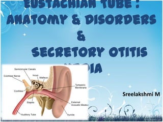
  • 1. Eustachian Tube : Anatomy & Disorders & Secretory Otitis Media Sreelakshmi M 1
  • 3. 3
  • 5. Lining of Eustachian Tube • Pseudostratified ciliated columnar epithelium interspersed with mucous secreting goblet cells • Submucosa of cartilagenous part rich in seromucinous gland • Cilia beat in direction of nasopharynx 5
  • 6. Nerve Supply • Sensory & parasympathetic : tympanic branch of glossopharyngeal N • Tensor veli palatini: V3 • Levator veli palatini pharyngeal plexus • Salpingopharyngeus (cranial part of XI N via vagus) 6
  • 7. Infant ET v/s Adult ET INFANT ADULT LENGTH 13-18 mm at birth 36 mm DIRECTION More horizontal Forms an angle of 45 with the horizontal ANGULATION AT ISTHMUS No angulation Angulation present BONY VERSUS Bony part> 1/3 of the total Bony part 1/3; cartilaginous CARTILAGINOUS PART length part2/3 TUBAL CARTILAGE flaccid Comparatively rigid DENSITY OF ELASTIN AT THE Less dense More dense HINGE OSTMANN’S PAD OF FAT Less in volume Large & helps to keep the tube closed 7
  • 8. 8
  • 9. Functions 1. Ventilation & regulation of ME pressure 2. Protective funtions – Nasopharyngeal sound pressure – Reflux of nasopharyngeal secretions 3. Clearance of ME secretions 9
  • 10. ET Function Tests • VALSALVA TEST – Principle: positive pressure in the nasopharynx causes air to enter the Eustachian tube 10
  • 11. – Tympanic membrane perforation- a hissing sound – Discharge in the middle ear- cracking sound – Only 65% of persons can do this test. – Contraindications: • Atrophic scar of tympanic membrane which can rupture • Infection of nose & nasopharynx 11
  • 12. • Politzer test – Done in children who are unable to perform valsalva test. – Olive shaped tip of the politzer’s bag is introduced into the patient’s nostril on the side of which the tubal function is desired to be tested – Other nostril closed & the bag compressed while at the same time the patient swallows or says “ik,ik,ik” 12
  • 13. – By means of an auscultation tube a hissing sound is heard. – Compressed air can also be used instead of politzer’s bag – Test is also therapeutically used to ventilate the middle ear. 13
  • 15. – Complications: • Injury to Eustachian tube opening • Bleeding from nose • Transmission of nasal & nasopharyngeal infection into middle ear • Rupture of atrophic area of tympanic membrane 15
  • 16. • Toynbee’s test – Uses negative pressure • Tympanometry (inflation-deflation test) – +Ve & -ve pressures are created in the external ear and the patient swallows repeatedly – in patients with perforated or intact tympanic membrane • Radiological Test • Saccharine/ Methylene blue Test – Saccharine solution – Methylene blue dye – Ear drops into ear with TM perforation • Sonotubometry 16
  • 18. Tubal Blockage ACUTE TUBAL BLOCKAGE ABSORPTION OF ME GASES -VE PRESSURE IN ME RETRACTION OF TM TRANSUDATE IN ME/HAEMORRHAGE PROLONGED TUBAL BLOCKAGE/DYSFUNCTION OME(THIN WATERY OR MUCOID DISCHARGE) ATELECTATIC EAR/PERFORATION RETRACTION POCKET/CHOLESTEATOMA EROSION OF INCUDOSTAPEDIAL JOINT 18
  • 19. mechani • intrinsic cal • Extrinsic Block functional •Collapse both 19
  • 20. • Symptoms of tubal occlusion – Otalgia – Hearing loss – Popping sensation – Tinnitus – Disturbances of equilibrium • Signs of tubal occlusion – Retracted TM – Congestion along the handle of malleus and pars tensa – Transudate behind TM 20
  • 21. • Clinical causes of ET obstruction – Upper respiratory tract infection – Allergy – Sinusitis – Nasal polypi – DNS – Hypertrophic adenoids – Nasopharyngeal tumour/ mass – Cleft palate – Submucous cleft palate – Down’s syndrome 21
  • 22. Adenoids • Adenoids cause tubal dysfunction by: – Mechanical obstruction of the tubal opening – Acting as reservoir for pathogenic organisms – Inflammatory mediators in allergy cause tubal blockage • Adenoids can cause otitis media with effusion or recurrent acute otitis media • Adenoidectomy 22
  • 23. 23
  • 25. Cleft palate • Tubal dysfunction due to: – Abnormalities of torus tubaris – Tensor veli palatini doe not insert into the torus tubaris • Otitis media with effusion is common in these patients 25
  • 26. Down’s syndrome • Dysfunction due to: – Poor tone of tensor veli palatini – Abnormal shape of nasopharynx 26
  • 28. • Any obstruction in the ventilation pathway retraction pockets or atelectasis of tympanic membrane – Obstruction of Eustachian tube  total atelectasis of tm – Obstruction at additus  cholesterol granuloma & collection of mucoid discharge in mastoid air cells 28
  • 29. 29
  • 30. • Other changes – Thin atrophic TM – Cholesteatoma – Ossicular necrosis – Tympanosclerotic changes • Management – Repair of irreversible pathologic processes – Establishment of ventilation 30
  • 31. 31
  • 32. Patulous Eustachian Tube • ET is abnormally patent • Causes: – Idiopathic, rapid weight loss, pregnancy (esp 3rd trim) & multiple sclerosis • Chief complaints – Autophony, hearing his own breath sounds • Pressure changes in the nasopharynx are easily transmitted to the ME • Movements of the TM can be seen with inspiration & expiration 32
  • 33. • Management – Acute cases Usually self-limiting – Weight gain & oral administration of KI – Long standing cases = cauterisation/ insertion of grommet 33
  • 34. EXAMINATION OF EUSTACHIAN TUBE Pharyngeal end of eustachian tube :posterior rhinoscopy, rigid nasal endoscope or flexible nasopharyngoscope Tympanic end :microscope or endoscope Simple examination of TM may reveal retraction pockets or fluid in the me Movements of TM with respiration point to patulous eustachian tube 34
  • 35. • Aetiologic causes of eustachian tube dysfunction assessed through: – Nasal examination – Endoscopy – Tests of allergy – CT scan of temporal bones – MRI to exclude multiple sclerosis 35
  • 36. Otitis Media with Effusion 36
  • 37. Serous otitis media Secretory otitis media Mucoid otitis media “Glue Ear” 37
  • 38. • Insidious condition characterized by accumulation of non purulent effusion in ME cleft • Effusion is thick & viscid. • Fluid is sterile 38
  • 39. Pathogenesis • Malfunctioning of Eustachian tube • Increased secretory activity of ME mucosa 39
  • 40. Aetiology 1. Malfunctioning of Eustachian tube – Adenoid hyperplasia – Chronic rhinosinusitis – Chronic tonsillitis – Tumors ( to be excluded in unilateral ser. OM in adults) 2. Allergy 3. Unresolved otitis media 4. Viral infections 40
  • 41. Clinical Features Symptoms : affects 5-8 yrs age gp Hearing loss Delayed & defective speech Mild earaches 41
  • 42. Otoscopic Findings – Dull & opaque TM – Loss of light reflex – TM: yellow grey or bluish – Fluid level & air bubbles may be seen – Restricted mobility of tm – Thin leash of vessels along malleus handle/ periphery of TM == differentiate from acute supp. Otitis media – TM: varying degree of retraction 42
  • 43. 43
  • 44. 44
  • 45. Hearing Tests • Tuning fork test-conductive hearing loss • Audiometry-conductive hearing loss of 20-40db • Impedance Audiometry-reduced compliance indicates presence of fluid • X-ray mastoid-clouding of air cells due to fluid. 45
  • 46. Treatment Medical Surgical Decongestants  Myringotomy & Aspiration  Grommet Insertion Antihistaminics  Tympanotomy/ cortical Steroids mastoidectomy( loculated thick fluid/ chol. granuloma) Antibiotics  Surgical treatment of causative factor 46
  • 47. 47
  • 48. 48
  • 49. Sequelae of Chronic Secretory Otitis Media • Atrophic TM & atelectasis of ME • Ossicular necrosis • Tympanosclerosis • Retraction pockets & cholesteatoma • Cholesterol granuloma 49
  • 50. The above picture shows a very thin or atelectatic eardrum (tympanic membrane) 50 which is draped over the promontory and round window nitch.
  • 52. Thank You 52