SlideShare a Scribd company logo
1 of 65
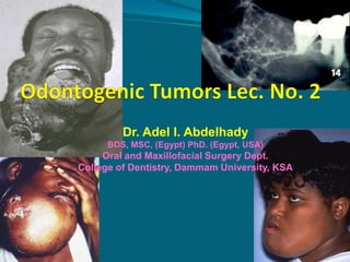
Dr. Adel I. Abdelhady
BDS, MSC, (Egypt) PhD. (Egypt, USA)

Oral and Maxillofacial Surgery Dept.
College of Dentistry, Dammam University, KSA
 By the end of the lecture(s) Students should display a






knowledge, information and understanding of :
1.Classification the odontogenic tumors
2.familiar with the different types of odontogenic tumor
according to its origin
3.Examine and diagnose patient’s complain from facial
swelling
4. Establish a Differential Diagnosis of the mandibular as will
as maxillary swelling
5. Differentiate between different types of tumor’s biological
behaviors and accordingly select the suitable management
technique (s)
Cementoblastoma
 This tumor typically occurs

around the roots of the lower
posterior teeth.
 Like virtually all odontogenic
tumors, it is benign but it
expands the jaw, causes pain and
requires surgical removal.
Radiographically it appears as a
ball of dense material attached to
the end of the root
(Central) Odontogenic Fibroma
Clinical Features
 Fewer than 50 cases have been reported in the
English literature.
 Patient Age: Patients have ranged in age from 980 years old with a mean of 40 years.
 Gender Predilection: Females, 7.4 : 1 in one study.
 Location: 60% Sixty percent occur in the maxilla
where most are located anterior to the first molar.
When in the mandible, approximately 50 % occur
in the posterior jaw.
Odontogenic Fibroma: Radiographic
Appearance
 The odontogenic fibroma usually appears as a well-

defined, unilocular radiolucency. It is often
associated with the apical area of an erupted tooth.
 Larger lesions are often multilocular.
 Many odontogenic fibromas have sclerotic borders.
root resorption is common.
Odontogenic Fibroma: Additional Features
 Small odontogenic fibromas are usually asymptomatic.
 The larger lesions may be associated with localized

bony expansion of the jaw or with the loosening of
adjacent teeth.
Odontogenic Fibroma: Histologic Features
 Some authors have described two separate types of
odontogenic fibromas.
 The simple odontogenic fibroma is composed
of stellate fibroblasts arranged in a whorled

pattern with fine collagen fibrils and a lot of
ground substance.
 Foci of odontogenic epithelium may or may not be
present.
 Occasionally, foci of dystrophic calcification may
be present.
Odontogenic Fibroma: Histological Features
 The WHO type odontogenic fibroma appears as a

fairly cellular fibrous connective tissue with collagen
fibers arranged in interlacing bundles.
 Odontogenic epithelium in the form of long strands or
isolated nests is present throughout the lesion.
 Calcifications composed of cementoid and/or
dentinoid may be present.
Odontogenic Fibroma: Treatment and
Prognosis
 The odontogenic fibroma is usually treated

by enucleation and curettage.
 There have been few recurrences, this the
prognosis is good.
Mixed Odontogenic Tumors
 Ameloblastic fibroma, ameloblastic fibrodentinoma,





ameloblastic fibro-odontoma, odontoma
Both epithelial and mesenchymal cells
Mimic differentiation of developing tooth
Treatment – enucleation, thorough curettage with
extraction of impacted tooth
Ameloblastic fibrosarcomas – malignant, treat with
aggressive en bloc resection
Related Jaw Lesions
 Giant Cell Lesions
 Central giant cell

 Fibroosseous lesions
 Fibrous dysplasia

granuloma
 Ossifying fibroma
 Brown tumor
 Condensing Osteitis
 Aneurysmal bone cyst
Central Giant Cell Granuloma
 This is a neoplastic-like reactive proliferation of

the jaws that accounts for less than 7% of all
benign lesions of the jaws in tooth-bearing
areas.
 It commonly occurs in children and young
adults with a slight female predilection.
 The lesion is more common in the mandible
than maxilla underlying anterior or premolar
teeth. Expansile lesions can cause root
divergence or resorption.
Central Giant Cell Granuloma
 The clinical features:

 Vary according to the type of development the lesion
assumes. Lesions may be slow-growing and
asymptomatic or rapidly expanding with pain, facial
swelling and root resorption.
 The fast growing variants have a high rate of
recurrence.
 Because of the higher incidence of these lesions
among girls and women of child-bearing years,
hormonal influences have been suggested as
influential in their development.
 Intraosseous destructive lesions of the








jaws
Far less common than peripheral giant
cell granuloma
10-30 yrs of age; Female > Male
Mandible > Maxilla; Ant. > Post.
Mandibular lesions frequently cross the
midline
Asymptomatic or painless expansion
Non-aggressive and aggressive lesions
Perforation of the cortical plates and
resorption of roots
Central Giant Cell Granuloma
 Neoplastic -like reactive proliferation

 Common in children and young adults
 Males > Females (hormonal?)
 Maxilla >Mandible
 Expansile lesions – root resorption
 Slow-growing – asymptomatic swelling
 Rapid-growing – pain, loose dentition (high rate of

recurrence)
Central Giant Cell Granuloma
 Radiographic findings
 Aggressive lesions show cortical perforation and root
 resorption
 Unilocular, multilocular radiolucencies
 Well-defined or irregular borders

 Histology
 Multinucleated giant cells, dispersed throughout a

fibrovascular stroma
Central Giant Cell Granuloma
Central Giant Cell Lesion
CGCL

 75% before the age of 30; female> male

 Anterior mandible to first molar; can cross the midline
 Non-aggressive vs aggressive:
 Aggressive lesions: larger on presentation, painful,

rapid growth, root resorption, cortical perforation,
younger patients
 Recurrence: 75% in aggressive lesions (11% in nonaggressive)
 Questionable minor histological differences
Central Giant Cell Granuloma
 Treatment
 Curettage, segmental resection
 Radiation – out of favor (risk of sarcoma)
 Intralesional steroids – younger patients, very

large lesions
 Individualized treatment depending on
characteristics and location of tumor
Brown tumors of
hyperparathyroidism
 Brown tumors are tumors of bone that arise in settings of

excess osteoclast activity, such as hyperparathyroidism and
consist of fibrous tissue, woven bone and supporting
vasculature, but no matrix. They are radiolucent on x-ray.
The osteoclasts consume the trabecular bone that
osteoblasts lay down and this front of reparative bone
deposition followed by additional resorption can expand
beyond the usual shape of the bone, involving the
periosteum thus causing bone pain. The characteristic
brown coloration results from hemosiderin deposition into
the osteolytic cysts. Also characteristic of giant cell tumors
of the bone.
 Brown tumor, an uncommon focal giant-cell lesion, arises

as a direct result of the effect of parathyroid hormone on
bone tissue in patients with hyperparathyroidism
 Laboratory evaluation revealed that the patient had
primary hyperparathyroidism, parathyroid hormone level
was 988 pg/ml (normal: 12 to 72).
 Other laboratory measurements were total serum calcium,
11.2 mg/dl (normal: 8.8 to 11.0); phosphorus, 2.0 mg/dl
(normal: 2.5 to 4.8); and alkaline phosphatase, 145 U/L
(normal: 32 to 104).
 Ultrasonography of the neck revealed an enlargement of
the lower left parathyroid gland (2.1 x 1.4 x 0.8 cm). The
finding of hyperparathyroidism confirmed the diagnosis of
brown tumor.
Brown Tumor
 Local manifestation of hyperparathyroidism
 Histologically identical to CGCG
 Serum calcium and phosphorus

 More likely in older patients
Cherubism
 Definition
 Genetic disorder characterized by

bilateral mandibular and maxillary
intraosseous fibrous swellings in
young individuals with autosomal
dominant inheritance.
Indistinguishable histologically
from central giant cell granuloma
 bilateral mandibular and maxillary
involvement
 young
 Histopathology
 Large number of osteoclast-like, multinucleated giant

cells
 Indistinguishable microscopically from central giant
cell granuloma
 sometimes more delicate fibrovascular stroma without

bone formation

 Differential Diagnosis
 Select up to 2 differential diagnoses to compare with

Cherubism
 Central Giant Cell Granuloma [Mandible and Maxilla]
 Primary Hyperparathyroidism
 Ossifying Fibroma
DD
 Large number of osteoclast-like, multinucleated giant

cells also in:
 central giant cell granuloma
 giant cell tumor (osteoclastoma)

 fibro-osseous lesions
 bone lesion of hyperparathyroidism
 aneurysmal bone cyst
Fibrous Dysplasia
 Monostotic vs. polystotic
 Monostotic
 More common in jaws
and cranium
 Polystotic
 McCune-Albright’s
syndrome
 Cutaneous pigmentation,
hyper-functioning
endocrine glands,
precocious puberty
Fibrous Dysplasia
 Painless expansile dysplastic process of






osteoprogenitor connective tissue
Site :Maxilla most common
Does not typically cross midline (one bone)
Antrum obliterated, orbital floor involvement
(globe displacement)
Radiology : ground-glass appearance
Fibrous dysplasia
 Fibrous dysplasia is a condition in which normal
 medullary bone is replaced by an abnormal fibrous
 connective tissue proliferation in which new,

nonmaturing bone is formed.
 A genetic defect involving Gs-alpha proteins appears to
underlie this process.
Etiology and Pathogenesis.
 The nature of this condition has not been firmly

established.
 The name given to fibrous dysplasia was originally
intended to indicate that the condition represented a
dysplastic growth resulting from derange
mesenchymal cell activity or a defect in the control of
bone cell activity.
 This genetic alteration may ultimately affect the
proliferation and differentiation of fibroblasts/
osteoblasts that make up these lesions.
Clinical Features.
 This disease most commonly

 The most commonly

presents as an asymptomatic, slow
enlargement of the involved bone.
 Fibrous dysplasia may involve a
single bone or several bones
concomitantly.
 Monostotic fibrous dysplasia is the
designation used to describe the
process in one bone.
 Polyostotic fibrous dysplasia applies
to cases in which more than one
bone is involved. McCune-Albright
syndrome consists of polyostotic
fibrous dysplasia, cutaneous
melanotic pigmentations (cafe-aulait macules), and endocrine
abnormalities.

reported endocrine
disorder consists of
precocious sexual
development in girls.
Acromegaly,
hyperthyroidism,
hyperparathyroidism, and
hyperprolactinemia have
also been described










Monostotic fibrous dysplasia is much more common
than the polyostotic form, accounting for as many as
80% of cases. Jaw involvement is common in this form
of the disease. Other bones that are commonly affected
are the ribs and femur.
Fibrous dysplasia occurs more often in the maxilla than
in the mandible
Maxillary lesions may extend to involve the maxillary
sinus, zygoma , sphenoid bone, and floor of the orbit.
This form of the disease, with involvement of several
adjacent bones, has been referred to as craniofacial
fibrous dysplasia.
The most common site of occurrence with mandibular
involvement is in the body portion.
 The slow, progressive enlargement of the affected

jaw is usually painless and typically presents as a
unilateral swelling.
 As the lesion grows, facial asymmetry becomes evident
and may be the initial presenting complaint.
 The dental arch is generally maintained, although
displacement of teeth, malocclusion, and interference
with tooth eruption may occasionally occur.
 Tooth mobility is not seen
 Age:
 This condition characteristically has its onset during

the first or second decade of life. Rarely, the lesion
presents later in life, although this may only reflect
the insidious, asymptomatic nature of fibrous
dysplasia.
 Gender :
 Monostotic fibrous dysplasia generally exhibits an
equal gender distribution, and the polyostotic form
tends to occur more commonly in females.
 Fibrous dysplasia has available radiographic appearance
 that ranges from a radiolucent lesion to a uniformly

radiopaque mass
 The classic lesion has been described as having a
radiopaque change that imparts a "ground glass" effect
 This characteristic image, which is most identifiable on
intraoral radiographs, is not, however, pathognomonic.
 Lesions of fibrous dysplasia may also present as
unilocular or multilocular radiolucencies, especially in
long bones.
 An important distinguishing feature of fibrous

dysplasia is the poorly defined radiographic and
clinical margins of the lesion. The process appears to
blend into the surrounding normal bone without
evidence of a circumscribed border.
 Laboratory values for patients with monostotic
fibrous dysplasia, specifically serum calcium,
phosphorus, and alkaline phosphatase, are usually
within normal
 However, these serum chemistry markers may be
altered in patients with McCune-Albright syndrome
Histopathology
 Fibrous dysplasia consists of a slight to moderate

cellular fibrous connective tissue stroma that
contains foci of irregularly shaped trabcculae of
immature bone
 A relatively constant ratio of fibrous tissue to bone
throughout a given lesion is characteristic.
Treatment and Prognosis
 Deferred, if possible until skeletal maturity
 Quarterly clinical and radiographic f/u
 If quiescent – contour excision (cosmesis or

function)
 Accelerated growth or disabling functional
impairment - surgical intervention
Treatment and Prognosis
 After a variable period of pre-pubertal






growth, fibrous dysplasia characteristically stabilizes,
although a slow advance may be noted into adulthood.
Small lesions may therefore require no treatment other
than biopsy confirmation and periodic follow-up.
Large lesions that have caused cosmetic or functional
deformity may be treated by surgical recontouring.
This procedure is generally deferred until after stabilization
of the disease process.
En bloc resections for complete removal are impractical
and unnecessary, because the lesions arc relatively large
and poorly delineated.
 Malignant transformation is a rare complication of
 fibrous dysplasia {fewer than 1% of cases) that has

been described, usually in patients with the
polyostotic type.
 Many of the patients reported on were treated with
radiation therapy, suggesting a role for radiation
in the transformation process, although malignant
change has been documented in the absence of
radiation treatment.
Fibrous Dysplasia
Clinical Features
 Age: Ossifying fibroma is an uncommon lesion that tends








to occur during the third and fourth decades of life
Sex :in women more than men. It is a slow-growing,
asymptomatic, and expansile lesion.
In the head and neck, ossifying fibroma may be seen in
the jaws and craniofacial bones.
Site: Lesions of the jaws characteristically arise in the
tooth-bearing regions, most often in the mandibular
premolar-molar area
The slow but persistent growth of the tumor may
ultimately produce expansion and thinning of the buccal
and lingual cortical plates, although perforation and
mucosal ulceration are rare
Most of these lesions are solitary
 Ossifying fibromas of the jaw are wellcircumscribed, slowly growing lesions. They are

often mentioned in the same differential diagnosis
as fibrous dysplasia, but it is important to make the
distinction because the former lends itself to ready
enucleation, while the latter can be admixed with
surrounding tissues, making surgery more
complicated.
 Patients generally present with a history of a painless

expansion of a tooth-bearing portion of the mandible.
Lesions of the maxilla are also encountered, but they
are less common.
 Radiographically, the lesions are typically 1 to 5 cm at
their greatest dimension. Well-defined areas of
osteolysis are noted radiographically, with varying
degrees of calcification and cortical thinning.
 These lesions can often be readily identified at the
time of surgery by noting the case with which they can
be separated from surrounding tissue.
radiographic features
 The most important

radiographic feature of
this lesion is the wellcircumscribed, sharply
defined border. Ossifying
fibroma otherwise present a
 variable appearance,
depending on the density of
 calcifications present.
 Lesions may be relatively
radiolucent because of evenly
dispersed, calcified new
bone.

 Lesions may also appear as

unilocular or multilocular
radiolucencies that bear a
resemblance to odontogenic
lesions.
 A mixed radiolucentradiopaque image is seen
when islands of tumor bone
are densely calcified.
 The roots of teeth may be
displaced and, less commonly
tooth resorption is seen.
 A variant of ossifying fibroma, juvenile (aggressive)

ossifying fibroma, has been described in children and
young adults .Most affected individuals are younger
than 15 years of age.
 This lesion most commonly involves the paranasal
sinuses and periorbital bones, where it may cause
exophthalmoses, proptosis , sinusitis, and nasal
symptoms.
 This rare tumor behaves in a more aggressive fashion
than does ossifying fibroma, and it may require more
extensive surgery when encountered.
 Microscopically, juvenile ossifying fibroma is highly
cellular and contains trabeculae or spheroids of new
bone.
 Cementifying fibroma, and cemento-ossifying
fibroma, are terms occasionally used when the bony
islands in these lesions have a round or spheroidal

shape.
 These tumors occur in similar age-groups and
locations, exhibit comparable clinical characteristics,
and have the same biologic behavior.
 They are, for all practical purposes, the same lesion as
ossifying fibroma.
 Other differential considerations are osteoblastoma,

focal cementoosseous dysplasia, and focal
osteomyelitis.
 Osteoblastoma is evident in a slightly younger agegroup and is often characterized by pain.
In addition, osseous trabeculae in these lesions are
rimmed by abundant plump osteoblasts, and a central
nidus may be evident.
 Periapical cemento-osseous dysplasia in posterior
teeth may appear radiographically similar and require
a biopsy to separate it from ossifying fibroma.
 Focal osteomyelitis is associated with a source of
inflammation and is possibly accompanied by pain
and swelling.
Treatment And Prognosis.
 Treatment of ossifying fibroma : is most often

accomplished by surgical removal using CURETTAGE
OR ENUCLEATION.
 The lesion can typically be separated easily from the
surrounding normal bone.
 Recurrence is described only rarely after removal.
Differential Diagnosis.
 The primary differential consideration

for fibrous dysplasia of the jaws is ossifying fibroma.
 As previously noted, clinical, radiographic, and
microscopic features must be considered together
in order to distinguish these processes.
 The well circumscribed ossifying fibroma as compared
with the diffuse fibrous dysplasia often serves as the
differentiating factor.
 Chronic osteomyelitis may occasionally mimic the

radiographic appearance of fibrous dysplasia.
 Inflammation, often mild, is present in osteomyelitis

and may be accompanied by symptoms that include

tenderness, pain, or drainage. The slowly progressive,
asymptomatic nature of fibrous dysplasia usually
allows differentiation from malignant tumors of bone.
Ossifying Fibroma
 True neoplasm of medullary jaws

 Elements of periodontal ligament
 Younger patients, premolar – mandible
 Frequently grow to expand jaw bone
 Radiology
 radiolucent lesion early, well-demarcated
 Progressive calcification (radiopaque – 6 yrs)
Ossifying Fibroma
 Distinguishing between









 Historically, differentiating

ossifying fibroma and fibrous
dysplasia is the primary
diagnostic challenge.
Both lesions may exhibit similar 
clinical, radiographic, and
microscopic features.
The most helpful feature in
distinguishing the two is the well
circumscribed radiographic

appearance of ossifying fibroma
and the ease with which it can be
separated from normal bone.
In most cases the well defined
appearance of ossifying fibroma
is evident radiographically.

the two lesions was based
primarily on histologic
criteria.
Fibrous dysplasia was
reported to contain only
woven bone, without
evidence of osteoblastic
rimming of bone.
The presence of more mature
lamellar bone was believed to
be characteristic of ossifying
fibroma
Types of Jaw Tumors and Primary Treatment Modalities
Odontogenic tumors II

More Related Content

What's hot

What's hot (20)

Leukemia and oral manifestations
Leukemia and oral manifestationsLeukemia and oral manifestations
Leukemia and oral manifestations
 
"GINGIVAL-ENLARGEMENT"
"GINGIVAL-ENLARGEMENT""GINGIVAL-ENLARGEMENT"
"GINGIVAL-ENLARGEMENT"
 
Oral Medicine :Burning mouth syndrome
Oral Medicine :Burning mouth syndromeOral Medicine :Burning mouth syndrome
Oral Medicine :Burning mouth syndrome
 
Pindborgs Tumour
Pindborgs TumourPindborgs Tumour
Pindborgs Tumour
 
Odontogeniccysts OKC
Odontogeniccysts OKCOdontogeniccysts OKC
Odontogeniccysts OKC
 
cysts of the oral and maxillofacial region
cysts of the oral and maxillofacial regioncysts of the oral and maxillofacial region
cysts of the oral and maxillofacial region
 
Diagnosis of cysts in oral cavity
Diagnosis of cysts in oral cavityDiagnosis of cysts in oral cavity
Diagnosis of cysts in oral cavity
 
SLOB Technique
SLOB TechniqueSLOB Technique
SLOB Technique
 
PULP AND PERIAPICAL LESIONS OF THE TOOTH ppt
PULP AND PERIAPICAL LESIONS OF THE TOOTH pptPULP AND PERIAPICAL LESIONS OF THE TOOTH ppt
PULP AND PERIAPICAL LESIONS OF THE TOOTH ppt
 
Adenomatoid odontogenic tumor
Adenomatoid odontogenic tumorAdenomatoid odontogenic tumor
Adenomatoid odontogenic tumor
 
Oral White lesions
Oral White lesionsOral White lesions
Oral White lesions
 
calcifying odontogenic cyst
calcifying odontogenic cyst calcifying odontogenic cyst
calcifying odontogenic cyst
 
Pyogenic Granuloma
Pyogenic GranulomaPyogenic Granuloma
Pyogenic Granuloma
 
Odontogenic Myxoma
Odontogenic MyxomaOdontogenic Myxoma
Odontogenic Myxoma
 
Oral pyogenic granuloma
Oral pyogenic granulomaOral pyogenic granuloma
Oral pyogenic granuloma
 
Central Ossifying Fibroma
Central Ossifying FibromaCentral Ossifying Fibroma
Central Ossifying Fibroma
 
Amelogenesis Imperfecta
Amelogenesis ImperfectaAmelogenesis Imperfecta
Amelogenesis Imperfecta
 
Inflammatory lesions
Inflammatory lesionsInflammatory lesions
Inflammatory lesions
 
Traumatic fibroma made easy
Traumatic fibroma  made easy Traumatic fibroma  made easy
Traumatic fibroma made easy
 
Giant cell lesion’s of jaw
Giant cell lesion’s of jawGiant cell lesion’s of jaw
Giant cell lesion’s of jaw
 

Viewers also liked

Ossifying fibroma vs fibrous dysplasia of the jaw/rotary endodontic courses b...
Ossifying fibroma vs fibrous dysplasia of the jaw/rotary endodontic courses b...Ossifying fibroma vs fibrous dysplasia of the jaw/rotary endodontic courses b...
Ossifying fibroma vs fibrous dysplasia of the jaw/rotary endodontic courses b...Indian dental academy
 
Cementoblastoma
CementoblastomaCementoblastoma
CementoblastomaLaTia Tuca
 
Fibro osseous lesions of jaw
Fibro osseous lesions of jawFibro osseous lesions of jaw
Fibro osseous lesions of jawShivani Shivu
 
Peripheral giant cell granuloma (giant cell epulis
Peripheral giant cell granuloma (giant cell epulisPeripheral giant cell granuloma (giant cell epulis
Peripheral giant cell granuloma (giant cell epulisKhin Soe
 
CENTRAL GIANT CELL GRANULOMA
CENTRAL GIANT CELL GRANULOMACENTRAL GIANT CELL GRANULOMA
CENTRAL GIANT CELL GRANULOMADr Faraz Mohammed
 
Pathology of fibro osseous lesions
Pathology of fibro osseous lesionsPathology of fibro osseous lesions
Pathology of fibro osseous lesionsMd Roohia
 
Juvenile aggressive ossifying fibroma /prosthodontic course
Juvenile aggressive ossifying fibroma /prosthodontic course   Juvenile aggressive ossifying fibroma /prosthodontic course
Juvenile aggressive ossifying fibroma /prosthodontic course Indian dental academy
 
Paget's disease of bone -
Paget's disease of bone - Paget's disease of bone -
Paget's disease of bone - vinod naneria
 
Odontogenic Tumors
Odontogenic TumorsOdontogenic Tumors
Odontogenic TumorsIAU Dent
 
Casos clinicos d td un poco
Casos clinicos d td un pocoCasos clinicos d td un poco
Casos clinicos d td un poco4paulo74
 
Freud and fight club
Freud and fight clubFreud and fight club
Freud and fight clublgoodhew
 
Human papillomavirus in the oral cavity of children
Human papillomavirus in the oral cavity of childrenHuman papillomavirus in the oral cavity of children
Human papillomavirus in the oral cavity of childrenGaurav Salunkhe
 
Tumor of oral cavity
Tumor of oral cavityTumor of oral cavity
Tumor of oral cavityshekhar star
 
Odontogenic tumours part 3
Odontogenic tumours part 3Odontogenic tumours part 3
Odontogenic tumours part 3Ali Tahir
 
Fibro osseous lesion
Fibro osseous lesionFibro osseous lesion
Fibro osseous lesionmemoalawad
 

Viewers also liked (20)

Ossifying fibroma vs fibrous dysplasia of the jaw/rotary endodontic courses b...
Ossifying fibroma vs fibrous dysplasia of the jaw/rotary endodontic courses b...Ossifying fibroma vs fibrous dysplasia of the jaw/rotary endodontic courses b...
Ossifying fibroma vs fibrous dysplasia of the jaw/rotary endodontic courses b...
 
Cementoblastoma
CementoblastomaCementoblastoma
Cementoblastoma
 
Fibro osseous lesions of jaw
Fibro osseous lesions of jawFibro osseous lesions of jaw
Fibro osseous lesions of jaw
 
Peripheral giant cell granuloma (giant cell epulis
Peripheral giant cell granuloma (giant cell epulisPeripheral giant cell granuloma (giant cell epulis
Peripheral giant cell granuloma (giant cell epulis
 
CENTRAL GIANT CELL GRANULOMA
CENTRAL GIANT CELL GRANULOMACENTRAL GIANT CELL GRANULOMA
CENTRAL GIANT CELL GRANULOMA
 
Pathology of fibro osseous lesions
Pathology of fibro osseous lesionsPathology of fibro osseous lesions
Pathology of fibro osseous lesions
 
Juvenile aggressive ossifying fibroma /prosthodontic course
Juvenile aggressive ossifying fibroma /prosthodontic course   Juvenile aggressive ossifying fibroma /prosthodontic course
Juvenile aggressive ossifying fibroma /prosthodontic course
 
Odontogenic tumors
Odontogenic tumorsOdontogenic tumors
Odontogenic tumors
 
Paget's disease of bone -
Paget's disease of bone - Paget's disease of bone -
Paget's disease of bone -
 
Odontogenic Tumors
Odontogenic TumorsOdontogenic Tumors
Odontogenic Tumors
 
Pul circulation
Pul circulationPul circulation
Pul circulation
 
Xpo pato odontomas
Xpo pato odontomasXpo pato odontomas
Xpo pato odontomas
 
Odontogenic tumors
Odontogenic tumorsOdontogenic tumors
Odontogenic tumors
 
Casos clinicos d td un poco
Casos clinicos d td un pocoCasos clinicos d td un poco
Casos clinicos d td un poco
 
Freud and fight club
Freud and fight clubFreud and fight club
Freud and fight club
 
Human papillomavirus in the oral cavity of children
Human papillomavirus in the oral cavity of childrenHuman papillomavirus in the oral cavity of children
Human papillomavirus in the oral cavity of children
 
Tumor of oral cavity
Tumor of oral cavityTumor of oral cavity
Tumor of oral cavity
 
Displacia osteocementaria florida
Displacia osteocementaria floridaDisplacia osteocementaria florida
Displacia osteocementaria florida
 
Odontogenic tumours part 3
Odontogenic tumours part 3Odontogenic tumours part 3
Odontogenic tumours part 3
 
Fibro osseous lesion
Fibro osseous lesionFibro osseous lesion
Fibro osseous lesion
 

Similar to Odontogenic tumors II

Primary bone tumors
Primary bone tumorsPrimary bone tumors
Primary bone tumorsDpt Memon
 
Odontogenic and Nonodontogenic Tumors of the Jaws
Odontogenic and Nonodontogenic Tumors of the JawsOdontogenic and Nonodontogenic Tumors of the Jaws
Odontogenic and Nonodontogenic Tumors of the JawsAndres Cardona
 
Odontogenic Tumors
Odontogenic TumorsOdontogenic Tumors
Odontogenic TumorsDrMuqheem
 
Giant Cell Lesions
Giant Cell LesionsGiant Cell Lesions
Giant Cell LesionsMsccMohamed
 
bone tumors 2.ppt
bone tumors 2.pptbone tumors 2.ppt
bone tumors 2.pptdrqazi7777
 
Benign connective tissue tumors 2/ dental implant courses by Indian dental ac...
Benign connective tissue tumors 2/ dental implant courses by Indian dental ac...Benign connective tissue tumors 2/ dental implant courses by Indian dental ac...
Benign connective tissue tumors 2/ dental implant courses by Indian dental ac...Indian dental academy
 
Radiological and pathological correlation of bone tumours Dr.Argha Baruah
Radiological and pathological correlation of bone tumours  Dr.Argha BaruahRadiological and pathological correlation of bone tumours  Dr.Argha Baruah
Radiological and pathological correlation of bone tumours Dr.Argha BaruahArgha Baruah
 
D. Firas lecture minimum muhadharaty require
D. Firas lecture minimum muhadharaty requireD. Firas lecture minimum muhadharaty require
D. Firas lecture minimum muhadharaty requirehussainAltaher
 
Benign connective tissue jaw tumors rl
Benign connective tissue jaw tumors rlBenign connective tissue jaw tumors rl
Benign connective tissue jaw tumors rlSsemagandaAddinan
 
Gaint cell lesions of bone/oral surgery courses by indian dental academy
Gaint cell lesions of bone/oral surgery courses by indian dental academyGaint cell lesions of bone/oral surgery courses by indian dental academy
Gaint cell lesions of bone/oral surgery courses by indian dental academyIndian dental academy
 
benign bone tumors contd...
benign bone tumors contd...benign bone tumors contd...
benign bone tumors contd...Anand Dev
 

Similar to Odontogenic tumors II (20)

Primary bone tumors
Primary bone tumorsPrimary bone tumors
Primary bone tumors
 
Odontogenic and Nonodontogenic Tumors of the Jaws
Odontogenic and Nonodontogenic Tumors of the JawsOdontogenic and Nonodontogenic Tumors of the Jaws
Odontogenic and Nonodontogenic Tumors of the Jaws
 
Odontogenic Tumors
Odontogenic TumorsOdontogenic Tumors
Odontogenic Tumors
 
Bone tumor dr patnaik
Bone tumor dr patnaikBone tumor dr patnaik
Bone tumor dr patnaik
 
Giant Cell Lesions
Giant Cell LesionsGiant Cell Lesions
Giant Cell Lesions
 
bone tumors 2.ppt
bone tumors 2.pptbone tumors 2.ppt
bone tumors 2.ppt
 
Tumors of bone
Tumors of boneTumors of bone
Tumors of bone
 
Benign connective tissue tumors 2/ dental implant courses by Indian dental ac...
Benign connective tissue tumors 2/ dental implant courses by Indian dental ac...Benign connective tissue tumors 2/ dental implant courses by Indian dental ac...
Benign connective tissue tumors 2/ dental implant courses by Indian dental ac...
 
FOLs
FOLs FOLs
FOLs
 
Radiological and pathological correlation of bone tumours Dr.Argha Baruah
Radiological and pathological correlation of bone tumours  Dr.Argha BaruahRadiological and pathological correlation of bone tumours  Dr.Argha Baruah
Radiological and pathological correlation of bone tumours Dr.Argha Baruah
 
D. Firas lecture minimum muhadharaty require
D. Firas lecture minimum muhadharaty requireD. Firas lecture minimum muhadharaty require
D. Firas lecture minimum muhadharaty require
 
Osteosarcoma ppt
Osteosarcoma pptOsteosarcoma ppt
Osteosarcoma ppt
 
Benign connective tissue jaw tumors rl
Benign connective tissue jaw tumors rlBenign connective tissue jaw tumors rl
Benign connective tissue jaw tumors rl
 
Jaw bone disaese ii
Jaw bone disaese iiJaw bone disaese ii
Jaw bone disaese ii
 
Gaint cell lesions of bone/oral surgery courses by indian dental academy
Gaint cell lesions of bone/oral surgery courses by indian dental academyGaint cell lesions of bone/oral surgery courses by indian dental academy
Gaint cell lesions of bone/oral surgery courses by indian dental academy
 
Gaint cell lesions of bone
Gaint cell lesions of boneGaint cell lesions of bone
Gaint cell lesions of bone
 
giant_cell_lesions.pptx
giant_cell_lesions.pptxgiant_cell_lesions.pptx
giant_cell_lesions.pptx
 
Sclerotic
ScleroticSclerotic
Sclerotic
 
benign bone tumors contd...
benign bone tumors contd...benign bone tumors contd...
benign bone tumors contd...
 
mixed tumors.pptx
mixed tumors.pptxmixed tumors.pptx
mixed tumors.pptx
 

More from IAU Dent

Odontogenic Infection
Odontogenic InfectionOdontogenic Infection
Odontogenic InfectionIAU Dent
 
Maxillofacial injuries
Maxillofacial injuriesMaxillofacial injuries
Maxillofacial injuriesIAU Dent
 
Impacted teeth
Impacted teethImpacted teeth
Impacted teethIAU Dent
 
Odontogenic Cysts
Odontogenic CystsOdontogenic Cysts
Odontogenic CystsIAU Dent
 
Chronic gingivitis
Chronic gingivitisChronic gingivitis
Chronic gingivitisIAU Dent
 
Plaque control
Plaque controlPlaque control
Plaque controlIAU Dent
 
8. hypotension & hypertension
8. hypotension & hypertension8. hypotension & hypertension
8. hypotension & hypertensionIAU Dent
 
8. Prescription Writing
8. Prescription Writing8. Prescription Writing
8. Prescription WritingIAU Dent
 
7. Adrenocorticosteriods
7. Adrenocorticosteriods7. Adrenocorticosteriods
7. AdrenocorticosteriodsIAU Dent
 
7.a. histamine & antihistaminics
7.a. histamine & antihistaminics7.a. histamine & antihistaminics
7.a. histamine & antihistaminicsIAU Dent
 
8 anticancer drugs
8  anticancer drugs8  anticancer drugs
8 anticancer drugsIAU Dent
 
7 antibiotic-dental
7 antibiotic-dental7 antibiotic-dental
7 antibiotic-dentalIAU Dent
 
7.b. sedative hypnotics
7.b. sedative hypnotics 7.b. sedative hypnotics
7.b. sedative hypnotics IAU Dent
 
6. peptic ulcer drugs 323
6. peptic ulcer drugs 3236. peptic ulcer drugs 323
6. peptic ulcer drugs 323IAU Dent
 
6. anti drenergic
6. anti drenergic 6. anti drenergic
6. anti drenergic IAU Dent
 
6 beta lactum drugs dental
6  beta lactum drugs dental6  beta lactum drugs dental
6 beta lactum drugs dentalIAU Dent
 
4.anti colinergic
4.anti colinergic 4.anti colinergic
4.anti colinergic IAU Dent
 
5 aminoglycosides,macrolides, anti tb dental
5 aminoglycosides,macrolides, anti tb dental5 aminoglycosides,macrolides, anti tb dental
5 aminoglycosides,macrolides, anti tb dentalIAU Dent
 
5. opioid analgesics
5. opioid analgesics5. opioid analgesics
5. opioid analgesicsIAU Dent
 
5. adrenergic drugs
5. adrenergic drugs5. adrenergic drugs
5. adrenergic drugsIAU Dent
 

More from IAU Dent (20)

Odontogenic Infection
Odontogenic InfectionOdontogenic Infection
Odontogenic Infection
 
Maxillofacial injuries
Maxillofacial injuriesMaxillofacial injuries
Maxillofacial injuries
 
Impacted teeth
Impacted teethImpacted teeth
Impacted teeth
 
Odontogenic Cysts
Odontogenic CystsOdontogenic Cysts
Odontogenic Cysts
 
Chronic gingivitis
Chronic gingivitisChronic gingivitis
Chronic gingivitis
 
Plaque control
Plaque controlPlaque control
Plaque control
 
8. hypotension & hypertension
8. hypotension & hypertension8. hypotension & hypertension
8. hypotension & hypertension
 
8. Prescription Writing
8. Prescription Writing8. Prescription Writing
8. Prescription Writing
 
7. Adrenocorticosteriods
7. Adrenocorticosteriods7. Adrenocorticosteriods
7. Adrenocorticosteriods
 
7.a. histamine & antihistaminics
7.a. histamine & antihistaminics7.a. histamine & antihistaminics
7.a. histamine & antihistaminics
 
8 anticancer drugs
8  anticancer drugs8  anticancer drugs
8 anticancer drugs
 
7 antibiotic-dental
7 antibiotic-dental7 antibiotic-dental
7 antibiotic-dental
 
7.b. sedative hypnotics
7.b. sedative hypnotics 7.b. sedative hypnotics
7.b. sedative hypnotics
 
6. peptic ulcer drugs 323
6. peptic ulcer drugs 3236. peptic ulcer drugs 323
6. peptic ulcer drugs 323
 
6. anti drenergic
6. anti drenergic 6. anti drenergic
6. anti drenergic
 
6 beta lactum drugs dental
6  beta lactum drugs dental6  beta lactum drugs dental
6 beta lactum drugs dental
 
4.anti colinergic
4.anti colinergic 4.anti colinergic
4.anti colinergic
 
5 aminoglycosides,macrolides, anti tb dental
5 aminoglycosides,macrolides, anti tb dental5 aminoglycosides,macrolides, anti tb dental
5 aminoglycosides,macrolides, anti tb dental
 
5. opioid analgesics
5. opioid analgesics5. opioid analgesics
5. opioid analgesics
 
5. adrenergic drugs
5. adrenergic drugs5. adrenergic drugs
5. adrenergic drugs
 

Recently uploaded

Call Girls Jayanagar Just Call 7001305949 Top Class Call Girl Service Available
Call Girls Jayanagar Just Call 7001305949 Top Class Call Girl Service AvailableCall Girls Jayanagar Just Call 7001305949 Top Class Call Girl Service Available
Call Girls Jayanagar Just Call 7001305949 Top Class Call Girl Service Availablenarwatsonia7
 
Housewife Call Girls Hoskote | 7001305949 At Low Cost Cash Payment Booking
Housewife Call Girls Hoskote | 7001305949 At Low Cost Cash Payment BookingHousewife Call Girls Hoskote | 7001305949 At Low Cost Cash Payment Booking
Housewife Call Girls Hoskote | 7001305949 At Low Cost Cash Payment Bookingnarwatsonia7
 
Call Girls Service Nandiambakkam | 7001305949 At Low Cost Cash Payment Booking
Call Girls Service Nandiambakkam | 7001305949 At Low Cost Cash Payment BookingCall Girls Service Nandiambakkam | 7001305949 At Low Cost Cash Payment Booking
Call Girls Service Nandiambakkam | 7001305949 At Low Cost Cash Payment BookingNehru place Escorts
 
Bangalore Call Girls Marathahalli 📞 9907093804 High Profile Service 100% Safe
Bangalore Call Girls Marathahalli 📞 9907093804 High Profile Service 100% SafeBangalore Call Girls Marathahalli 📞 9907093804 High Profile Service 100% Safe
Bangalore Call Girls Marathahalli 📞 9907093804 High Profile Service 100% Safenarwatsonia7
 
High Profile Call Girls Jaipur Vani 8445551418 Independent Escort Service Jaipur
High Profile Call Girls Jaipur Vani 8445551418 Independent Escort Service JaipurHigh Profile Call Girls Jaipur Vani 8445551418 Independent Escort Service Jaipur
High Profile Call Girls Jaipur Vani 8445551418 Independent Escort Service Jaipurparulsinha
 
VIP Call Girls Lucknow Nandini 7001305949 Independent Escort Service Lucknow
VIP Call Girls Lucknow Nandini 7001305949 Independent Escort Service LucknowVIP Call Girls Lucknow Nandini 7001305949 Independent Escort Service Lucknow
VIP Call Girls Lucknow Nandini 7001305949 Independent Escort Service Lucknownarwatsonia7
 
VIP Call Girls Pune Vrinda 9907093804 Short 1500 Night 6000 Best call girls S...
VIP Call Girls Pune Vrinda 9907093804 Short 1500 Night 6000 Best call girls S...VIP Call Girls Pune Vrinda 9907093804 Short 1500 Night 6000 Best call girls S...
VIP Call Girls Pune Vrinda 9907093804 Short 1500 Night 6000 Best call girls S...Miss joya
 
Book Call Girls in Kasavanahalli - 7001305949 with real photos and phone numbers
Book Call Girls in Kasavanahalli - 7001305949 with real photos and phone numbersBook Call Girls in Kasavanahalli - 7001305949 with real photos and phone numbers
Book Call Girls in Kasavanahalli - 7001305949 with real photos and phone numbersnarwatsonia7
 
Glomerular Filtration and determinants of glomerular filtration .pptx
Glomerular Filtration and  determinants of glomerular filtration .pptxGlomerular Filtration and  determinants of glomerular filtration .pptx
Glomerular Filtration and determinants of glomerular filtration .pptxDr.Nusrat Tariq
 
See the 2,456 pharmacies on the National E-Pharmacy Platform
See the 2,456 pharmacies on the National E-Pharmacy PlatformSee the 2,456 pharmacies on the National E-Pharmacy Platform
See the 2,456 pharmacies on the National E-Pharmacy PlatformKweku Zurek
 
Low Rate Call Girls Mumbai Suman 9910780858 Independent Escort Service Mumbai
Low Rate Call Girls Mumbai Suman 9910780858 Independent Escort Service MumbaiLow Rate Call Girls Mumbai Suman 9910780858 Independent Escort Service Mumbai
Low Rate Call Girls Mumbai Suman 9910780858 Independent Escort Service Mumbaisonalikaur4
 
Aspirin presentation slides by Dr. Rewas Ali
Aspirin presentation slides by Dr. Rewas AliAspirin presentation slides by Dr. Rewas Ali
Aspirin presentation slides by Dr. Rewas AliRewAs ALI
 
Artifacts in Nuclear Medicine with Identifying and resolving artifacts.
Artifacts in Nuclear Medicine with Identifying and resolving artifacts.Artifacts in Nuclear Medicine with Identifying and resolving artifacts.
Artifacts in Nuclear Medicine with Identifying and resolving artifacts.MiadAlsulami
 
College Call Girls Pune Mira 9907093804 Short 1500 Night 6000 Best call girls...
College Call Girls Pune Mira 9907093804 Short 1500 Night 6000 Best call girls...College Call Girls Pune Mira 9907093804 Short 1500 Night 6000 Best call girls...
College Call Girls Pune Mira 9907093804 Short 1500 Night 6000 Best call girls...Miss joya
 
Asthma Review - GINA guidelines summary 2024
Asthma Review - GINA guidelines summary 2024Asthma Review - GINA guidelines summary 2024
Asthma Review - GINA guidelines summary 2024Gabriel Guevara MD
 
Book Call Girls in Yelahanka - For 7001305949 Cheap & Best with original Photos
Book Call Girls in Yelahanka - For 7001305949 Cheap & Best with original PhotosBook Call Girls in Yelahanka - For 7001305949 Cheap & Best with original Photos
Book Call Girls in Yelahanka - For 7001305949 Cheap & Best with original Photosnarwatsonia7
 
Russian Call Girls in Pune Riya 9907093804 Short 1500 Night 6000 Best call gi...
Russian Call Girls in Pune Riya 9907093804 Short 1500 Night 6000 Best call gi...Russian Call Girls in Pune Riya 9907093804 Short 1500 Night 6000 Best call gi...
Russian Call Girls in Pune Riya 9907093804 Short 1500 Night 6000 Best call gi...Miss joya
 
Call Girls Frazer Town Just Call 7001305949 Top Class Call Girl Service Avail...
Call Girls Frazer Town Just Call 7001305949 Top Class Call Girl Service Avail...Call Girls Frazer Town Just Call 7001305949 Top Class Call Girl Service Avail...
Call Girls Frazer Town Just Call 7001305949 Top Class Call Girl Service Avail...narwatsonia7
 
Call Girls ITPL Just Call 7001305949 Top Class Call Girl Service Available
Call Girls ITPL Just Call 7001305949 Top Class Call Girl Service AvailableCall Girls ITPL Just Call 7001305949 Top Class Call Girl Service Available
Call Girls ITPL Just Call 7001305949 Top Class Call Girl Service Availablenarwatsonia7
 

Recently uploaded (20)

Call Girls Jayanagar Just Call 7001305949 Top Class Call Girl Service Available
Call Girls Jayanagar Just Call 7001305949 Top Class Call Girl Service AvailableCall Girls Jayanagar Just Call 7001305949 Top Class Call Girl Service Available
Call Girls Jayanagar Just Call 7001305949 Top Class Call Girl Service Available
 
Housewife Call Girls Hoskote | 7001305949 At Low Cost Cash Payment Booking
Housewife Call Girls Hoskote | 7001305949 At Low Cost Cash Payment BookingHousewife Call Girls Hoskote | 7001305949 At Low Cost Cash Payment Booking
Housewife Call Girls Hoskote | 7001305949 At Low Cost Cash Payment Booking
 
Call Girls Service Nandiambakkam | 7001305949 At Low Cost Cash Payment Booking
Call Girls Service Nandiambakkam | 7001305949 At Low Cost Cash Payment BookingCall Girls Service Nandiambakkam | 7001305949 At Low Cost Cash Payment Booking
Call Girls Service Nandiambakkam | 7001305949 At Low Cost Cash Payment Booking
 
Bangalore Call Girls Marathahalli 📞 9907093804 High Profile Service 100% Safe
Bangalore Call Girls Marathahalli 📞 9907093804 High Profile Service 100% SafeBangalore Call Girls Marathahalli 📞 9907093804 High Profile Service 100% Safe
Bangalore Call Girls Marathahalli 📞 9907093804 High Profile Service 100% Safe
 
High Profile Call Girls Jaipur Vani 8445551418 Independent Escort Service Jaipur
High Profile Call Girls Jaipur Vani 8445551418 Independent Escort Service JaipurHigh Profile Call Girls Jaipur Vani 8445551418 Independent Escort Service Jaipur
High Profile Call Girls Jaipur Vani 8445551418 Independent Escort Service Jaipur
 
VIP Call Girls Lucknow Nandini 7001305949 Independent Escort Service Lucknow
VIP Call Girls Lucknow Nandini 7001305949 Independent Escort Service LucknowVIP Call Girls Lucknow Nandini 7001305949 Independent Escort Service Lucknow
VIP Call Girls Lucknow Nandini 7001305949 Independent Escort Service Lucknow
 
sauth delhi call girls in Bhajanpura 🔝 9953056974 🔝 escort Service
sauth delhi call girls in Bhajanpura 🔝 9953056974 🔝 escort Servicesauth delhi call girls in Bhajanpura 🔝 9953056974 🔝 escort Service
sauth delhi call girls in Bhajanpura 🔝 9953056974 🔝 escort Service
 
VIP Call Girls Pune Vrinda 9907093804 Short 1500 Night 6000 Best call girls S...
VIP Call Girls Pune Vrinda 9907093804 Short 1500 Night 6000 Best call girls S...VIP Call Girls Pune Vrinda 9907093804 Short 1500 Night 6000 Best call girls S...
VIP Call Girls Pune Vrinda 9907093804 Short 1500 Night 6000 Best call girls S...
 
Book Call Girls in Kasavanahalli - 7001305949 with real photos and phone numbers
Book Call Girls in Kasavanahalli - 7001305949 with real photos and phone numbersBook Call Girls in Kasavanahalli - 7001305949 with real photos and phone numbers
Book Call Girls in Kasavanahalli - 7001305949 with real photos and phone numbers
 
Glomerular Filtration and determinants of glomerular filtration .pptx
Glomerular Filtration and  determinants of glomerular filtration .pptxGlomerular Filtration and  determinants of glomerular filtration .pptx
Glomerular Filtration and determinants of glomerular filtration .pptx
 
See the 2,456 pharmacies on the National E-Pharmacy Platform
See the 2,456 pharmacies on the National E-Pharmacy PlatformSee the 2,456 pharmacies on the National E-Pharmacy Platform
See the 2,456 pharmacies on the National E-Pharmacy Platform
 
Low Rate Call Girls Mumbai Suman 9910780858 Independent Escort Service Mumbai
Low Rate Call Girls Mumbai Suman 9910780858 Independent Escort Service MumbaiLow Rate Call Girls Mumbai Suman 9910780858 Independent Escort Service Mumbai
Low Rate Call Girls Mumbai Suman 9910780858 Independent Escort Service Mumbai
 
Aspirin presentation slides by Dr. Rewas Ali
Aspirin presentation slides by Dr. Rewas AliAspirin presentation slides by Dr. Rewas Ali
Aspirin presentation slides by Dr. Rewas Ali
 
Artifacts in Nuclear Medicine with Identifying and resolving artifacts.
Artifacts in Nuclear Medicine with Identifying and resolving artifacts.Artifacts in Nuclear Medicine with Identifying and resolving artifacts.
Artifacts in Nuclear Medicine with Identifying and resolving artifacts.
 
College Call Girls Pune Mira 9907093804 Short 1500 Night 6000 Best call girls...
College Call Girls Pune Mira 9907093804 Short 1500 Night 6000 Best call girls...College Call Girls Pune Mira 9907093804 Short 1500 Night 6000 Best call girls...
College Call Girls Pune Mira 9907093804 Short 1500 Night 6000 Best call girls...
 
Asthma Review - GINA guidelines summary 2024
Asthma Review - GINA guidelines summary 2024Asthma Review - GINA guidelines summary 2024
Asthma Review - GINA guidelines summary 2024
 
Book Call Girls in Yelahanka - For 7001305949 Cheap & Best with original Photos
Book Call Girls in Yelahanka - For 7001305949 Cheap & Best with original PhotosBook Call Girls in Yelahanka - For 7001305949 Cheap & Best with original Photos
Book Call Girls in Yelahanka - For 7001305949 Cheap & Best with original Photos
 
Russian Call Girls in Pune Riya 9907093804 Short 1500 Night 6000 Best call gi...
Russian Call Girls in Pune Riya 9907093804 Short 1500 Night 6000 Best call gi...Russian Call Girls in Pune Riya 9907093804 Short 1500 Night 6000 Best call gi...
Russian Call Girls in Pune Riya 9907093804 Short 1500 Night 6000 Best call gi...
 
Call Girls Frazer Town Just Call 7001305949 Top Class Call Girl Service Avail...
Call Girls Frazer Town Just Call 7001305949 Top Class Call Girl Service Avail...Call Girls Frazer Town Just Call 7001305949 Top Class Call Girl Service Avail...
Call Girls Frazer Town Just Call 7001305949 Top Class Call Girl Service Avail...
 
Call Girls ITPL Just Call 7001305949 Top Class Call Girl Service Available
Call Girls ITPL Just Call 7001305949 Top Class Call Girl Service AvailableCall Girls ITPL Just Call 7001305949 Top Class Call Girl Service Available
Call Girls ITPL Just Call 7001305949 Top Class Call Girl Service Available
 

Odontogenic tumors II

  • 1. Dr. Adel I. Abdelhady BDS, MSC, (Egypt) PhD. (Egypt, USA) Oral and Maxillofacial Surgery Dept. College of Dentistry, Dammam University, KSA
  • 2.  By the end of the lecture(s) Students should display a      knowledge, information and understanding of : 1.Classification the odontogenic tumors 2.familiar with the different types of odontogenic tumor according to its origin 3.Examine and diagnose patient’s complain from facial swelling 4. Establish a Differential Diagnosis of the mandibular as will as maxillary swelling 5. Differentiate between different types of tumor’s biological behaviors and accordingly select the suitable management technique (s)
  • 3. Cementoblastoma  This tumor typically occurs around the roots of the lower posterior teeth.  Like virtually all odontogenic tumors, it is benign but it expands the jaw, causes pain and requires surgical removal. Radiographically it appears as a ball of dense material attached to the end of the root
  • 4.
  • 5. (Central) Odontogenic Fibroma Clinical Features  Fewer than 50 cases have been reported in the English literature.  Patient Age: Patients have ranged in age from 980 years old with a mean of 40 years.  Gender Predilection: Females, 7.4 : 1 in one study.  Location: 60% Sixty percent occur in the maxilla where most are located anterior to the first molar. When in the mandible, approximately 50 % occur in the posterior jaw.
  • 6. Odontogenic Fibroma: Radiographic Appearance  The odontogenic fibroma usually appears as a well- defined, unilocular radiolucency. It is often associated with the apical area of an erupted tooth.  Larger lesions are often multilocular.  Many odontogenic fibromas have sclerotic borders. root resorption is common.
  • 7.
  • 8. Odontogenic Fibroma: Additional Features  Small odontogenic fibromas are usually asymptomatic.  The larger lesions may be associated with localized bony expansion of the jaw or with the loosening of adjacent teeth.
  • 9. Odontogenic Fibroma: Histologic Features  Some authors have described two separate types of odontogenic fibromas.  The simple odontogenic fibroma is composed of stellate fibroblasts arranged in a whorled pattern with fine collagen fibrils and a lot of ground substance.  Foci of odontogenic epithelium may or may not be present.  Occasionally, foci of dystrophic calcification may be present.
  • 10. Odontogenic Fibroma: Histological Features  The WHO type odontogenic fibroma appears as a fairly cellular fibrous connective tissue with collagen fibers arranged in interlacing bundles.  Odontogenic epithelium in the form of long strands or isolated nests is present throughout the lesion.  Calcifications composed of cementoid and/or dentinoid may be present.
  • 11. Odontogenic Fibroma: Treatment and Prognosis  The odontogenic fibroma is usually treated by enucleation and curettage.  There have been few recurrences, this the prognosis is good.
  • 12.
  • 13. Mixed Odontogenic Tumors  Ameloblastic fibroma, ameloblastic fibrodentinoma,     ameloblastic fibro-odontoma, odontoma Both epithelial and mesenchymal cells Mimic differentiation of developing tooth Treatment – enucleation, thorough curettage with extraction of impacted tooth Ameloblastic fibrosarcomas – malignant, treat with aggressive en bloc resection
  • 14. Related Jaw Lesions  Giant Cell Lesions  Central giant cell  Fibroosseous lesions  Fibrous dysplasia granuloma  Ossifying fibroma  Brown tumor  Condensing Osteitis  Aneurysmal bone cyst
  • 15. Central Giant Cell Granuloma  This is a neoplastic-like reactive proliferation of the jaws that accounts for less than 7% of all benign lesions of the jaws in tooth-bearing areas.  It commonly occurs in children and young adults with a slight female predilection.  The lesion is more common in the mandible than maxilla underlying anterior or premolar teeth. Expansile lesions can cause root divergence or resorption.
  • 16. Central Giant Cell Granuloma  The clinical features:  Vary according to the type of development the lesion assumes. Lesions may be slow-growing and asymptomatic or rapidly expanding with pain, facial swelling and root resorption.  The fast growing variants have a high rate of recurrence.  Because of the higher incidence of these lesions among girls and women of child-bearing years, hormonal influences have been suggested as influential in their development.
  • 17.  Intraosseous destructive lesions of the        jaws Far less common than peripheral giant cell granuloma 10-30 yrs of age; Female > Male Mandible > Maxilla; Ant. > Post. Mandibular lesions frequently cross the midline Asymptomatic or painless expansion Non-aggressive and aggressive lesions Perforation of the cortical plates and resorption of roots
  • 18. Central Giant Cell Granuloma  Neoplastic -like reactive proliferation  Common in children and young adults  Males > Females (hormonal?)  Maxilla >Mandible  Expansile lesions – root resorption  Slow-growing – asymptomatic swelling  Rapid-growing – pain, loose dentition (high rate of recurrence)
  • 19. Central Giant Cell Granuloma  Radiographic findings  Aggressive lesions show cortical perforation and root  resorption  Unilocular, multilocular radiolucencies  Well-defined or irregular borders  Histology  Multinucleated giant cells, dispersed throughout a fibrovascular stroma
  • 20. Central Giant Cell Granuloma
  • 21. Central Giant Cell Lesion CGCL  75% before the age of 30; female> male  Anterior mandible to first molar; can cross the midline  Non-aggressive vs aggressive:  Aggressive lesions: larger on presentation, painful, rapid growth, root resorption, cortical perforation, younger patients  Recurrence: 75% in aggressive lesions (11% in nonaggressive)  Questionable minor histological differences
  • 22.
  • 23. Central Giant Cell Granuloma  Treatment  Curettage, segmental resection  Radiation – out of favor (risk of sarcoma)  Intralesional steroids – younger patients, very large lesions  Individualized treatment depending on characteristics and location of tumor
  • 24. Brown tumors of hyperparathyroidism  Brown tumors are tumors of bone that arise in settings of excess osteoclast activity, such as hyperparathyroidism and consist of fibrous tissue, woven bone and supporting vasculature, but no matrix. They are radiolucent on x-ray. The osteoclasts consume the trabecular bone that osteoblasts lay down and this front of reparative bone deposition followed by additional resorption can expand beyond the usual shape of the bone, involving the periosteum thus causing bone pain. The characteristic brown coloration results from hemosiderin deposition into the osteolytic cysts. Also characteristic of giant cell tumors of the bone.
  • 25.  Brown tumor, an uncommon focal giant-cell lesion, arises as a direct result of the effect of parathyroid hormone on bone tissue in patients with hyperparathyroidism  Laboratory evaluation revealed that the patient had primary hyperparathyroidism, parathyroid hormone level was 988 pg/ml (normal: 12 to 72).  Other laboratory measurements were total serum calcium, 11.2 mg/dl (normal: 8.8 to 11.0); phosphorus, 2.0 mg/dl (normal: 2.5 to 4.8); and alkaline phosphatase, 145 U/L (normal: 32 to 104).  Ultrasonography of the neck revealed an enlargement of the lower left parathyroid gland (2.1 x 1.4 x 0.8 cm). The finding of hyperparathyroidism confirmed the diagnosis of brown tumor.
  • 26. Brown Tumor  Local manifestation of hyperparathyroidism  Histologically identical to CGCG  Serum calcium and phosphorus  More likely in older patients
  • 27. Cherubism  Definition  Genetic disorder characterized by bilateral mandibular and maxillary intraosseous fibrous swellings in young individuals with autosomal dominant inheritance. Indistinguishable histologically from central giant cell granuloma  bilateral mandibular and maxillary involvement  young
  • 28.  Histopathology  Large number of osteoclast-like, multinucleated giant cells  Indistinguishable microscopically from central giant cell granuloma  sometimes more delicate fibrovascular stroma without bone formation  Differential Diagnosis  Select up to 2 differential diagnoses to compare with Cherubism  Central Giant Cell Granuloma [Mandible and Maxilla]  Primary Hyperparathyroidism  Ossifying Fibroma
  • 29. DD  Large number of osteoclast-like, multinucleated giant cells also in:  central giant cell granuloma  giant cell tumor (osteoclastoma)  fibro-osseous lesions  bone lesion of hyperparathyroidism  aneurysmal bone cyst
  • 30. Fibrous Dysplasia  Monostotic vs. polystotic  Monostotic  More common in jaws and cranium  Polystotic  McCune-Albright’s syndrome  Cutaneous pigmentation, hyper-functioning endocrine glands, precocious puberty
  • 31. Fibrous Dysplasia  Painless expansile dysplastic process of     osteoprogenitor connective tissue Site :Maxilla most common Does not typically cross midline (one bone) Antrum obliterated, orbital floor involvement (globe displacement) Radiology : ground-glass appearance
  • 32. Fibrous dysplasia  Fibrous dysplasia is a condition in which normal  medullary bone is replaced by an abnormal fibrous  connective tissue proliferation in which new, nonmaturing bone is formed.  A genetic defect involving Gs-alpha proteins appears to underlie this process.
  • 33. Etiology and Pathogenesis.  The nature of this condition has not been firmly established.  The name given to fibrous dysplasia was originally intended to indicate that the condition represented a dysplastic growth resulting from derange mesenchymal cell activity or a defect in the control of bone cell activity.  This genetic alteration may ultimately affect the proliferation and differentiation of fibroblasts/ osteoblasts that make up these lesions.
  • 34. Clinical Features.  This disease most commonly  The most commonly presents as an asymptomatic, slow enlargement of the involved bone.  Fibrous dysplasia may involve a single bone or several bones concomitantly.  Monostotic fibrous dysplasia is the designation used to describe the process in one bone.  Polyostotic fibrous dysplasia applies to cases in which more than one bone is involved. McCune-Albright syndrome consists of polyostotic fibrous dysplasia, cutaneous melanotic pigmentations (cafe-aulait macules), and endocrine abnormalities. reported endocrine disorder consists of precocious sexual development in girls. Acromegaly, hyperthyroidism, hyperparathyroidism, and hyperprolactinemia have also been described
  • 35.         Monostotic fibrous dysplasia is much more common than the polyostotic form, accounting for as many as 80% of cases. Jaw involvement is common in this form of the disease. Other bones that are commonly affected are the ribs and femur. Fibrous dysplasia occurs more often in the maxilla than in the mandible Maxillary lesions may extend to involve the maxillary sinus, zygoma , sphenoid bone, and floor of the orbit. This form of the disease, with involvement of several adjacent bones, has been referred to as craniofacial fibrous dysplasia. The most common site of occurrence with mandibular involvement is in the body portion.
  • 36.  The slow, progressive enlargement of the affected jaw is usually painless and typically presents as a unilateral swelling.  As the lesion grows, facial asymmetry becomes evident and may be the initial presenting complaint.  The dental arch is generally maintained, although displacement of teeth, malocclusion, and interference with tooth eruption may occasionally occur.  Tooth mobility is not seen
  • 37.
  • 38.  Age:  This condition characteristically has its onset during the first or second decade of life. Rarely, the lesion presents later in life, although this may only reflect the insidious, asymptomatic nature of fibrous dysplasia.  Gender :  Monostotic fibrous dysplasia generally exhibits an equal gender distribution, and the polyostotic form tends to occur more commonly in females.
  • 39.
  • 40.  Fibrous dysplasia has available radiographic appearance  that ranges from a radiolucent lesion to a uniformly radiopaque mass  The classic lesion has been described as having a radiopaque change that imparts a "ground glass" effect  This characteristic image, which is most identifiable on intraoral radiographs, is not, however, pathognomonic.  Lesions of fibrous dysplasia may also present as unilocular or multilocular radiolucencies, especially in long bones.
  • 41.  An important distinguishing feature of fibrous dysplasia is the poorly defined radiographic and clinical margins of the lesion. The process appears to blend into the surrounding normal bone without evidence of a circumscribed border.  Laboratory values for patients with monostotic fibrous dysplasia, specifically serum calcium, phosphorus, and alkaline phosphatase, are usually within normal  However, these serum chemistry markers may be altered in patients with McCune-Albright syndrome
  • 42. Histopathology  Fibrous dysplasia consists of a slight to moderate cellular fibrous connective tissue stroma that contains foci of irregularly shaped trabcculae of immature bone  A relatively constant ratio of fibrous tissue to bone throughout a given lesion is characteristic.
  • 43. Treatment and Prognosis  Deferred, if possible until skeletal maturity  Quarterly clinical and radiographic f/u  If quiescent – contour excision (cosmesis or function)  Accelerated growth or disabling functional impairment - surgical intervention
  • 44. Treatment and Prognosis  After a variable period of pre-pubertal     growth, fibrous dysplasia characteristically stabilizes, although a slow advance may be noted into adulthood. Small lesions may therefore require no treatment other than biopsy confirmation and periodic follow-up. Large lesions that have caused cosmetic or functional deformity may be treated by surgical recontouring. This procedure is generally deferred until after stabilization of the disease process. En bloc resections for complete removal are impractical and unnecessary, because the lesions arc relatively large and poorly delineated.
  • 45.  Malignant transformation is a rare complication of  fibrous dysplasia {fewer than 1% of cases) that has been described, usually in patients with the polyostotic type.  Many of the patients reported on were treated with radiation therapy, suggesting a role for radiation in the transformation process, although malignant change has been documented in the absence of radiation treatment.
  • 47.
  • 48. Clinical Features  Age: Ossifying fibroma is an uncommon lesion that tends      to occur during the third and fourth decades of life Sex :in women more than men. It is a slow-growing, asymptomatic, and expansile lesion. In the head and neck, ossifying fibroma may be seen in the jaws and craniofacial bones. Site: Lesions of the jaws characteristically arise in the tooth-bearing regions, most often in the mandibular premolar-molar area The slow but persistent growth of the tumor may ultimately produce expansion and thinning of the buccal and lingual cortical plates, although perforation and mucosal ulceration are rare Most of these lesions are solitary
  • 49.  Ossifying fibromas of the jaw are wellcircumscribed, slowly growing lesions. They are often mentioned in the same differential diagnosis as fibrous dysplasia, but it is important to make the distinction because the former lends itself to ready enucleation, while the latter can be admixed with surrounding tissues, making surgery more complicated.
  • 50.
  • 51.
  • 52.  Patients generally present with a history of a painless expansion of a tooth-bearing portion of the mandible. Lesions of the maxilla are also encountered, but they are less common.  Radiographically, the lesions are typically 1 to 5 cm at their greatest dimension. Well-defined areas of osteolysis are noted radiographically, with varying degrees of calcification and cortical thinning.  These lesions can often be readily identified at the time of surgery by noting the case with which they can be separated from surrounding tissue.
  • 53. radiographic features  The most important radiographic feature of this lesion is the wellcircumscribed, sharply defined border. Ossifying fibroma otherwise present a  variable appearance, depending on the density of  calcifications present.  Lesions may be relatively radiolucent because of evenly dispersed, calcified new bone.  Lesions may also appear as unilocular or multilocular radiolucencies that bear a resemblance to odontogenic lesions.  A mixed radiolucentradiopaque image is seen when islands of tumor bone are densely calcified.  The roots of teeth may be displaced and, less commonly tooth resorption is seen.
  • 54.  A variant of ossifying fibroma, juvenile (aggressive) ossifying fibroma, has been described in children and young adults .Most affected individuals are younger than 15 years of age.  This lesion most commonly involves the paranasal sinuses and periorbital bones, where it may cause exophthalmoses, proptosis , sinusitis, and nasal symptoms.  This rare tumor behaves in a more aggressive fashion than does ossifying fibroma, and it may require more extensive surgery when encountered.  Microscopically, juvenile ossifying fibroma is highly cellular and contains trabeculae or spheroids of new bone.
  • 55.  Cementifying fibroma, and cemento-ossifying fibroma, are terms occasionally used when the bony islands in these lesions have a round or spheroidal shape.  These tumors occur in similar age-groups and locations, exhibit comparable clinical characteristics, and have the same biologic behavior.  They are, for all practical purposes, the same lesion as ossifying fibroma.
  • 56.
  • 57.  Other differential considerations are osteoblastoma, focal cementoosseous dysplasia, and focal osteomyelitis.  Osteoblastoma is evident in a slightly younger agegroup and is often characterized by pain. In addition, osseous trabeculae in these lesions are rimmed by abundant plump osteoblasts, and a central nidus may be evident.  Periapical cemento-osseous dysplasia in posterior teeth may appear radiographically similar and require a biopsy to separate it from ossifying fibroma.  Focal osteomyelitis is associated with a source of inflammation and is possibly accompanied by pain and swelling.
  • 58. Treatment And Prognosis.  Treatment of ossifying fibroma : is most often accomplished by surgical removal using CURETTAGE OR ENUCLEATION.  The lesion can typically be separated easily from the surrounding normal bone.  Recurrence is described only rarely after removal.
  • 59. Differential Diagnosis.  The primary differential consideration for fibrous dysplasia of the jaws is ossifying fibroma.  As previously noted, clinical, radiographic, and microscopic features must be considered together in order to distinguish these processes.  The well circumscribed ossifying fibroma as compared with the diffuse fibrous dysplasia often serves as the differentiating factor.
  • 60.  Chronic osteomyelitis may occasionally mimic the radiographic appearance of fibrous dysplasia.  Inflammation, often mild, is present in osteomyelitis and may be accompanied by symptoms that include tenderness, pain, or drainage. The slowly progressive, asymptomatic nature of fibrous dysplasia usually allows differentiation from malignant tumors of bone.
  • 61. Ossifying Fibroma  True neoplasm of medullary jaws  Elements of periodontal ligament  Younger patients, premolar – mandible  Frequently grow to expand jaw bone  Radiology  radiolucent lesion early, well-demarcated  Progressive calcification (radiopaque – 6 yrs)
  • 63.  Distinguishing between       Historically, differentiating ossifying fibroma and fibrous dysplasia is the primary diagnostic challenge. Both lesions may exhibit similar  clinical, radiographic, and microscopic features. The most helpful feature in distinguishing the two is the well circumscribed radiographic  appearance of ossifying fibroma and the ease with which it can be separated from normal bone. In most cases the well defined appearance of ossifying fibroma is evident radiographically. the two lesions was based primarily on histologic criteria. Fibrous dysplasia was reported to contain only woven bone, without evidence of osteoblastic rimming of bone. The presence of more mature lamellar bone was believed to be characteristic of ossifying fibroma
  • 64. Types of Jaw Tumors and Primary Treatment Modalities