

Recommandé
Contenu connexe
Tendances
Tendances (20)
En vedette
En vedette (20)
Similaire à Pedia caries prevention
Similaire à Pedia caries prevention (20)
Plus de IAU Dent
Plus de IAU Dent (20)
Dernier
Dernier (20)
Pedia caries prevention
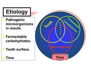
- 1. 1.Pathogenic microorganisms in mouth. 2.Fermentable carbohydrates. 3.Tooth surface. 4.Time Time Etiology Microorganism
- 2. Dental caries is an infectious, communicable disease, which causes destruction of teeth by acid-forming bacteria found in dental plaque. The most important concept to remember is that caries is a dynamic disease process, and not a static problem. Secondly, before a cavity is formed in the tooth, the caries infection can actually be reversed! Caries progression or reversal is determined by the balance between protective and pathological factors in the mouth. The development of dental caries is a dynamic process: Demineralization of the hard dental tissue by the acidic products of bacterial metabolism – alternating with periods of remineralization. The development of the carious lesion is episodic, with periods of demineralization alternating with periods of remineralization The lactic acid produced by the cariogenic bacterial dissolve the calcium phosphate mineral of the tooth enamel in a process call demineralization. Baby teeth have thinner enamel than permanent teeth, making them very susceptible to caries.
- 3. Streptococcus mutans is the major cariogenic bacterium. S. mutans forms glucan and levan polymers that are adhesive. The bacteria, along with the polymers, work together to form a biofilm – called dental plaque. The bacteria use a substrate (sugar) to produce acids that dissolve dental enamel. Repeated demineralization by these acids leads to dental cavities. S. mutans has been highly associated with dental caries. The proportion of S. mutans in plaque associated with ECC can be 30% to 50% of the total viable bacterial counts in dental plaque. In contrast, S. mutans usually constitutes less than 1% of the plaque flora in non-caries active children. Lactobacilli are highly acidogenic microorganisms, associated more with deep cavities in dentin than with the initiation of the disease. Lactobacilli counts alone are not considered reliable enough in predicting dental caries activity, however.
- 4. The Process of Demineralization and Remineralization A tooth enamel is covered by plaque, which consists mainly of bacteria. Plaque is often found close to the gum, in between teeth, in fissures and at other "hidden" sites. Demineralization: When sugar and other fermentable carbohydrates reaches the bacteria, they form acids which start to dissolve the enamel - an early caries lesion occurs due to loss of Calcium and Phosphates Remineralization: When sugar consumption has ceased, saliva can wash away sugars and buffer the acids. Calcium and Phosphates can again enter the tooth. The process is strongly facilitated by fluorides
- 5. •Frequent intake of carbohydrate-rich or sugary foods enables the cariogenic bacteria to maintain a low pH on the surfaces of the teeth. •Night- time bottle feeding, or prolonged use of a sippy cup, can lead to early childhood caries. The flow of saliva is decreased during sleep, so clearance of the sugary liquid from the oral cavity is slowed down. •The earlier that a child’s mouth is infected with Mutans streptococci, the greater the risk for future caries development. The causes of caries are multifactorial, and the individual risk factors associated with ECC are therefore not necessarily causative
- 6. •Children who already have one or more dental cavities are considered high risk for developing more. •A low fluoride level on the surface of the teeth reduces the remineralization process and increases the risk for caries. •When the saliva flow is below 0.7 ml/minute, the saliva cannot wash carbohydrates off the dental surface. In addition, low salivary buffering capacity, low salivary IgA, low salivary calcium, and low salivary phosphate reduce the potential for neutralization of acids in the dental plaque. •Finally, a low socioeconomic status can reduce interest in oral hygiene and a healthy diet.
- 7. Biochemical factors Factors to which the tooth surface is directly exposed, and which contributes to the development of the lesion:- 1. Amount of plaque 2. Type of bacteria 3. Type of diet 4. Frequency of carbohydrates 5. Saliva secretion 6. Saliva buffer capacity 7. Fluorides Saliva has a critical role in the prevention of dental caries. Saliva provides calcium, phosphate, proteins, lipids, antibacterial substances, and buffers. Saliva buffering can reverse the low pH in plaque, and with a higher pH, calcium and phosphate can be driven back into the tooth enamel. One factor that lowers the risk of cavity formation is normal salivary flow. Anything less than 0.7 ml/minute increases the risk for cavity development.
- 8. The general diagnostic principles of caries detection have always been clinical evaluation consisting of tactile probing and visual identification of carious tooth structure, radiographic evidence and clinical symptoms of sensitivity. Caries detection
- 9. CARIES DETECTION METHODS 1. CLINICAL OBSERVATIONS The oldest and most used method for the detection of caries is visual. The clinical look of tooth structure along with a feel of the tooth surface using probes combines to form the basic clinical evaluation method. Today, more and more clinicians are enhancing their vision with magnification. 2. RADIOGRAPHIC DIAGNOSIS For the ages, dental radiographs have been the one diagnostic tool which dentists have used as their secret weapon. If we couldn't see it clinically and the tooth hurt, then..." there must be something on this x-ray I'm looking at".
- 10. The new direction of caries detection has taken a path towards three basic areas:- electrical resistance, light illumination and digital imaging. Another useful, older diagnostic tool, risk assessment, is making a comeback of sorts in the literature. 1.Electrical resistance measurements such as a.c. impedance spectroscopy, electrical conductance measurements (ECMs), electrical resistance monitors; 2.light illumination such as laser fluorescence, stereomicroscopy, fiberoptic transillumination; 3.strip mutans test for risk measurement, and ultrasonic imaging have all shown promise if not singly, surely in concert.
- 11. ELECTRICAL CONDUCTANCE is based on the simple theory that when an electrical current is passed through a whole, sound, caries-free tooth it will have a differing resistance reading than a decayed, unsound or fracture-filled tooth. KaVo DIAGNOdent®: Laser Cavity Detection
- 12. CARIES RISK ASSESSMENT The ability to identify caries-susceptible individuals has always been a goal of research. Most studies have focused on the direct cause of caries, bacteria, and also some of the mitigating factors such as salivary pH, protein makeup or home care
- 13. DIGITALLY-ENHANCED RADIOGRAPHY Though most practitioners are well aware of the technology, most feel that the radiographs produced by the new digital radiographic systems are inferior to conventional radiographs. This is a misrepresentation of both technologies. Digital radiographic systems, has abilities to modify and enhance the original image, become far more accurate in the caries diagnosis field.
- 14. LIGHT ILLUMINATION This method of caries detection uses a light source, preferably bright, to illuminate the tooth. This then shows internal variations of color, shading and morphology which goes undetected when the tooth is exposed to only low, mainly reflective light. Caries or demineralized areas in dentin or enamel show up as darkened areas with this technique. You can do this simply with a fiber-optic hand piece and illuminate the tooth from inside to outside the mouth.
- 15. Caries detector dyes • Acid red • Basic fuchsin • Carbolan green • Coomassie blue • Lissamin blue • Providone iodine
- 18. F A C T O R S I N F L U E N C I N G C A R I E S I N T E R P R E T A T I O N 1. Errors in technique may result in non-diagnostic films. For example, a bite-wing film that is used to detect dental caries must be free of overlapped contacts. Improper horizontal angulation causes overlapped contact areas ( red arrows) and makes it impossible to interpret the interproximal regions for dental caries. 2. Errors in exposure may also result in non-diagnostic films. For example, a dental radiograph used to detect dental caries must exhibit proper contrast and density. Incorrect exposure factors result in films that are too dark or too light and are useless in the detection of caries. Restorative materials, such as composites, silicates and acrylics, may appear radiolucent and resemble dental caries on a radiograph. The appearance of an anterior cavity preparation restored with these materials differs from the appearance of interproximal caries and can be identified by the well-defined, smooth outline.
- 19. Abrasion refers to the wearing away of tooth structure from the friction of a foreign object. The surface of the tooth affected depends on the causative factor. The most frequent type of abrasion is caused by tooth brushing and is seen at the cervical margin of the teeth. Tooth brush abrasion affects the root surface of a tooth and may be confused with root surface caries. Attrition, or the mechanical wearing down of teeth, may be mistaken for dental caries on a radiograph. Attrition may be seen on the incisal or occlusal surfaces of deciduous or permanent teeth. When the incisal or occlusal enamel is worn away, the underlying dentin wears away rapidly, and shallow concavities may form (red arrows). These concavities may resemble occlusal or incisal caries on a dental radiograph. Clinical examination enables the dental professional to distinguish attrition from caries. Cervical burnout, a radiolucent artifact seen on dental radiographs, may also be confused with dental caries. Cervical burnout appears as a collar or wedge-shaped radiolucency on the mesial and distal root surfaces near the CEJ of a tooth (red arrows).
- 20. Sequella of Preventive Strategies for caries • Identify the individuals caries risk and activity by a complete assessment procedure • Control oral bacterial levels - Restorations as needed ("incision and drainage"). - Chemotherapeutics (refers to use of fluoride varnish, Chlorhexidine rinse, Xylitol and MI Paste). • Identify measures which will shift the patient at risk to a low risk category. • Identify how non-cavitated lesions should be treated for remineralization and reversal. • Cavitated lesions will be treated in the traditional manner. • Home and office maintenance procedures
- 21. Assessment of dental caries risk and caries activity must involve the following:- 1. Host risk factors: • Medical history • Dental caries status • Fluoride use • Salivary assessment 2. Diet assessment 3. Bacterial assessment 4. Tooth surface activity assessment
- 22. 1. COLLECTING RELEVANT BACKGROUND DATA • General diseases of importance • Medicines • Social situation • Dietary habits 2. CLINICAL INVESTIGATION • Estimation of caries prevalence (DMFT & DMFS) and incidence • Checking for aggravating factors • Factors immediately involved in the caries process 3. TEST METHODS: ORAL MICROBIOLOGY, SALIVA, PLAQUE Mutans streptococci • Description of the Strip mutans test . Lactobacilli. • Chair side test method: Dentocult. Saliva • Saliva flow - measurement of saliva secretion rate • Saliva buffer capacity
- 23. Socio-economic factors or circumstances which may indicate increased caries risk, examples: •Socially deprived, no work, bad economy •Low knowledge, low education of parents •No regular dental check-up Epidemiological factors or circumstances which may indicate increased caries risk, examples: •Living in high DMF country •Living in high DMF area •Member of high DMF family •High past caries experience •Intra-oral distribution of earlier lesions/fillings Clinical findings which may indicate increased caries risk, examples Early signs of the disease (for example white spot lesions) Newly erupted teeth •Exposed root surfaces •Crowded teeth •Deep fissures or other "natural" retentive sites •Retentive sites caused by dental treatment
- 24. 1) Environmental factors are noted: Existing dental caries, estimated sugar intake, estimated fluoride exposure, socioeconomic status, oral hygiene practices, and dietary habits are recorded 2) Infectious agents can be assessed using microbial sampling, such as the Dentocult SM Strip for assaying MS levels in the mouth. 3) Genetic factors can be evaluated, including: Salivary flow, salivary buffering capacity, and tooth morphology disorders. THE FIRST DENTAL VISIT
- 25. The purpose of the first dental visit is to assess individual risk, and to educate the parent or caregiver about reducing such risk. A clinical examination is an essential part of a child’s first dental visit, and as an important part of risk assessment. A correct and efficient diagnostic investigation must include the identification and evaluation of risk factors. THE FIRST DENTAL VISIT
- 27. Healthy Tips: 1. Shop smart!! Do not routinely stock your pantry with sugary or starch snacks. But “fun foods” just for special times 2. Limit the number of snacks; choose nutritious snacks 3. Provide a balanced diet, and save foods with sugar or starch for mealtimes 4. Don’t put your young child to bed with a bottle of milk, formula, or juice 5. If your child chews gum or sips soda, choose those without sugar
- 28. FOR PARENTS The American Academy of Pediatric Dentistry, the American Dental Association, and the Academy of General Dentistry recommend that children visit a dentist within six months of the eruption of the first tooth, and no later than 12 months of age. •Infants should not be put to sleep with a bottle. Breast- feeding at night should be avoided after 12 months of age. •Infants should be weaned from the bottle at 12-14 months of age. •Consumption of juice from a bottle or sippy cup should be avoided. Juice should be offered to a child only in a cup. Infants and toddlers should drink no more than 6 ounces of juice per day. •Cleansing of the baby teeth should be started by the time of eruption of the first primary tooth. A small piece of clean gauze or a small toothbrush can be used.
- 29. HOW CAN PARENTS PREVENT DENTAL CARIES? 1. Parents can modify oral hygiene techniques, depending on the child's age. For small infants, the gums need to be cleaned once or twice a day with a piece of clean gauze. This will help to establish a healthy oral environment for the baby teeth. Infants should be introduced to the toothbrush around the age of one. 2. Parents should not put children to sleep with a bottle containing any liquid other than water. Parents should encourage their infants to begin drinking from a cup around their first birthday. 3. Parents should help brush their children's teeth every day, after every meal. 4. Parents should not let their children drink fruit juice or sweetened drinks from a bottle or "tippy" cup, since this prolongs the exposure of teeth to harmful sugar. 5. Parents should provide healthy, balanced meals for children. They should limit the amount of sugar-laden foods and snacks in their diet. Plenty of healthy snacks should be available for children. Cheese products actually fight dental caries.
- 30. 6. Parents can help make children's teeth more decay-resistant by using an ADA-approved children's toothpaste. Place only a pea-sized drop of toothpaste on the toothbrush. Until a child is 3 years old, parents should only use baby tooth cleanser - to avoid causing fluorosis discoloration of the adult teeth. 7. Children taking oral medications should have their teeth cleansed after each dose of medication. Nearly 100% of children's medications contain sucrose, which can increase the risk of developing dental caries. 8. Children should have their first oral/dental health evaluation by the age of 12 months, or within 6 months of the eruption of the first tooth . 9. Parents should consider providing children with xylitol- containing chewing gum, which can help prevent dental caries.
- 31. 1) Gaining control of the bacterial infection: The control of S. mutans is accomplished in two phases: Caries control, followed by chemotherapeutic medication. We will start with caries control – treating the cavitated lesions with glass ionomer cements. Caries control: Minimally invasive caries control, also called Atraumatic Restorative Treatment, Chemotherapeutic medication: A combination of fluoride varnish and Chlorhexidine application is used to lower the Mutans streptococci count. - The varnishes contain 5% sodium fluoride (NaF) at 22,600 ppm of fluoride. There is a mean caries reduction of 38% when fluoride varnish is used in caries prevention. In an aggressive preventive program, varnish can be applied 3 times within a 10 day period. This is followed by another varnish application every 3 months for the first year. - The 0.12% Chlorhexidine gluconate can be applied to toddlers’ teeth twice a day for 14 days. It is applied at least 30 minutes after the use of toothpaste because the sodium lauryl sulfate contained in most toothpastes will neutralize chlohexidine gluconate
- 32. 2) Reduction of risk levels: Step two in the medical model is reduction of the risk levels for patients. First, sugar intake must be reduced. A dietary assessment can identify when sugar consumption needs to decreased . Increasing fluoride use at home will also reduce the risk of dental caries. 3) Remineralization of teeth: Step three in the medical model of caries management is the reversal of active caries site by remineralization. There are four parts to this step: a) Fluoride varnish is applied 3 times in a 10 day period. b) Fluoride is applied at home. A fluoridated dentifrice is used twice daily. Application of 1.1% NaF gel by toothbrush is recommended for very high risk children with dentin caries. c) Xylitol gum is recommended. d) A source of calcium, such as cheese, is also recommended. 4) Long term follow-up: The last step in the medical model is long term follow-up at home and in the dental office. The office recall frequency should be every 3 months for high risk patients and every six months for low risk cases. Caries activity and risk are re- evaluated at the dental recall visits.
- 33. There are three principal ways to prevent dental caries or ECC: Community-based programs Home-care methods Professional dental measures
- 35. Enhancing Remineralization Home Fluoride rinses (0.05%) Builds CaF2 layer at enamel/cuticle interface. Home fluoride at night for 1 year. Increases remineralization and decreases deminerealization. Decreases acid production. Inhibits mutans streptococci (ms) growth. Inhibits plaque growth.
- 36. 1. Systemic fluoride • Community water fluoridation • School water fluoridation • Fluoride tables 2. Topical fluoride • Solution • Gel • Varnish • Foam 3. Fluoride supplements • Dental material with fluoride - amalgam - glass ionomer - impression materials • Fluoridated milk • Fluoridated salts • Fluoridated tooth pastes Fluoride protocol
- 37. Water Fluoridation: decrease in caries: primary = 40%, permanent = 50-60% Mechanism of action of fluorides: - decreases enamel solubility. - improves crystalinity. - promotes remineralization. - decreases free surface energy of bacteria so it cannot stick on tooth. - bacteriocidal or bacteriostatic. - causes developing crystal to get bigger, less soluble Systemic Fluoride Supplements • No evidence to support prenatal benefit • Benefit to mother is primarily topical
- 38. Fluoride Facts Fluorine, from which fluoride is derived, is the 13th most abundant element and is released into the environment naturally in both water and air. Fluoride is naturally present in all water. Community water fluoridation is the addition of fluoride to adjust the natural fluoride concentration of a community's water supply to the level recommended for optimal dental health, approximately 1.0 ppm . One ppm is the equivalent of 1 mg/L, or 1 inch in 16 miles. Community water fluoridation is an effective, safe, and inexpensive way to prevent tooth decay. Children and adults who are at low risk of dental decay can stay cavity-free through frequent exposure to small amounts of fluoride. This is best gained by drinking fluoridated water and using a fluoride toothpaste twice daily. Children and adults at high risk of dental decay may benefit from using additional fluoride products, including dietary supplements (for children who do not have adequate levels of fluoride in their drinking water), mouth rinses, and professionally applied gels and varnishes. Fluoride was first added to drinking water to prevent tooth decay in Grand Rapids, Michigan. Fluoridation of drinking water has been used successfully in the United States for more than 50 years.
- 39. •Fluoridation of community water has been credited with reducing tooth decay by 50 - 60 in the United States since World War II. •Fluoride's main effect occurs after the tooth has erupted above the gum. This topical effect happens when small amounts of fluoride are maintained in the mouth in saliva and dental plaque. •Fluoride works by stopping or even reversing the tooth decay process. It keeps the tooth enamel strong and solid by preventing the loss of (and enhancing the reattachment of) important minerals from the tooth enamel. •Water fluoridation costs, on average, 72 cents per person per year in U.S. communities (1999 dollars). •Consumption of fluids--water, soft drinks, and juice--accounts for approximately 75 percent of fluoride intake in the United States. •Children under age six years may develop enamel fluorosis if they ingest more fluoride than needed. Enamel fluorosis is a chalk-like discoloration (white spots) of tooth enamel. A common source of extra fluoride is unsupervised use of toothpaste in very young children.
- 42. Fluoride is needed regularly throughout life to protect teeth against caries. Fluoride in solution, from topical sources, enhances remineralization by speeding up the growth of a new surface in the partially demineralized subsurface crystals in the carious dental lesion. Recommendations for fluoride supplementation can be made based on 1. the fluoride content of the water, 2. the child’s age, and 3. the child’s caries risk. The risk of dental fluorosis is highest during the period of enamel maturation, from 1 to 3 years of age.
- 43. Cariostatic Action of Topical Fluoride Fluoride in the liquid phase around the apatite crystal is today believed to be more important in decreasing dissolution of crystal than fluoride incorporation into the crystal lattice. Very low liquid fluoride concentrations under low pH conditions can block crystal dissolution and reduce the rate of demineralization
- 44. High fluoride concentrations on the tooth surface results in Ca(F)2 like deposits. This is deposited on the surface and into porous enamel at decalcified sites. As pH drops the globular Ca(F)2 dissociates into Ca++ and F-, it provides both ions for remineralization. Some ions will also diffuse into the plaque. It is important to maintain these ions in plaque. Cariostatic Action of Topical Fluoride
- 45. Enhancing Remineralization Saliva is not as good in remineralization as is a calcifying solutions (with Ca & HPO4 ions). There are fewer free ions in saliva due to such factors as proteins interference. •Using Ca(F)2 and Na2HPO4 rinses in sprays enhance remineralization. •Also using Ca++ salt + buffer + Na2SiF6 is good. This provides Ca(F)2 for the production of amorphous CaHPO4 which in turn induces rapid enamel remineralizatiton. •Researchers are using CO2 lasers to enhance remineralization. •Remineralization does not usually replace the old carbonated hydroxy-apatite. Fluorapatite formed as a veneer over old crystals and is more most insoluble.
- 47. Topical Fluoride Therapy: 1. Efficacy depends on potency, frequency, duration a. Low doses at high frequency is currently believed to be the best method 2. Routine office applications: high potency & low frequency a. 2% Na F: 4 applications over 2 weeks at 2-3 yr intervals (10 g F/ml) b. 8% SnF2 for 4 min Gel or Powder (20 mg F/ml) c. 1.23% APF for 4 min (thixotropic gels) (12.3 mg F/ml) d. Foam vs Gel (0.6-0.8 mg/tray vs 3-4 mg/tray) 3. Home or school programs: high frequency & low potency a. 0.2% Na F weekly 1 min rinse (1 mgF/ml)-most rinses are with 10 ml b. 0.05% Na F daily 1 min rinse (0.25 mg F/ml) high alcohol concentration (+20%) 0.2 mgF/ml bid offers better protection c. 0.4% SnF2 gel, daily in trays or brush (1.0 mg F/ml =1000 ppm) d. Toothpaste (1000ppm is 0.1% F): one 9oz tube has 270 mg F
- 51. Low strength fluoride mouthwashes For children with very active caries, a fluoride mouthwash for home use may be useful provided that the patient is motivated to use it. There are two options:- 1. The Principal Dental Officer can provide a prescription for a three- month supply of a 0.05% sodium fluoride mouthwash. 2. Colgate and Macleans market low strength (0.05 percent) fluoride mouthwashes (Fluorigard and Coolmint with fluoride, respectively). 3. Pre-school children who cannot spit well may have fluoride solution applied by parents using a cotton bud.
- 52. Fluoride-Containing Dentifrices Most toothpastes at 1,000 ppm or 0.1% F. - During brushing F may go to the following forms: • Fluoroaptite. • Calcium F. • Free F. - Between brushing saliva F levels from 0.02-0.08 ppm. This helps to transfer Ca++ and HPO4= into enamel. - Three types of F toothpastes FDA approved: • Sodium Fluoride:- provides free F. Does not use Ca++ abrasives. • Sodium Monofluorophosphate:- This F complex releases free F when in contact with salivary phosphotases. Can use Ca++ abrasives. • Stannous Fluoride:- Aids in controlling gingivitis. Not as popular as other two.
- 53. Brushing with the new toothpaste (Enamelon) provides fluoride, which will facilitates uptake of the ions, as well as adequate concentrations of calcium and phosphate to remineralize and strengthen the tooth enamel. This presumably will aid in the reversal and healing of this lesion. Studies are currently underway to determine the ability of this new toothpaste to do this.
- 55. Clinical application of fluoride The selective use of professionally applied topical fluorides is encouraged in the school dental service. The decision to use topical fluorides should be based on the assessment of the caries risk of the child. There are two types of professionally applied topical fluoride preparation in common use in school dental. These are: a gel of either neutral or acidulated fluoride and a fluoride varnish. 1. Topical fluoride gel Professionally applied topical fluoride gel is an effective way of preventing dental decay in patients with moderate to high caries activity. It is particularly useful in controlling smooth surface proximal caries, and has its maximum effect on newly erupted teeth.
- 56. 1. Check that the tray fits in the child’s mouth and covers all teeth. 2. Limit the amount of gel placed in each tray to no more than 2 mls (5 cm), or a smear over the inner surface of the tray. 3. Seat the patient in an upright position during application. 4. Leave the fluoride gel in place for the time specified by the manufacturer. 5. Use suction to remove excess saliva/gel mixture during and after the procedure. 6. Instruct the patient to spit out as much of the material as possible following the removal of trays and not to rinse, eat or drink for 30 minutes after the procedure. Topical fluoride gel technique
- 57. PreviDent®-Self applied topical neutral fluoride containing 1.1% sodium fluoride for use as a dental caries preventive in adults and children. This prescription product is not a dentifrice and is used in cases where caries activity is high. It increases tooth resistance to acid dissolution and enhance penetration of the fluoride ion into tooth enamel. . Apply a thin ribbon of gel to the teeth with a toothbrush or mouthtrays for at least one minute, preferably at bedtime. After use, adults expectorate gel. For best results, do not eat, drink orrinse for 30 minutes. Children expectorate gel after use and rinse mouth thoroughly.
- 58. 2.Fluoride foams Fluoride foams for topical fluoride application are also available and have some advantages over fluoride gels. Fluoride foams provide better control and are less likely to overflow the applicator tray, thus reducing gagging and ingestion. The consistency of the foam is also claimed to provide better interproximal coverage.
- 59. 3. Fluoride varnish The fluoride varnish currently in use in the school dental services is Duraphat. Each ml of Duraphat contains 50 mg of sodium fluoride (22.6 mg of available fluoride) in a suspension of natural resins. Each tube of Duraphat contains 10 ml of varnish or 226 mg of available fluoride. Duraphat fluoride varnish reduces decay into dentine by 20–50 percent depending on the surfaces it is applied to and their state of demineralisation. Applications to smooth surfaces showed the greatest reduction. Fluoride varnish is used in • the school dental services principally for spot applications of fluoride on enamel caries diagnosed visually or by radiograph, rather than for whole mouth applications. • the occlusal surfaces of partly erupted permanent molars and hypoplastic areas of teeth. For maximal effect the varnish should be applied to demineralised surfaces more than once during a year (several applications over several weeks were more effective than annual or bi-annual applications). Such repeat applications can often be carried out when the child comes to the clinic for restorative appointments.
- 60. Fluoride varnish contains 2.26% fluoride ion. The actual amount of fluoride used per treatment is 5-11 mg. The volume of fluoride varnish per treatment (0.2 – 0.5 ml) is significantly less than the probable toxic value for a 10 kg child (2.0 ml). The plasma fluoride concentration after varnish application is barely measurable. Therefore, fluoride varnish is very safe for use on infants’ teeth! The varnish is applied with a small soft brush, and reapplication is recommended every 3 to 6 months. Data show an overall reduction of caries incidence after fluoride varnish applications, ranging from 18% to 70%, compared with untreated control subjects.
- 61. Dose and safety for fluoride varnish Pre-school children The maximum usage of Duraphat on pre-school children should be no more than 0.25 ml (5.65 mg of available fluoride). Mixed dentition For the mixed dentition (5–11 years) the maximum usage should be no more than 0.40 ml (9.04 mg of available fluoride). Permanent dentition For the permanent dentition maximum usage should be no more than 0.75 ml (16.95 mg of available fluoride).
- 62. • Deeper enamel penetration. • Better localization, less systemic uptake. • Good over incipient lesion especially if there is a question about patient compliance. • No prophy required. Have a patient brush and apply after drying area. • Currently, the dose and frequency needed to arrest lesions and initiate remineralization Advantage of fluoride varnish
- 63. Procedure 1. Dispense the appropriate quantity of varnish on to a pad. 2. Remove any gross plaque from the surface(s) to be treated. 3. Isolate the tooth or teeth if possible, using cotton rolls. 4. Dry the enamel surface(s) to be treated using compressed air. 5. Apply fluoride varnish in a thin film to the specific surfaces to be treated, using a suitable instrument. To ensure approximal surfaces are treated, fluoride varnish can be placed above the contact and flossed into the contact area. 6. Dry the varnish for 30 seconds if possible, using a gentle flow of compressed air. 7. Ask the child not to eat or drink for 30 minutes after the application and not to brush their teeth until the next day.
- 64. FUTURE TRENDS IN CARIES PREVENTION: 1. Chemotheraputic methods of fighting the caries- causing bacteria will be used. These methods include the application of fluoride varnish and chlorhexidine gel on teeth. 2. Vaccines against dental caries. The vaccines would function by giving a child an improved IgA or IgG immune response to cariogenic bacteria. 3. Molecular probes to measure the level of cariogenic bacteria in a child's mouth. 4. Earlier caries detection, including fluorescence and ultrasonography. 5. Laser treatment of teeth. This technique would inhibit the progression of dental caries by making the enamel surface highly resistant to acid attack.
- 65. As the amount of fluoride is absorbed in the plasma and then distributed to the various organs of the body the concentration of fluoride in muscle, liver, heart and all soft tissues begins to rise. After the fluoride has been absorbed from the stomach and the intestines and is absorbed into the other soft tissues and other organ systems the plasma level will decrease, primarily because of the decrease by excretion of the kidneys and that of bone uptake. Usually the plasma level will reach the pre-ingestion level in three to six hours. The excretion pattern of fluoride is interesting because it is influenced by several factors -- the primary one being age. The age of an individual will determine how much fluoride can be eliminated from the body. The excreted percentage of fluoride varies as a direct function of age. Obviously, the younger an individual, the less fluoride is excreted and the older, the more rapidly a given does is excreted. This is primarily due to the age of the skeleton and the surface area of bone mineral available for fluoride uptake. Therefore, it is quite obvious that in a growing individual - early childhood to adolescent years - those factors will favor the enhanced fluoride uptake so that relatively less is excreted in the urine.
- 66. Chronic fluorosis- a. Dean's Classification: 0-4 scale 0- (None)59% of population at optimal fluoride level will exhibit no fluorosis. 1- (Very mild) 7.4% of population at optimal dose 2- (Mild) Greater than 25%, less than 50% of surface with opaque defects greater than 1.2 ppm 3- (Moderate) Brown stains present ( greater than 2 ppm) 4- (Severe) Pitting and attrition problems ( reater than 2-3 ppm ) b. Fluorosis occurs during maturation process. For incisors, age 2-3 years is the critical period. Improper use of dietary fluoride supplements and ingestion of fluoride dentifrices by small children, particularly in fluoridated communities, may result in dental fluorosis. Dental fluorosis is defined as hypoplasia or hypomaturation of tooth enamel produced by chronic ingestion of excessive amounts of fluoride as the teeth are developing, and are manifested as whitish opacities on the teeth. In severe cases, mottled enamel may occur.
- 67. 2. Acute a. CLD ( certain lethal dose ): 32-64 mg/kg in one source, 71-140 mg/kg in another source. A death has been reported at 17 mg/kg. b. PTD ( Probable toxic dose ) 5mg/kg i.e. 50 mg for the average 10 kg 2 year old. c. STD ( Safely tolerated dose ) 8-16 mg/kg d. Toxicity at low doses is due to gastric irritation. Hypocalcemia and hyperkalemia at high doses leads to cardiovascular collapse and death. e. Treatment for overdose: - less than 5mg/kg: oral calcium ( milk) and observe - More than 5 but less than 15 mg/kg: induce vomiting, oral calcium (milk, calcium gluconate, or calcium lactate), admit to hospital - More than 15 mg/kg: admit to hospital immediately, induce vomiting, monitor cardiac function, 10% calcium gluconate solution IV, monitor electrolytes.
- 68. Common signs and symptoms of acute fluoride toxicity: low dosages high dosages nausea 3-C's vomiting convulsions hypersalivation cardiac arrhythmia abdominal pain comatose diarrhea - pt becomes hypocalcaemia due to Ca binding with F
- 69. Lethal Dosage: - Adult = 1.25 gms F or 2.5 gms of NaF is lethal. (8-16 mg/kg of body weight). Best to consider 8 mg of F/kg body weight as lethal adult dose. e.g. 80Kg adult x 8mg/Kg =640 mg of F. - For a child 5 mg of F/kg body weight considered toxic. e.g. 20 Kg child X 5 mg/Kg body weight= 100 mg of F toxic. Small F tray may hold 2 ml each. APF = 12.3 mg F/ml, 2 trays = 4 ml x 12.3 mg= about 50 mg. This is half the toxic dose
- 70. It is possible to prevent the adhesion of the micro-organisms to the tooth surface by mechanical means which quite simply expressed means to complete tooth brushing and dental flossing to break up the plaque matrix. The use of plaque dissolving agents such as Chlorhexidine or Dextranase to break up the plaque matrix. Or alter the production of dextran as a byproduct of certain micro organisms which allow the micro- organisms to attach and adhere to the tooth surface. Dextran is an extra polysaccharide produced by specific organisms metabolizing specific types of metabolites. Chlorhexidine (CHX) • Best for ms control. • Can be used with fluoride. • Will not penetrate enamel. • Will not suppress lactobacillus. • Suppression will last 3-5 months.
- 71. The Role of Xylitol in Caries Prevention • Xylitol is a sugar alcohol produced from xylose (Wood Sugar) , obtained mainly from the white birch tree. Corn cobs are another source of xylose and recently it has been produced by fermentation. • Sorbitol is also a sugar alcohol and is produced primarily from corn starch in the U.S., but can be made from other sources of starch, such as potatoes. • Due to the increase of sugar consumption, sugar substitutes are essential in caries prevention programs. • Xylitol has good sweetening qualities and caries protective properties. Use 2 sticks, 5x/day for 5 minutes. • Its consumption does not promote low pH in dental plaque probably by enhancing salivary flow and buffer capacity.
- 72. •It inhibits adhesion, growth and metabolism of oral microorganisms. Suppresses ms even with sucrose intake. •It allows remineralization of initial enamel lesions. Enhances reversals (Turku study). •When it is incorporated to chewing-gum, its action is increased due to the stimulation of salivary secretion. •In association with fluoride, a synergic action is observed. The Role of Xylitol in Caries Prevention
- 73. A sealant is a plastic material that is bonded onto the chewing surfaces of the back teeth. The sealant acts as a barrier protecting this part of the tooth from decay- causing bacterial plaque. When are sealants necessary? Depressions and grooves form in the enamel of the back teeth as they develop. When these fissures are deep they are difficult to clean and keep free of plaque. A sealant is a very effective plaque barrier. Unfortunately this material is not effective on the smooth surfaces of the teeth, which encompass the rest of the visible tooth structure. These surfaces can be kept free of plaque by good brushing and flossing habits.
- 74. Factors In Using Sealants: - • Caries risk is the major consideration, not age. • Caries in pit and fissure extends beyond adolescence. • Moderate to high risk patient having pit and fissure morphology which would increase caries. • Sealants can be used as a preventive measure. • Incipient pit and fissure lesions limited to enamel are good candidates for therapeutic sealants. If x-rays show caries penetration into dentin is less than 1/3 and there is no cavitation, fluoride release pit and fissure sealant is the choice. The literature has shown that sealing frank caries terminates its progression. • Teeth with well-coalesced pit and fissures and with wide, easily cleaned groves do not require sealants.
- 75. The greatest benefits of sealants are their application in children soon after the erruption of a tooth that is a candidate for sealant application. Who should have sealants placed? The teeth are cleaned with an abrasive to remove all the plaque, then they are dried and isolated to keep them free of moisture. A material is placed on the teeth to allow the sealant to bond and adhere to the tooth. The sealant is then placed on the tooth and bonded to place with a high intensity white light. How are sealants applied?