

Recommended
More Related Content
What's hot
What's hot (20)
Viewers also liked
Viewers also liked (16)
Similar to Radiographic Differential Diagnosis 2009
Similar to Radiographic Differential Diagnosis 2009 (20)
More from IAU Dent
More from IAU Dent (20)
Recently uploaded
Recently uploaded (20)
Radiographic Differential Diagnosis 2009
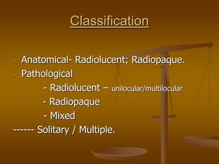
- 1. Classification - Anatomical- Radiolucent; Radiopaque. - Pathological - Radiolucent – unilocular/multilocular - Radiopaque - Mixed ------ Solitary / Multiple.
- 2. Classification-pathological Radiolucent – Contacting tooth - Not contacting tooth Mixed – Contacting tooth - Not contacting tooth Radiopaque – Contacting tooth - Not contacting tooth
- 3. Classification-pathological Radiolucent – Contacting tooth 1. Periapical - Usually sequelae of pulpitis. Periapical granuloma; Radicular cyst; Abcess; Osteomyelitis, Periapical cementoma. 2. Pericoronal Follicular spaces; Dentigerous cysts; Ameloblastoma; Adeno ameloblastoma.
- 4. Periapical-radiolucent Periapical granuloma – well circumscribed, rounded - around apex. May’ve thin radiopaque border. Tooth may have deep caries/restorations. Tooth-non-vital.
- 5. Periapical-radiolucent Radicular cyst – involve apex of perm.tooth. Untreated cyst slowly enlarge, expand and thin cortex – crackling sound (crepitus). If infected, all painful symptoms of an abscess develops.
- 6. Periapical-radiolucent C/c & a/c Dento alveolar abscess: Small/large radiolucencies. May have cortical expansion. Associated tooth- non vital. Teeth with a/c abscess –pain to percussion (high to bite on ) - Pd’l abcess originating in deep pd’l pkt – PA radiolucency + intra bony pkt; pulp vital usually.
- 8. Periapical-radiolucent Osteomyelitis: Seen seldom in maxilla (due to rich bld supply). Non vital pulp, sensitive to percussion,h/o assoc.a/c or c/c PA abscess. Borders –poorly defined and ragged. Sinus tract – if present, appears as a radiolucency from PA radiolucency through the cortical plate opening on the skin / mucosa. Is sequestrum seen (segment of dead bone) & large enough, it appears radiopaque within a radiolucency.
- 9. Osteomyelitis (Oral Surg Oral Med Oral Pathol Oral Radiol Endod 2006;102:94-8)
- 10. Periapical-radiolucent - PACOD (Periapical cementoma): Its early stage- round well defined borders, assoc. with vital tooth. Usually in MND incisors; asymptomatic. If a pulpo periapical lesion – non vital pulp.
- 12. Pericoronal - radiolucent Follicular space: Surrounding crowns of unerupted teeth. Homogenous radiolucent halo with a thin outer radiopaque border, that is continuous with LD. Normal follicular spaces usually decrease in size with age.
- 13. Pericoronal - radiolucent Dentigerous cyst: Mostly MNDlr 3rd M, Mx- C, MND- PM, Mx- 3rd M. . Ameloblastoma – Infiltrate bone
- 14. Pericoronal - radiolucent Adeno ameloblastoma – (AOT)- Benign & non-invasive. It differs from ameloblastoma. AOT-slow growing tr, doesn’t infiltrate bone. Displace teeth but no root resorption. Antr maxilla. Expand cortical plate, produces clinical swelling, no soft tissue invasion.
- 15. Radiolucencies- not contacting teeth - Inter radicular - Solitary cyst – like - Multilocular - Solitary, ragged, poorly defined borders - Multiple separate - Generalized rarefaction
- 16. Radiolucencies- not contacting teeth Inter radicular radiolucencies: That occur b/w roots of teeth. Pd’l pkts: Pd’l bone loss (H/V) appear on films. Occurs closer to involved tooth contacting its surface. Confirmed diagnosis by placing a pd’l probe into the defect.
- 17. Radiolucencies- not contacting teeth Furcation involvement: Seen in advanced Pd’l disease. Produces furcation involvement. Usually seen in MNDlr M, where bifurcation is devoid of bone & shows a radiolucency. Usually a probe can be introd. into bifurc area from the B/L aspect. LD remains intact in furcation in normal furcation.
- 18. Radiolucencies- not contacting teeth Lat. Radicular cyst: Assoc with non vital pulp.(near to lat. accessory canal opening). If infec. pain, swelling occur on offending tooth; sensitive to percussion
- 19. Radiolucencies- not contacting teeth Primordial cyst: Cyst like radiolucencies, not contacting tooth. May occur in a region where a tooth may’ve failed to develop. Odontogenic trs: Usually Odontomas. Freq seen as inter radicular radiolucencies. In its radiolucent stage- cyst like with a well defined border. Odontomas
- 20. Radiolucencies- not contacting teeth Globulomaxillary cyst: Asymptomatic. If large, expands cortical plate buccally; if sec. infected, pain. Inverted tear shaped radiolucency.
- 21. Radiolucencies- not contacting teeth Incisive canal cyst: Cyst like radiolucency. Mx CI. Often – antr nasal spine is seen over the supr portion of the cyst as a radiopaque shadow, thus producing a Heart- shaped radiolucency.
- 22. Radiolucencies- not contacting teeth Malignancies: May begin in inter septal bone and usually present as radiolucencies with poorly marginated borders. If they involve PDL early in their development, charact. pds a band like widening image of PDL. Lat’l PDL cyst: More in Mndlr C & PM. Adjacent teeth’ve vital pulps. Round/oval, well defined, often with sclerotic border. .
- 23. Radiolucencies- not contacting teeth Median mandibular cyst: Occurs in symphyseal region of L/jaw. If the adj teeth are non-vital, it is usually a radicular cyst.
- 24. Radiolucencies- not contacting teeth Solitary cyst like: Post extraction socket: sometimes show cyst like radiolucency after extn. H/o extn exists. Residual cyst: Is a radicular/ another cyst that’as remained after its assoc: tooth has been lost. Usually over 20 yrs & more in Mx.
- 25. Radiolucencies- not contacting teeth Lingual MNDlr bone defect (Stafne’s cyst): Invagination in the median surface of MND, Usually 3rd M, angle area. Located infr to MNDlr canal in 3rd molar area. Asymptomatic, Unilocular/multilocular, lined by cortex
- 26. Radiolucencies- not contacting teeth Odontogenic Keratocyst (OKC):Usually in 2nd & 3rd decades. Findings suggestive of OKC: 1. Cyst like radiolucency in MNDlr 3rd M region/ ramus. 2. A diameter of > 3 cm. 3. Unilocular cyst like radiolucency with scalloped margins. 4. Multilocular cyst. 5. Odorless, creamy or caseous contents on aspiration.
- 27. Radiolucencies- not contacting teeth OKC
- 28. Radiolucencies- not contacting teeth Primordial cyst: B/w 10 & 30 yrs. MNDlr 3rd M. Seldom produces cortical expansion. Usually in areas where a tooth failed to develop.
- 29. Radiolucencies- not contacting teeth Ameloblastoma: Asymptomatic initially. Expands, perforates cortical plates. Feels firm if it is of solid type. Cystic type is soft & fluctuant and straw colored fluid can be aspirated in some cases.
- 30. Radiolucencies- not contacting teeth Multilocular type: Terms – Soap bubble, honey comb and tennis racket – used to describe the various radiographic images of multilocular lesions.
- 31. Radiolucencies- not contacting teeth Soap bubble appearance
- 32. Radiolucencies- not contacting teeth Ameloblastoma: Multilocular type may be of soap bubble/honey comb variety.
- 33. Radiolucencies- not contacting teeth Solitary radiolucencies with ragged & poorly defined borders: Chronic osteitis & osteomyelitis: Inflammation of bone caused by pathogenic micro org, called Osteitis, when just alveolar bone is affected. If basal bone of jaws is involved, this process is Osteomyelitis.
- 34. Osteomyelitis (Oral Surg Oral Med Oral Pathol Oral Radiol Endod 2006;102:94-8)
- 35. Radiolucencies- not contacting teeth Multiple separate, well defined radiolucencies: - Multiple cysts/granulomas
- 36. Radiolucencies- not contacting teeth Gen: rarefactions of jaw bones: -Hyperparathyroidism: -Osteoporosis:
- 37. Radiolucencies- not contacting teeth Osteoporosis
- 38. Mixed lesions – contacting tooth Mixed lesions assoc: with teeth: 1. Peri apical mixed lesions. 2. Pericoronal mixed lesions. Peri apical mixed lesions: Calcifying crown of developing tooth:
- 39. Mixed lesions – contacting tooth Calcifying crowns of - developing teeth
- 40. Mixed lesions – contacting tooth Calcified material with in an intermediate stage odontoma
- 41. Mixed lesions – contacting tooth Pericoronal mixed lesions: Odontoma – intermediate stage.
- 42. Mixed lesions – not contacting tooth 1. Healing surgical site: h/o Surgery. 2. C/c Osteomyelitis:
- 43. Osteomyelitis (Oral Surg Oral Med Oral Pathol Oral Radiol Endod 2006;102:94-8)
- 44. Radiopacities Contacting tooth – Periapical radiopacities Not contacting tooth – - Solitary - Multiple separate - Generalized opacification
- 45. Radiopacities contacting tooth Periapical radiopacities: 1. Condensing or sclerosing osteitis: Sclerosis of bone induced by an inflamm. or infec. that most often occurs as a pulpo periapical lesion. Non-vital teeth. Usually in MND – 1st M & PM.
- 46. Radiopacities contacting tooth 2. Mature periapical cemental dysplasia
- 47. Radiopacities contacting tooth Periapical Cemental Dysplasia
- 48. Solitary radiopacities not contacting tooth True intra bony radiopacities: a. Tori. b. Unerupted, impacted & supernumerary teeth. c. Retained roots. d. Focal & diffuse sclerosing osteomyelitis
- 49. Multiple separate radiopacities not contacting tooth 1. Tori & exostoses 2. Multiple retained roots Mandibular tori
- 50. Multiple separate radiopacities not contacting tooth 3. Multiple hypercementosis 4. Multiple embedded/impacted teeth Hypercementosis
- 51. Generalized radiopacities D/d of Gen. radiopacities of jaw bones: Osteopetrosis Normal variations in form & density.
- 52. Generalized radiopacities Dense radiographic images of the jaw bones may be seen in patients who have heavy jaw bones or are over weight. -----------------------