SPE-LEEM Studies of MoS2 and WSe2 Surface Structure
•Télécharger en tant que PPSX, PDF•
2 j'aime•4,838 vues

A brief introduction to 1-4ML MoS2 ARPES study - background, motivation, tools, measurement, results.

Recommandé
Contenu connexe
Tendances
Tendances (20)
En vedette
En vedette (18)
Similaire à SPE-LEEM Studies of MoS2 and WSe2 Surface Structure
Similaire à SPE-LEEM Studies of MoS2 and WSe2 Surface Structure (20)
Dernier
Dernier (20)
SPE-LEEM Studies of MoS2 and WSe2 Surface Structure
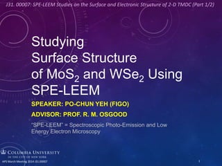
- 1. Studying Surface Structure of MoS2 and WSe2 Using SPE-LEEM SPEAKER: PO-CHUN YEH (FIGO) ADVISOR: PROF. R. M. OSGOOD “SPE-LEEM” = Spectroscopic Photo-Emission and Low Energy Electron Microscopy 1APS March Meeting 2014 J31.00007 J31. 00007: SPE-LEEM Studies on the Surface and Electronic Structure of 2-D TMDC (Part 1/2)
- 2. MANY THANKS! Also, credits and thanks to: Jonathan Liou, XiaoXiao Zhang, and YuMeng You Jurek Sadowski DaTong Zhang Arend van der Zande Abdullah Al-Mahboob Prof. James Hone Prof. Irving Herman Daniel A. Chenet Prof. R. M. Osgood WenCan Jin Jerry Dadap Nader Zaki Peter Sutter 2APS March Meeting 2014 J31.00007
- 3. WHY WE WANT TO STUDY THIS? • Spin-orbit coupling • It has a bandgap! • Photoluminescence • WSe2: both p- and n- type FET are fabricated Strong PL in monolayer MoS2 Nano. Lett. 10, 1271-1275 (2010) High quantum efficiency 1000 times stronger PL in ML WS2, WSe2 than in bulk ACS Nano 7 (1), 791–797 (2013) Direct bandgap in ML Thin, flexible devices E.g. Li-ion battery and transistors Nano Lett., 11 (9), pp 3768– 3773 (2011) Chem. Commun. , 47, 4252-4254 (2011) Enhanced spin lifetimes Large spin Hall angles VBM S-O splitting up to 456meV in WSe2 PRB 84, 153402 (2011) Nano Lett. 13 (7), pp 3106–3110 (2013) Nano Lett. 13(5), pp 1983– 1990 (2013) http://meetings.aps.org/Meeting/MAR14/Session/D51.1 Nano Lett., 2012, 12 (7), pp 3788–3792 3 Our aim: Study sample quality: CVD vs. exfoliated MoS2 Find an ideal substrate for studying MoS2 EM and PE Determine the surface corrugation and structure Measure the electronic structure directly
- 4. WHY SPE-LEEM? Micron-size spot, Direct band structure, fast real time imaging, large area mapping, UHV, clean, surface doping, depth profile. NSLS I NSLS II 1. LEED – Crystal orientation. 2. LEEM – Surface corrugation, quality probe. 3. ARPES – Energy resolved k space mapping. 4. XPEEM – study ionization, core level orbitals. Now Future 4 All in one!
- 5. SPE-LEEM: HOW DOES IT WORK? Main Chamber Objective Illumination Column Imaging Column Sample Detector Energy Analyzer Electron Gun UV beam Synchrotron Beam Splitter 5APS March Meeting 2014 J31.00007
- 6. SPE-LEEM - PERSPECTIVES ELMITEC SPLEEM Energy Analyzer Manipulator. Grounded. (High voltage @ 2kV) Preparation chambers Photon energy: 15-150eV Good energy resolution: 100meV Good spatial resolution: 8nm Large mapping area: d = 100µm Thermal coupler Sample holder d ~ 10mm 6APS March Meeting 2014 J31.00007
- 7. LEEM – EXFOLIATED MoS2 ON SiO2 2ML 1ML 10 µm a. b. 10 µm Charging hinders LEEM to extract information on: • Depth profile • Layer number • Surface corrugation/defects 7APS March Meeting 2014 J31.00007 Charged up in 5min!
- 8. 5 150 10 µm Photoluminescence (Courtesy of the Hone group) 20 µm Grain Boundaries LEEM – CVD MoS2 ON SiO2 Similar charging effect hinders imaging Precise surface doping: SEAS Getter dispenser, 730C, Y = 0.2 mg/cm, adhesion rate: 0.7 Surface corrugation Periodicity: sub-micron Potassium doping LEEM imageOptical image 8 In collaboration with the Hone group APS March Meeting 2014 J31.00007 Nature Materials 12, 554–561 (2013) Need a conducting substrate! Use native oxide silicon
- 9. SAMPLE PREPARATION - TRANSFER Si Flakes on PMMA layer Water soluble PVA layer DI water Scoop PMMA Sample on PMMA film suspended on scoop Si, patterned Wet transfer process for exfoliated MoS2 Rinsed in Acetone for 24hr + Anneal at 350C in UHV chamber for 12hr + 9 In collaboration with the Herman group APS March Meeting 2014 J31.00007
- 10. TRANSFER MoS2 onto Silicon SUBSTRATE 10 µm 15 µm 10 µm 15 µm 10 µm 15 µm With LEEM, flakes are visible on native oxide silicon substrate Transfer preserves the sample morphology Wrinkles (growth-induced, strain) removed Doping changes work function a lot, but a little to Fermi level 10APS March Meeting 2014 J31.00007 Single domain Mirror- Twin* domains *Ref: Nature Materials 12, 554–561 (2013)
- 11. LEED ANALYSIS 1ML 2ML 3ML 4ML LEED (00) spot Correlation length 𝜉 (in) Roughness 𝛼 (in) Substrate scattering (ex) Bulk Compare to Graphene LEED 1st order spots Correlation length 𝜉 (in) Roughness 𝛼 (in) Substrate scattering (ex) 11APS March Meeting 2014 J31.00007 00 spot 42eV 𝐹𝑊𝐻𝑀 ∝ 𝑘⊥ 𝜔 1/𝛼 /𝜉 Exfoliated on SiO2, at 42eV FWHM in Å-1 1st order Graphene 1ML 1.20 Graphene 2ML 0.51 Graphene 3ML 0.37 MoS2 1ML, exfoliated 0.34 0.30 MoS2 1ML, CVD 0.52 0.34 MoS2 1ML, transferred on Si 0.67 0.50
- 12. A SNEAK PEEK ON MoS2 ARPES Monolayer MoS2 12 Phys. Rev. Lett. 111, 106801 (2013) Band structure EDCs MDCs 4:30 PM Session J31. 00009 by WenCan Jin.
- 13. ON GOING - WSe2 STUDIES Optical image 5µm 20µm 1ML 2ML 3ML 2ML LEEM image WSe2 transferred on Si substrate. Contrast between layers. Wrinkles and cracks. LEED 1-3ML 13APS March Meeting 2014 J31.00007
- 14. CONCLUSION SPE-LEEM is a strong and versatile tool for layered material. LEEM gives good depth profile, layer number, and surface images if there is no charging effect. LEED reflects sample quality via careful analysis on spot width; it’s layer dependent. CVD MoS2 has a comparable quality to exfoliated MoS2. Transfer onto native oxide Si substrate made doing ARPES possible. It also removes the strain-induced wrinkles. 14 Preview – Part 2/2 ARPES result of MoS2 1-4ML and bulk. Substrate-induced compression and effective mass. Session J31. 00009 by WenCan Jin. APS March Meeting 2014 J31.00007 J31. 00007: SPE-LEEM Studies on the Surface and Electronic Structure of 2-D TMDC (Part 1/2)
- 15. 1-4ML + bulk, CVD + exfo IV analysis Simplify this and bring up the multi-scattering theory 15APS March Meeting 2014 J31.00007
- 16. LEEM IV ANALYSIS – DIFFERENT ORIGINS AND LAYER DEPENDENCE Among 1ML CVD samples (magenta curve is exfo) 1-4ML + bulk exfoliated samples 16APS March Meeting 2014 J31.00007
- 17. LEED ANALYSIS 1ML 2ML 3ML 4ML LEED (00) spot Surface corrugation (in) Number of defects (in) Substrate (ex) Bulk Compare to Graphene Exfoliated on SiO2, at 42eV FWHM in Å-1 1st order Graphene 1ML 1.20 Graphene 2ML 0.51 Graphene 3ML 0.37 MoS2 1ML 0.34 0.30 MoS2 1ML, CVD 0.52 0.34 MoS2 1ML, transferred 0.67 0.50 LEED 1st order spots Surface corrugation (in) Number of defects (in) Substrate (ex) 17APS March Meeting 2014 J31.00007
- 18. ARPES BY SPE-LEEM ARPES in SPE-LEEM Bulk WSe2 Normal incident light; without out-of-plane polarization Photoelectron k-space mapping ARPES: Angle-resolved photoemission spectroscopy Photons in, electrons out. Direct measurement of band dispersion ML WSe2 With DFT LDA bands 18APS March Meeting 2014 J31.00007
- 19. ARPES AND CURVATURE ANALYSIS Uses 2nd order derivatives of the ARPES intensity mapping. Separates band dispersion from linear background and detector artifacts. VBM transits from K to Γ. A strong evidence for indirect to direct bandgap transition. 19APS March Meeting 2014 J31.00007 Low 2ML 3ML Bulk1ML High
- 20. EFFECTIVE MASS AT K POINT a: experimental lattices, ref Phys. Rev. B 85 (2012). b: optimized lattices from calculation Hole effective mass agrees well with the calculations, for both 1ML and 2ML 20 Thickness Electron Mass Hole Mass Method Reference Lattice Constant ML N/A 0.435 LDA Our results. 3.28 ML N/A 0.564c Experiment Our results. 3.28 ML 0.53 0.52 DFT-GW-BSE A. Ramasubramanim, PRB 2012 3.32 ML 0.29a/0.26b 0.34a/0.33b DFT-GW-BSE Hongliang Shi, PRB 2013 3.286 ML 0.23 0.41 LDA A. Kumar, EPJB 2012 3.282 ML 0.19 0.4 FLAPW-GGA W. S. Yun. PRB 2012 3.286 2ML N/A 0.545 LDA Our results. 3.28 2ML N/A 0.432 Experiment Our results. 3.28 2ML 0.3 0.49 LDA A. Kumar, EPJB 2012 3.282 2ML 0.3 0.3 FLAPW-GGA W. S. Yun. PRB 2012 3.286
- 21. CONCLUSION SPE-LEEM is a strong and versatile tool for layered material. LEEM gives good depth profile, layer number, and surface images. LEED reflects sample quality via careful analysis on spot width; it’s layer dependent. Transfer onto native oxide Si substrate made doing ARPES possible. It also removes the strain-induced wrinkles. CVD is as good as exfoliated MoS2. Si serves as a 21 Preview – Part 2/2 ARPES result of MoS2 1-4ML and bulk. Substrate-induced compression and effective mass. Session J31. 00009 by WenCan Jin. APS March Meeting 2014 J31.00007 J31. 00007: SPE-LEEM Studies on the Surface and Electronic Structure of 2-D TMDC (Part 1/2)
- 22. Figure 2 Angle‐integrated photoemission spectra of exfoliated monolayer WSe2 extracted from high- ‐symmetry directions. (determine EF) At 33eV, the cross section between W 5d and Se 4p has an order of magnitude difference. (a) (c)(b) Γ K K’ M High symmetry points
- 23. 23APS March Meeting 2014 J31.00007
- 24. CVD MOS2 – A STAR MoS2 on SiO2 with K doping 24APS March Meeting 2014 J31.00007
- 25. diffraction contrast sample contrast aperture objective [0,0] [h,j] SURFACE STRUCTURE Au+O/Rh(110) quantum size contrast d FILM THICKNESS Co/W(110) geometric phase contrast MORPHOLOGY Mo(110) WHAT CAN BE MEASURED WITH LEEM? 25APS March Meeting 2014 J31.00007
- 26. 26APS March Meeting 2014 J31.00007
- 27. • Talk about exfo MoS2 after transfer and the comparable quality to CVD? APS March Meeting 2014 J31.00007 27
Notes de l'éditeur
- Hi, everyone. My name is Po-Chun. I am from Prof. Osgood’s lab. Today, I will be talking about (title). Here, SPE-LEEM is stands for …
- (Fast) First, I would like to thank our collaborators from Columbia and from Brookhaven National lab.
- Why we want to study MoS2 and WSe2? We already know that graphene is the super star now and has drawn a lot of attention. Why we want to study this? Because compare to graphene, MoS2 and WSe2 have a bandgap, which makes many application and devices possible such as Li-ion battery and transistors. Also, they have strong PL. In ML. Moreover, a strong spin-orbit coupling and splitting makes it valuable in Spintronics and Valleytronics applications. Last-but-not-the-least, people have fabricated both n type and p type FET by using WSe2, which can be make into p-n junction devices. In our study, we aim to …
- Then, why choose SPE-LEEM? It has many merits, such as … Basically, it’s a combination of XX, XX, XX, and XX. For example, …(3 examples) To sum up, it’s an all-in-one system. [Cut](It is located in NSLS I in BNL, and will be upgraded and moved to NSLS II in the near future.) *XPEEM. laterally resolved version of x-ray photoelectron spectroscopy (XPS)
- Here’s a cartoon to show you how it works. Here is the electron beam. The electrons make a normal incidence on the sample and are collected for imaging. For ARPES, the UV comes from synchrotron and reaches the sample surface normally. Photoemission happens and we collect the electrons for mapping.
- (Fast) Here’s a layout for SPELEEM.
- In our sample preparation, part of it requires using wet transfer process. CVD MoS2 was transferred via a PDS-based lift-off process.
- Shape preserved. Surface is clean and flat. The transfer was successful. The web-like wrinkles disappeared. *Workfunction of Si drops 2.1eV to the level of MoS2. The Fermi level shifts very little. Annealing can swipe away the K ions. The workfunction restores to pre-doping level. This is a reversible process.
- Maybe put the chart on the next slide and add some more nice graphs?
- (Fast) Here’s a preview of ML MoS2 band dispersion.
- (Fast) Ok, so now I will move to our current study on WSe2. Here’s the optical, LEEM, and LEED image of exfoliated WSe2. The transfer was successful.
- (Stay on this page for Q&A)
- *Only orbits from in-plane polarization survives. The interesting VBM is from W 5d orbits.
- Here it’s a curvature analysis of the bands using the second derivative method. These are the WSe2 band dispersion of 1-3ML and bulk; the red lines are from DFT calculation. I just want to make one point here (click out the animation) The highest energy point of VB is at gamma in ML and shifts to K in 2ML and more. This VBM transition from K to G is a strong evidence for indirect to direct bandgap transition.
- (Quick) This is our study of the effective mass at K point. The hole effective mass from our measurement agrees well with the theoretical calculations, in ML and bilayer cases.
- (Stay on this page for Q&A)