SlideShare une entreprise Scribd logo
1  sur  1
Poster produced by Faculty & Curriculum Support (FACS), Georgetown University School of Medicine
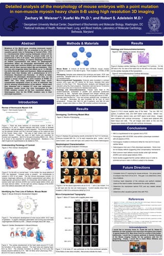
Detailed analysis of the morphology of mouse embryos with a point mutation
in non-muscle myosin heavy chain II-B using high resolution 3D imaging
Zachary M. Weisner1,2
; Xuefei Ma Ph.D.2
; and Robert S. Adelstein M.D.2
Abstract
1
Georgetown University Medical Center, Department of Biochemistry and Molecular Biology, Washington, DC
2
National Institutes of Health, National Heart, Lung, and Blood Institute, Laboratory of Molecular Cardiology,
Bethesda, Maryland
Introduction
Methods & Materials
Conclusions
Future Directions
References
Acknowledgments
Mutations of the Myh10 gene, encoding nonmuscle myosin
heavy chain II-B (NMHC II-B), are proposed to be one of the
causative genetic links to Pentaology of Cantrell (POC). POC
is a developmental defect causing inadequate closure of the
ventral body wall often resulting in ectopia cordis and an
omphalocele in 63% of all cases. POC presents with a set of
five phenotypes including: (1) anterior diaphragm deficiency,
(2) midline supraumbilical wall defect, (3) diaphragmatic
pericardium defect, (4) intracardiac abnormalities such as
double outlet right ventricle (DORV) and ventral septal defect
(VSD), and (5) lower sternum defect (Ma and Adelstein). POC
is a very rare congenital defect that affects up to 1 in 200,000
live births (Harmath, Hajdu and Hauzman). Males are generally
affected more than females with a predominance of 2.7:1
(Pakdaman, Woodward and Kennedy). In efforts to better
understand the phenotypic development over time, our lab
utilized a mouse model with a point mutation at R709C on
NMHC II-B. This study performed whole body micro-
computerized topography scans on mice at embryonic day
(E)14.5, allowing characterization of the defects. Hearts were
then dissected and a detailed CT scan was performed.
Preliminary results reveal that mice homozygous for the
R709C mutation contain all the fore- mentioned defects.
Heterozygous mice for the R709C mutation vary in their
phenotype expression.
Review of Nonmuscle Myosin II-B:
I would like to sincerely thank Dr. Xuefei Ma and Dr. Robert S.
Adelstein for their guidance and instruction throughout this project.
A special thanks to Dr. Mary Anne Conti for a review of this poster.
Additionally, I would like to express gratitude to Danielle Donahue
of the Mouse Imaging Facility for her guidance with the CT
programing. Finally, I would like to thank Milton Williams and
Mohamed Elshazzly for their support.
Figure 1: Non-muscle myosin II-B
Genotyping: Confirming Mutant Mice
Figure 5: Results of Genotyping
Figure 5 displays the genotyping results conducted for the E14.5 embryos.
Embryos included MA-134, 1-8, for each respective lane. Lanes 1 and 2
were mutant, Lane 3 was wild type, and Lanes 4 thru 8 were heterozygous.
• NM-II is hypothesized to be a genetic link to POC
• Homozygous NM II-B R709C mice exhibit a phenotype consistent
with all aspects of POC
• Homozygous mutation is embryonic lethal for mice at E14.5 due to
cardiac failure
• Heterozygous mice vary in their phenotype expression. Some mice
do not display defects suggesting other compensatory mechanisms.
• Most common heart defects include double outlet right ventricle
(DORV) and ventral septal defects (VSD).
• Some studies suggest that the cardiac defects occur in order to
promote survival in utero in efforts to adapt to the disease.
Figure 6: Microscope Evaluation of Embryos:
Figure 6: All the above specimens are at E14.5. 1 and 2 are mutant, 3 is
a wild type and the rest are heterozygous. Current studies show that a
mutation in NM II-B is embryonic lethal by E14.5.
Understanding Pentalogy of Cantrell:
Identifying the Time Line of Defects: Mouse Model
Figure 2: POC Clinical Development
Figure 2: On the left is a normal heart. In the middle, the visual defects of
POC are displayed. Ectopia cordis is present. An omphalocele is
present in 63% of all cases. The five clinical phenotypes include: (1)
anterior diaphragm deficiency, (2) midline supraumbilical wall defect, (3)
diaphragmatic pericardium defect, (4) intracardiac abnormalities such as
double outlet right ventricle (DORV) and ventral septal defect (VSD), and
(5) lower sternum defect. On the right are cartoon displays of the DORV
and VSD defects associated with the heart.
Results
Results
Figure 1: There are three isoforms of nonmuscle myosin II (NM II),
including: A, B, and C. Myosin interacts with actin in order to facilitate
cell division, cell-cell adhesion, and cell migration. The N-terminal makes
up the globular domain and the C-terminal makes up the coiled-coil end
of the myosin heavy chain. Two sets of light chains bind near the
globular domain. These two pairs of light chains are essential for
function and regulation. Myh10 is the gene encoding NMHC II-B. NM II-
B is shown to be essential for cardiac development. Mutations in the
Myh10 gene are hypothesized to be related to POC.
Figure 3: Mouse Embryonic Development Diagram
Figure 4: Cardiac Development in Mouse Embryos
Figure 3: The embryonic development of mice occurs within 18-21 days
of gestation. A zygote undergoes gastrulation and turning and eventually
organogenesis followed by birth. Lifespan averages about 867 days.
Figure 4: The cardiac development of the heart starts around E7.5 with
the formation of the cardiac crescent. The heart tube and looping heart
form by E8.5. The chamber formation is defined by E10.5 and the
tissues become defined by E15. NM II-B Mice which phenocopy POC,
die at E14.5 due to cardiac failure.
Micro-Computerized Topography:
Figure 7: Micro-CT Scans with a sagittal plane view:
Histology and Immunohistochemistry:
Figure 8: H&E staining on Cardiac Structures
Figure 9: Immunofluorescence Confocal Microscopy
Figure 8 displays cardiac histology of a wild type E14.5 embryo. On the
left side is the 10x view of the heart and on the left is a 40x view, focused
on the cardiac myocytes of the myocardium.
Figure 7: A full body CT was performed on the fore-mentioned samples.
Three fields of view were included. Red arrows indicate the heart.
Figure 9: E14.5 mouse sagittal view of the heart. Top row, NM II-A
(green), E-cadherin (red), and DAPI stains nuclei (blue). Bottom row,
NM II-B (green), desmin (red), and DAPI stains nuclei (blue). Images
were obtained with confocal microscopy. Z stacks were obtained with
heart tissue and cells. The cell images were taken at 63x and the
tissues were viewed at 20x both with confocal microscopy.
• Complete micro-CT scanning for mouse embryos. Our group plans
to analyze mice from E10 to E14. The goal is to determine a time
line of defects.
• Continue heart dissection of the embryos and perform detailed
scans of the heart to better characterize the cardiac defects.
• Determine the mechanism behind POC and any related cellular
pathways.
• Determine other causative genetic links with POC.
1. Alghamdi, A. A., & Odim, J. (2015, January 30). Double Outlet Right Ventricle Surgery. Retrieved from Medscape
Refrence: http://emedicine.medscape.com/article/904397-overview#a04
2. Harmath, Agnes, et al. "Surgical and Dysmorphological Aspects of Abdominal Wall Defects." Donald School Journal of
Ultrasound in Obstetrics and Gynecology (2009): 38-47. Document.
3. Kim, A. J., Francis, R., Liu, X., Devine, W. A., Ramirez, R., Anderton, S. J., . . . Lo, C. W. (2013, July). Micro-computed
Tomography Provides High Accuaracy Congenital Heart Disease Diagnosis in Neonatal and Fetal Mice. Circ Cardiovasc
Imaging, 6(4), 551-559.
4. Liu, X., Tobita, K., Francis, R. J., & Lo, C. W. (2013, June ). Imaging Techniques for Visualizing and Phenotyping
Conenital Heart Defects in Murine Models. Birth Defects Res C Embryo Today, 93-105.
5. Ma, X., & Adelstein, R. S. (2014, May/June). The role of vertebrate nonmuscle Myosin II in development and human
disease. BioArchitecture, 4(3), 88-102. Retrieved 11 2014
6. Pakdaman, R., Woodward, P., & Kennedy, A. (2015). Complex Abdominal Wall Defects: Appearances at Prenatal
Imaging. Radio Graphics, 35, 636-649.
7. Vicente-Manzanares, M., Ma, X., Adelstein, R. S., & Horwitz, A. R. (2009, November). Non-muscle myosin II takes
centre stage in cell adhesion and migration. Nature Review Molecular Cell Biology, 10, 778-790.
Morphological Characterization:
Mouse Model: A mixture of SV129 and C57BL/6J mouse models
contained a mutation in the Myh10 gene. Point mutation in R709C of NM
II-B.
Genotyping: Samples were obtained from embryos and lysed. PCR was
performed. Samples were run on a 1.0% gel and photos were taken on a
UV-transilluminator.
Micro-Computerized Topography: Embryos were incubated in contrast
solution (25% lugol and 5% iodine) for 48 hours. Samples then were
placed in a Sky Scan 1172. Reconstruction, analysis, and histogram was
adjusted using Sky Scan programs. Resolution went as low as 13 microns
for full body scans and 4 microns for detailed heart scans.
Histology and Immunofluroescence: Tissues were fixed in 4%
paraformaldehyde, embedded in paraffin and sectioned (preformed
previously by XM). Samples were dewaxed and antigen retrieval was
performed. Blocking buffer was 1% BSA/10% normal goat serum in PBS.
Samples were blocked for 1 hour and primary antibody was incubated at
4˚C overnight. Secondary Ab was incubated for an hour at room
temperature. Primary Abs: NM II-A with E-cadherin, and NM II-B with
desmin. Images were taken on Zeiss 780 inverted confocal microscope.
Ao
SVC
LV RA
Harmath, Agnes, et al. "Surgical and Dysmorphological Aspects of Abdominal Wall Defects." Donald School Journal of Ultrasound in Obstetrics and Gynecology (2009): 38-47. Document.
DORV
VSD

Contenu connexe

Tendances

Stem cell therapy in neurological disorder
Stem cell therapy in neurological disorderStem cell therapy in neurological disorder
Stem cell therapy in neurological disorderNeurologyKota
 
Role of Stem Cells in Neurosurgery
Role of Stem Cells in NeurosurgeryRole of Stem Cells in Neurosurgery
Role of Stem Cells in NeurosurgeryAmit Agrawal
 
Cns degeneration, demyelination and tumors
Cns degeneration, demyelination and tumorsCns degeneration, demyelination and tumors
Cns degeneration, demyelination and tumorsraj kumar
 
Stem Cells in the Treatment of Alzheimer's Disease
Stem Cells in the Treatment of Alzheimer's DiseaseStem Cells in the Treatment of Alzheimer's Disease
Stem Cells in the Treatment of Alzheimer's DiseaseNatasha Delgado
 
Mouse retina count poster draft Final
Mouse retina count poster draft FinalMouse retina count poster draft Final
Mouse retina count poster draft FinalRichard Anderson III
 
App neural function
App neural functionApp neural function
App neural functionprvnpatel
 
Strauss_et_al_2015_with_supp (1)
Strauss_et_al_2015_with_supp (1)Strauss_et_al_2015_with_supp (1)
Strauss_et_al_2015_with_supp (1)Abigail Benkert
 
Zeine et al. J. Neuroscience Research 2001
Zeine et al. J. Neuroscience Research 2001Zeine et al. J. Neuroscience Research 2001
Zeine et al. J. Neuroscience Research 2001Rana ZEINE, MD, PhD, MBA
 
Seminario Biologia Molecular
Seminario Biologia Molecular Seminario Biologia Molecular
Seminario Biologia Molecular katerinaruiz
 
Stem cell therapy for spinal cord injuries
Stem cell therapy for spinal cord injuriesStem cell therapy for spinal cord injuries
Stem cell therapy for spinal cord injuriesVishnu Ambareesh
 
Alzforum Berislav Zlokovic
Alzforum Berislav ZlokovicAlzforum Berislav Zlokovic
Alzforum Berislav ZlokovicAlzforum
 
Abdulla ieee embc 2011
Abdulla ieee embc 2011Abdulla ieee embc 2011
Abdulla ieee embc 2011Tariq Abdulla
 
Abdulla compucell3d workshop
Abdulla compucell3d workshopAbdulla compucell3d workshop
Abdulla compucell3d workshopTariq Abdulla
 
Loss of photoreceptor potential from retinal progenitor cell cultures, despit...
Loss of photoreceptor potential from retinal progenitor cell cultures, despit...Loss of photoreceptor potential from retinal progenitor cell cultures, despit...
Loss of photoreceptor potential from retinal progenitor cell cultures, despit...Dr Reaz Vawda, MSc PhD
 
Stem cells in CNS disorders
Stem cells in CNS disordersStem cells in CNS disorders
Stem cells in CNS disordersdevang084
 
Prevalence of Gene Polymorphisms in Intervertebral Disc Degenerative Diseases
Prevalence of Gene Polymorphisms in Intervertebral Disc Degenerative DiseasesPrevalence of Gene Polymorphisms in Intervertebral Disc Degenerative Diseases
Prevalence of Gene Polymorphisms in Intervertebral Disc Degenerative Diseasesijtsrd
 

Tendances (20)

Stem cell therapy in neurological disorder
Stem cell therapy in neurological disorderStem cell therapy in neurological disorder
Stem cell therapy in neurological disorder
 
Role of Stem Cells in Neurosurgery
Role of Stem Cells in NeurosurgeryRole of Stem Cells in Neurosurgery
Role of Stem Cells in Neurosurgery
 
Cns degeneration, demyelination and tumors
Cns degeneration, demyelination and tumorsCns degeneration, demyelination and tumors
Cns degeneration, demyelination and tumors
 
Stem Cells in the Treatment of Alzheimer's Disease
Stem Cells in the Treatment of Alzheimer's DiseaseStem Cells in the Treatment of Alzheimer's Disease
Stem Cells in the Treatment of Alzheimer's Disease
 
Mouse retina count poster draft Final
Mouse retina count poster draft FinalMouse retina count poster draft Final
Mouse retina count poster draft Final
 
Restorative neurology
Restorative neurologyRestorative neurology
Restorative neurology
 
App neural function
App neural functionApp neural function
App neural function
 
Strauss_et_al_2015_with_supp (1)
Strauss_et_al_2015_with_supp (1)Strauss_et_al_2015_with_supp (1)
Strauss_et_al_2015_with_supp (1)
 
Virador fast 102913
Virador fast 102913Virador fast 102913
Virador fast 102913
 
Semana 15 2010
Semana 15 2010Semana 15 2010
Semana 15 2010
 
Zeine et al. J. Neuroscience Research 2001
Zeine et al. J. Neuroscience Research 2001Zeine et al. J. Neuroscience Research 2001
Zeine et al. J. Neuroscience Research 2001
 
Seminario Biologia Molecular
Seminario Biologia Molecular Seminario Biologia Molecular
Seminario Biologia Molecular
 
Stem cell therapy for spinal cord injuries
Stem cell therapy for spinal cord injuriesStem cell therapy for spinal cord injuries
Stem cell therapy for spinal cord injuries
 
Alzforum Berislav Zlokovic
Alzforum Berislav ZlokovicAlzforum Berislav Zlokovic
Alzforum Berislav Zlokovic
 
Abdulla ieee embc 2011
Abdulla ieee embc 2011Abdulla ieee embc 2011
Abdulla ieee embc 2011
 
Abdulla compucell3d workshop
Abdulla compucell3d workshopAbdulla compucell3d workshop
Abdulla compucell3d workshop
 
Loss of photoreceptor potential from retinal progenitor cell cultures, despit...
Loss of photoreceptor potential from retinal progenitor cell cultures, despit...Loss of photoreceptor potential from retinal progenitor cell cultures, despit...
Loss of photoreceptor potential from retinal progenitor cell cultures, despit...
 
Stem cells in CNS disorders
Stem cells in CNS disordersStem cells in CNS disorders
Stem cells in CNS disorders
 
Stem cells & Neurodegenerative diseases And Some clinical CNS
Stem cells & Neurodegenerative diseases And Some clinical CNSStem cells & Neurodegenerative diseases And Some clinical CNS
Stem cells & Neurodegenerative diseases And Some clinical CNS
 
Prevalence of Gene Polymorphisms in Intervertebral Disc Degenerative Diseases
Prevalence of Gene Polymorphisms in Intervertebral Disc Degenerative DiseasesPrevalence of Gene Polymorphisms in Intervertebral Disc Degenerative Diseases
Prevalence of Gene Polymorphisms in Intervertebral Disc Degenerative Diseases
 

En vedette

Inside option how to trade binary options
Inside option   how to trade binary optionsInside option   how to trade binary options
Inside option how to trade binary optionsJames Tenney
 
Formulas 131115120704-phpapp01
Formulas 131115120704-phpapp01Formulas 131115120704-phpapp01
Formulas 131115120704-phpapp01Nicole Ramon
 
The success of chocolate
The success of chocolateThe success of chocolate
The success of chocolatePauline Lemaire
 
Visual Impact: Photography and Video for Websites, Social Media and More
Visual Impact: Photography and Video for Websites, Social Media and MoreVisual Impact: Photography and Video for Websites, Social Media and More
Visual Impact: Photography and Video for Websites, Social Media and MoreKristin Kurzawa
 
Thoai Hoa đot Song Co
Thoai Hoa đot Song CoThoai Hoa đot Song Co
Thoai Hoa đot Song Coleif372
 
D2 2 2 national ict regulatory report morocco v1.2_final
D2 2 2 national ict regulatory report morocco v1.2_finalD2 2 2 national ict regulatory report morocco v1.2_final
D2 2 2 national ict regulatory report morocco v1.2_finalDalila Achour Limam
 
Cach Tri Gai Cot Song
Cach Tri Gai Cot SongCach Tri Gai Cot Song
Cach Tri Gai Cot Songbud449
 
Power point yrene
Power point yrenePower point yrene
Power point yreneYrene Lopes
 
кл.часправа
кл.часправакл.часправа
кл.часправаstepasha
 
Psiq ad-esquizofrenia-1com-2014-29
Psiq ad-esquizofrenia-1com-2014-29Psiq ad-esquizofrenia-1com-2014-29
Psiq ad-esquizofrenia-1com-2014-29Carlos Mantilla
 
Geo5 manual-para-ingenieros-mpi1
Geo5 manual-para-ingenieros-mpi1Geo5 manual-para-ingenieros-mpi1
Geo5 manual-para-ingenieros-mpi1elpapiriky7
 
Opinion TIC
Opinion TICOpinion TIC
Opinion TICdanielzb
 
Rcv dislipidemia-3pc-2014-27
Rcv dislipidemia-3pc-2014-27Rcv dislipidemia-3pc-2014-27
Rcv dislipidemia-3pc-2014-27Carlos Mantilla
 
Concepto de distribución
Concepto de distribuciónConcepto de distribución
Concepto de distribuciónmoctezuma_miguel
 
Bác Sĩ Hoàng Xuân Ba
Bác Sĩ Hoàng Xuân BaBác Sĩ Hoàng Xuân Ba
Bác Sĩ Hoàng Xuân Baazucena430
 
đau Khớp Gối Người Già
đau Khớp Gối Người Giàđau Khớp Gối Người Già
đau Khớp Gối Người Giàenedina382
 

En vedette (20)

Inside option how to trade binary options
Inside option   how to trade binary optionsInside option   how to trade binary options
Inside option how to trade binary options
 
estadistica
estadisticaestadistica
estadistica
 
Formulas 131115120704-phpapp01
Formulas 131115120704-phpapp01Formulas 131115120704-phpapp01
Formulas 131115120704-phpapp01
 
The success of chocolate
The success of chocolateThe success of chocolate
The success of chocolate
 
삼성제약 (001360) 알고리즘 기업분석 보고서 Plus
삼성제약 (001360) 알고리즘 기업분석 보고서 Plus삼성제약 (001360) 알고리즘 기업분석 보고서 Plus
삼성제약 (001360) 알고리즘 기업분석 보고서 Plus
 
Visual Impact: Photography and Video for Websites, Social Media and More
Visual Impact: Photography and Video for Websites, Social Media and MoreVisual Impact: Photography and Video for Websites, Social Media and More
Visual Impact: Photography and Video for Websites, Social Media and More
 
Cirsoc 105
Cirsoc 105Cirsoc 105
Cirsoc 105
 
Thoai Hoa đot Song Co
Thoai Hoa đot Song CoThoai Hoa đot Song Co
Thoai Hoa đot Song Co
 
D2 2 2 national ict regulatory report morocco v1.2_final
D2 2 2 national ict regulatory report morocco v1.2_finalD2 2 2 national ict regulatory report morocco v1.2_final
D2 2 2 national ict regulatory report morocco v1.2_final
 
Cach Tri Gai Cot Song
Cach Tri Gai Cot SongCach Tri Gai Cot Song
Cach Tri Gai Cot Song
 
Power point yrene
Power point yrenePower point yrene
Power point yrene
 
кл.часправа
кл.часправакл.часправа
кл.часправа
 
Psiq ad-esquizofrenia-1com-2014-29
Psiq ad-esquizofrenia-1com-2014-29Psiq ad-esquizofrenia-1com-2014-29
Psiq ad-esquizofrenia-1com-2014-29
 
Geo5 manual-para-ingenieros-mpi1
Geo5 manual-para-ingenieros-mpi1Geo5 manual-para-ingenieros-mpi1
Geo5 manual-para-ingenieros-mpi1
 
Opinion TIC
Opinion TICOpinion TIC
Opinion TIC
 
Rcv dislipidemia-3pc-2014-27
Rcv dislipidemia-3pc-2014-27Rcv dislipidemia-3pc-2014-27
Rcv dislipidemia-3pc-2014-27
 
Concepto de distribución
Concepto de distribuciónConcepto de distribución
Concepto de distribución
 
Ativ3.2
Ativ3.2Ativ3.2
Ativ3.2
 
Bác Sĩ Hoàng Xuân Ba
Bác Sĩ Hoàng Xuân BaBác Sĩ Hoàng Xuân Ba
Bác Sĩ Hoàng Xuân Ba
 
đau Khớp Gối Người Già
đau Khớp Gối Người Giàđau Khớp Gối Người Già
đau Khớp Gối Người Già
 

Similaire à Mouse Model Identifies Timeline of Pentalogy of Cantrell Defects

Congenital sideroblastic anemic asociated with germline polimorphisms reducin...
Congenital sideroblastic anemic asociated with germline polimorphisms reducin...Congenital sideroblastic anemic asociated with germline polimorphisms reducin...
Congenital sideroblastic anemic asociated with germline polimorphisms reducin...MEDICINE VALE´S
 
Stem Cells: Minireview Presentation
Stem Cells: Minireview PresentationStem Cells: Minireview Presentation
Stem Cells: Minireview PresentationJoshua Mendoza-Elias
 
HemangioEndotelioma epiteloide de la silla turca
HemangioEndotelioma epiteloide de la silla turcaHemangioEndotelioma epiteloide de la silla turca
HemangioEndotelioma epiteloide de la silla turcaCarlos Casallo
 
Urbach-Wiethe Syndrome Associated Fear Processing Defect
Urbach-Wiethe Syndrome Associated Fear Processing DefectUrbach-Wiethe Syndrome Associated Fear Processing Defect
Urbach-Wiethe Syndrome Associated Fear Processing DefectShalimar Shadeed
 
Menke’s Kinky Hair Syndrome - A Spectrum of Clinico-Radiological findings
Menke’s Kinky Hair Syndrome - A Spectrum of Clinico-Radiological findingsMenke’s Kinky Hair Syndrome - A Spectrum of Clinico-Radiological findings
Menke’s Kinky Hair Syndrome - A Spectrum of Clinico-Radiological findingsApollo Hospitals
 
Primary Small Cell Neuroendocrine Carcinoma of the Petrous Apex: A Report of ...
Primary Small Cell Neuroendocrine Carcinoma of the Petrous Apex: A Report of ...Primary Small Cell Neuroendocrine Carcinoma of the Petrous Apex: A Report of ...
Primary Small Cell Neuroendocrine Carcinoma of the Petrous Apex: A Report of ...IJBNT Journal
 
Cell cycle (Without Mieosis)
Cell cycle (Without Mieosis)Cell cycle (Without Mieosis)
Cell cycle (Without Mieosis)Dion Orquia
 
Cavernous Malformation of the Optic Chiasm with Multiples Cerebral Cavernous ...
Cavernous Malformation of the Optic Chiasm with Multiples Cerebral Cavernous ...Cavernous Malformation of the Optic Chiasm with Multiples Cerebral Cavernous ...
Cavernous Malformation of the Optic Chiasm with Multiples Cerebral Cavernous ...CrimsonPublishersTNN
 
Menkes Disease A Differential Diagnosis To Child Abuse
Menkes Disease A Differential Diagnosis To Child AbuseMenkes Disease A Differential Diagnosis To Child Abuse
Menkes Disease A Differential Diagnosis To Child Abusealisonegypt
 
Menkes disease a differential diagnosis to child abuse
Menkes disease a differential diagnosis to child abuseMenkes disease a differential diagnosis to child abuse
Menkes disease a differential diagnosis to child abuseAlison Stevens
 
Martin Pera stem cells and the future of medicine
Martin Pera stem cells and the future of medicineMartin Pera stem cells and the future of medicine
Martin Pera stem cells and the future of medicineigorod
 
Congenital Agenesis Of The Corpus Callosum With Intracerebral Lipoma And Fron...
Congenital Agenesis Of The Corpus Callosum With Intracerebral Lipoma And Fron...Congenital Agenesis Of The Corpus Callosum With Intracerebral Lipoma And Fron...
Congenital Agenesis Of The Corpus Callosum With Intracerebral Lipoma And Fron...iosrphr_editor
 
Stem cell therapy in neurological disorder
Stem cell therapy in neurological disorder  Stem cell therapy in neurological disorder
Stem cell therapy in neurological disorder NeurologyKota
 
Paludisme grave : pourquoi doit-on développer des modèles in vitro sur le ter...
Paludisme grave : pourquoi doit-on développer des modèles in vitro sur le ter...Paludisme grave : pourquoi doit-on développer des modèles in vitro sur le ter...
Paludisme grave : pourquoi doit-on développer des modèles in vitro sur le ter...Institut Pasteur de Madagascar
 
Stem Cells Trans Med-2016-Caron-sctm.2015-0224
Stem Cells Trans Med-2016-Caron-sctm.2015-0224Stem Cells Trans Med-2016-Caron-sctm.2015-0224
Stem Cells Trans Med-2016-Caron-sctm.2015-0224Leslie Caron, PhD
 

Similaire à Mouse Model Identifies Timeline of Pentalogy of Cantrell Defects (20)

Congenital sideroblastic anemic asociated with germline polimorphisms reducin...
Congenital sideroblastic anemic asociated with germline polimorphisms reducin...Congenital sideroblastic anemic asociated with germline polimorphisms reducin...
Congenital sideroblastic anemic asociated with germline polimorphisms reducin...
 
Stem Cells: Minireview Presentation
Stem Cells: Minireview PresentationStem Cells: Minireview Presentation
Stem Cells: Minireview Presentation
 
HemangioEndotelioma epiteloide de la silla turca
HemangioEndotelioma epiteloide de la silla turcaHemangioEndotelioma epiteloide de la silla turca
HemangioEndotelioma epiteloide de la silla turca
 
Urbach-Wiethe Syndrome Associated Fear Processing Defect
Urbach-Wiethe Syndrome Associated Fear Processing DefectUrbach-Wiethe Syndrome Associated Fear Processing Defect
Urbach-Wiethe Syndrome Associated Fear Processing Defect
 
Menke’s Kinky Hair Syndrome - A Spectrum of Clinico-Radiological findings
Menke’s Kinky Hair Syndrome - A Spectrum of Clinico-Radiological findingsMenke’s Kinky Hair Syndrome - A Spectrum of Clinico-Radiological findings
Menke’s Kinky Hair Syndrome - A Spectrum of Clinico-Radiological findings
 
PMID 23759365
PMID 23759365PMID 23759365
PMID 23759365
 
Primary Small Cell Neuroendocrine Carcinoma of the Petrous Apex: A Report of ...
Primary Small Cell Neuroendocrine Carcinoma of the Petrous Apex: A Report of ...Primary Small Cell Neuroendocrine Carcinoma of the Petrous Apex: A Report of ...
Primary Small Cell Neuroendocrine Carcinoma of the Petrous Apex: A Report of ...
 
Cell cycle (Without Mieosis)
Cell cycle (Without Mieosis)Cell cycle (Without Mieosis)
Cell cycle (Without Mieosis)
 
Cavernous Malformation of the Optic Chiasm with Multiples Cerebral Cavernous ...
Cavernous Malformation of the Optic Chiasm with Multiples Cerebral Cavernous ...Cavernous Malformation of the Optic Chiasm with Multiples Cerebral Cavernous ...
Cavernous Malformation of the Optic Chiasm with Multiples Cerebral Cavernous ...
 
Primary CNS lymphoma
Primary CNS lymphomaPrimary CNS lymphoma
Primary CNS lymphoma
 
Menkes Disease A Differential Diagnosis To Child Abuse
Menkes Disease A Differential Diagnosis To Child AbuseMenkes Disease A Differential Diagnosis To Child Abuse
Menkes Disease A Differential Diagnosis To Child Abuse
 
Menkes disease a differential diagnosis to child abuse
Menkes disease a differential diagnosis to child abuseMenkes disease a differential diagnosis to child abuse
Menkes disease a differential diagnosis to child abuse
 
The Role of EPC in Old Age and Atherosclerosis
The Role of EPC in Old Age and AtherosclerosisThe Role of EPC in Old Age and Atherosclerosis
The Role of EPC in Old Age and Atherosclerosis
 
Martin Pera stem cells and the future of medicine
Martin Pera stem cells and the future of medicineMartin Pera stem cells and the future of medicine
Martin Pera stem cells and the future of medicine
 
Congenital Agenesis Of The Corpus Callosum With Intracerebral Lipoma And Fron...
Congenital Agenesis Of The Corpus Callosum With Intracerebral Lipoma And Fron...Congenital Agenesis Of The Corpus Callosum With Intracerebral Lipoma And Fron...
Congenital Agenesis Of The Corpus Callosum With Intracerebral Lipoma And Fron...
 
Stem cell therapy in neurological disorder
Stem cell therapy in neurological disorder  Stem cell therapy in neurological disorder
Stem cell therapy in neurological disorder
 
8
88
8
 
Paludisme grave : pourquoi doit-on développer des modèles in vitro sur le ter...
Paludisme grave : pourquoi doit-on développer des modèles in vitro sur le ter...Paludisme grave : pourquoi doit-on développer des modèles in vitro sur le ter...
Paludisme grave : pourquoi doit-on développer des modèles in vitro sur le ter...
 
nature
naturenature
nature
 
Stem Cells Trans Med-2016-Caron-sctm.2015-0224
Stem Cells Trans Med-2016-Caron-sctm.2015-0224Stem Cells Trans Med-2016-Caron-sctm.2015-0224
Stem Cells Trans Med-2016-Caron-sctm.2015-0224
 

Mouse Model Identifies Timeline of Pentalogy of Cantrell Defects

  • 1. Poster produced by Faculty & Curriculum Support (FACS), Georgetown University School of Medicine Detailed analysis of the morphology of mouse embryos with a point mutation in non-muscle myosin heavy chain II-B using high resolution 3D imaging Zachary M. Weisner1,2 ; Xuefei Ma Ph.D.2 ; and Robert S. Adelstein M.D.2 Abstract 1 Georgetown University Medical Center, Department of Biochemistry and Molecular Biology, Washington, DC 2 National Institutes of Health, National Heart, Lung, and Blood Institute, Laboratory of Molecular Cardiology, Bethesda, Maryland Introduction Methods & Materials Conclusions Future Directions References Acknowledgments Mutations of the Myh10 gene, encoding nonmuscle myosin heavy chain II-B (NMHC II-B), are proposed to be one of the causative genetic links to Pentaology of Cantrell (POC). POC is a developmental defect causing inadequate closure of the ventral body wall often resulting in ectopia cordis and an omphalocele in 63% of all cases. POC presents with a set of five phenotypes including: (1) anterior diaphragm deficiency, (2) midline supraumbilical wall defect, (3) diaphragmatic pericardium defect, (4) intracardiac abnormalities such as double outlet right ventricle (DORV) and ventral septal defect (VSD), and (5) lower sternum defect (Ma and Adelstein). POC is a very rare congenital defect that affects up to 1 in 200,000 live births (Harmath, Hajdu and Hauzman). Males are generally affected more than females with a predominance of 2.7:1 (Pakdaman, Woodward and Kennedy). In efforts to better understand the phenotypic development over time, our lab utilized a mouse model with a point mutation at R709C on NMHC II-B. This study performed whole body micro- computerized topography scans on mice at embryonic day (E)14.5, allowing characterization of the defects. Hearts were then dissected and a detailed CT scan was performed. Preliminary results reveal that mice homozygous for the R709C mutation contain all the fore- mentioned defects. Heterozygous mice for the R709C mutation vary in their phenotype expression. Review of Nonmuscle Myosin II-B: I would like to sincerely thank Dr. Xuefei Ma and Dr. Robert S. Adelstein for their guidance and instruction throughout this project. A special thanks to Dr. Mary Anne Conti for a review of this poster. Additionally, I would like to express gratitude to Danielle Donahue of the Mouse Imaging Facility for her guidance with the CT programing. Finally, I would like to thank Milton Williams and Mohamed Elshazzly for their support. Figure 1: Non-muscle myosin II-B Genotyping: Confirming Mutant Mice Figure 5: Results of Genotyping Figure 5 displays the genotyping results conducted for the E14.5 embryos. Embryos included MA-134, 1-8, for each respective lane. Lanes 1 and 2 were mutant, Lane 3 was wild type, and Lanes 4 thru 8 were heterozygous. • NM-II is hypothesized to be a genetic link to POC • Homozygous NM II-B R709C mice exhibit a phenotype consistent with all aspects of POC • Homozygous mutation is embryonic lethal for mice at E14.5 due to cardiac failure • Heterozygous mice vary in their phenotype expression. Some mice do not display defects suggesting other compensatory mechanisms. • Most common heart defects include double outlet right ventricle (DORV) and ventral septal defects (VSD). • Some studies suggest that the cardiac defects occur in order to promote survival in utero in efforts to adapt to the disease. Figure 6: Microscope Evaluation of Embryos: Figure 6: All the above specimens are at E14.5. 1 and 2 are mutant, 3 is a wild type and the rest are heterozygous. Current studies show that a mutation in NM II-B is embryonic lethal by E14.5. Understanding Pentalogy of Cantrell: Identifying the Time Line of Defects: Mouse Model Figure 2: POC Clinical Development Figure 2: On the left is a normal heart. In the middle, the visual defects of POC are displayed. Ectopia cordis is present. An omphalocele is present in 63% of all cases. The five clinical phenotypes include: (1) anterior diaphragm deficiency, (2) midline supraumbilical wall defect, (3) diaphragmatic pericardium defect, (4) intracardiac abnormalities such as double outlet right ventricle (DORV) and ventral septal defect (VSD), and (5) lower sternum defect. On the right are cartoon displays of the DORV and VSD defects associated with the heart. Results Results Figure 1: There are three isoforms of nonmuscle myosin II (NM II), including: A, B, and C. Myosin interacts with actin in order to facilitate cell division, cell-cell adhesion, and cell migration. The N-terminal makes up the globular domain and the C-terminal makes up the coiled-coil end of the myosin heavy chain. Two sets of light chains bind near the globular domain. These two pairs of light chains are essential for function and regulation. Myh10 is the gene encoding NMHC II-B. NM II- B is shown to be essential for cardiac development. Mutations in the Myh10 gene are hypothesized to be related to POC. Figure 3: Mouse Embryonic Development Diagram Figure 4: Cardiac Development in Mouse Embryos Figure 3: The embryonic development of mice occurs within 18-21 days of gestation. A zygote undergoes gastrulation and turning and eventually organogenesis followed by birth. Lifespan averages about 867 days. Figure 4: The cardiac development of the heart starts around E7.5 with the formation of the cardiac crescent. The heart tube and looping heart form by E8.5. The chamber formation is defined by E10.5 and the tissues become defined by E15. NM II-B Mice which phenocopy POC, die at E14.5 due to cardiac failure. Micro-Computerized Topography: Figure 7: Micro-CT Scans with a sagittal plane view: Histology and Immunohistochemistry: Figure 8: H&E staining on Cardiac Structures Figure 9: Immunofluorescence Confocal Microscopy Figure 8 displays cardiac histology of a wild type E14.5 embryo. On the left side is the 10x view of the heart and on the left is a 40x view, focused on the cardiac myocytes of the myocardium. Figure 7: A full body CT was performed on the fore-mentioned samples. Three fields of view were included. Red arrows indicate the heart. Figure 9: E14.5 mouse sagittal view of the heart. Top row, NM II-A (green), E-cadherin (red), and DAPI stains nuclei (blue). Bottom row, NM II-B (green), desmin (red), and DAPI stains nuclei (blue). Images were obtained with confocal microscopy. Z stacks were obtained with heart tissue and cells. The cell images were taken at 63x and the tissues were viewed at 20x both with confocal microscopy. • Complete micro-CT scanning for mouse embryos. Our group plans to analyze mice from E10 to E14. The goal is to determine a time line of defects. • Continue heart dissection of the embryos and perform detailed scans of the heart to better characterize the cardiac defects. • Determine the mechanism behind POC and any related cellular pathways. • Determine other causative genetic links with POC. 1. Alghamdi, A. A., & Odim, J. (2015, January 30). Double Outlet Right Ventricle Surgery. Retrieved from Medscape Refrence: http://emedicine.medscape.com/article/904397-overview#a04 2. Harmath, Agnes, et al. "Surgical and Dysmorphological Aspects of Abdominal Wall Defects." Donald School Journal of Ultrasound in Obstetrics and Gynecology (2009): 38-47. Document. 3. Kim, A. J., Francis, R., Liu, X., Devine, W. A., Ramirez, R., Anderton, S. J., . . . Lo, C. W. (2013, July). Micro-computed Tomography Provides High Accuaracy Congenital Heart Disease Diagnosis in Neonatal and Fetal Mice. Circ Cardiovasc Imaging, 6(4), 551-559. 4. Liu, X., Tobita, K., Francis, R. J., & Lo, C. W. (2013, June ). Imaging Techniques for Visualizing and Phenotyping Conenital Heart Defects in Murine Models. Birth Defects Res C Embryo Today, 93-105. 5. Ma, X., & Adelstein, R. S. (2014, May/June). The role of vertebrate nonmuscle Myosin II in development and human disease. BioArchitecture, 4(3), 88-102. Retrieved 11 2014 6. Pakdaman, R., Woodward, P., & Kennedy, A. (2015). Complex Abdominal Wall Defects: Appearances at Prenatal Imaging. Radio Graphics, 35, 636-649. 7. Vicente-Manzanares, M., Ma, X., Adelstein, R. S., & Horwitz, A. R. (2009, November). Non-muscle myosin II takes centre stage in cell adhesion and migration. Nature Review Molecular Cell Biology, 10, 778-790. Morphological Characterization: Mouse Model: A mixture of SV129 and C57BL/6J mouse models contained a mutation in the Myh10 gene. Point mutation in R709C of NM II-B. Genotyping: Samples were obtained from embryos and lysed. PCR was performed. Samples were run on a 1.0% gel and photos were taken on a UV-transilluminator. Micro-Computerized Topography: Embryos were incubated in contrast solution (25% lugol and 5% iodine) for 48 hours. Samples then were placed in a Sky Scan 1172. Reconstruction, analysis, and histogram was adjusted using Sky Scan programs. Resolution went as low as 13 microns for full body scans and 4 microns for detailed heart scans. Histology and Immunofluroescence: Tissues were fixed in 4% paraformaldehyde, embedded in paraffin and sectioned (preformed previously by XM). Samples were dewaxed and antigen retrieval was performed. Blocking buffer was 1% BSA/10% normal goat serum in PBS. Samples were blocked for 1 hour and primary antibody was incubated at 4˚C overnight. Secondary Ab was incubated for an hour at room temperature. Primary Abs: NM II-A with E-cadherin, and NM II-B with desmin. Images were taken on Zeiss 780 inverted confocal microscope. Ao SVC LV RA Harmath, Agnes, et al. "Surgical and Dysmorphological Aspects of Abdominal Wall Defects." Donald School Journal of Ultrasound in Obstetrics and Gynecology (2009): 38-47. Document. Harmath, Agnes, et al. "Surgical and Dysmorphological Aspects of Abdominal Wall Defects." Donald School Journal of Ultrasound in Obstetrics and Gynecology (2009): 38-47. Document. DORV VSD