Histology ospe
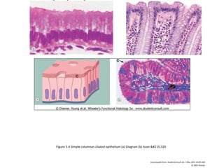
- 1. Figure 5.4 Simple columnar ciliated epithelium (a) Diagram (b) Azan ×320
Downloaded from: StudentConsult (on 7 May 2013 10:04 AM)
© 2005 Elsevier
- 2. Figure 5.5 Pseudostratified columnar ciliated epithelium (a) Diagram (b) H & E ×200
Downloaded from: StudentConsult (on 7 May 2013 10:04 AM)
© 2005 Elsevier
- 3. Figure 5.9 Transitional epithelium (a) Diagram (b) H & E ×320; BM basement membrane CJ communicating junctions D desmosome HD hemidesmosome IF intermediate filaments
Mf microfilaments Mv microvillus TJ tight junction TW terminal web ZA zonula adherens
Downloaded from: StudentConsult (on 7 May 2013 10:04 AM)
© 2005 Elsevier
- 4. Figure 5.1 Simple squamous epithelium (a) Diagram (b) H & E ×800 (c) Spread preparation, silver method/neutral red ×320; BM basement membrane E epithelial lining cells N
nucleus
Downloaded from: StudentConsult (on 7 May 2013 10:04 AM)
© 2005 Elsevier
HISTOLOGY O.S.P.E:
Epithelium
- 5. Figure 5.2 Simple cuboidal epithelium (a) Diagram (b) Azan ×400
Downloaded from: StudentConsult (on 7 May 2013 10:04 AM)
© 2005 Elsevier
- 6. Figure 5.4 Simple columnar ciliated epithelium (a) Diagram (b) Azan ×320
Downloaded from: StudentConsult (on 7 May 2013 10:04 AM)
© 2005 Elsevier
- 7. Figure 5.8 Stratified cuboidal epithelium H & E ×320
Downloaded from: StudentConsult (on 7 May 2013 10:04 AM)
© 2005 Elsevier
- 8. Figure 5.5 Pseudostratified columnar ciliated epithelium (a) Diagram (b) H & E ×200
Downloaded from: StudentConsult (on 7 May 2013 10:04 AM)
© 2005 Elsevier
- 9. Figure 5.6 Stratified squamous epithelium (a) Diagram (b) H & E ×100 (c) H & E ×200 (d) Papanicolaou ×400; BM basement membrane K keratin layer Ke abnormal
keratinisation U umbrella cell
Downloaded from: StudentConsult (on 7 May 2013 10:04 AM)
© 2005 Elsevier
- 10. Figure 5.9 Transitional epithelium (a) Diagram (b) H & E ×320; BM basement membrane CJ communicating junctions D desmosome HD hemidesmosome IF intermediate filaments
Mf microfilaments Mv microvillus TJ tight junction TW terminal web ZA zonula adherens
Downloaded from: StudentConsult (on 7 May 2013 10:04 AM)
© 2005 Elsevier
- 13. Figure 10.1 Hyaline cartilage (a) H & E ×150 (b) Thin epoxy resin section, toluidine blue ×1200; Cb chondroblasts Cc chondrocytes L lipid droplet M cartilage matrix N nucleus
P perichondrium
Downloaded from: StudentConsult (on 11 May 2013 12:38 PM)
© 2005 Elsevier
Bones and Cartilages
- 15. Figure 10.9 Cortical (compact) bone
Downloaded from: StudentConsult (on 11 May 2013 12:38 PM)
© 2005 Elsevier
- 17. Figure 6.3 Skeletal muscle (a) H & E TS ×300 (b) H & E LS ×310 (c) H & E LS ×500
Downloaded from: StudentConsult (on 12 May 2013 09:42 AM)
© 2005 Elsevier
- 18. Figure 6.22 Cardiac muscle (a) LS, H & E ×198 (b) TS, H & E ×480 (c) LS, H & E polarised light ×480
Downloaded from: StudentConsult (on 12 May 2013 09:42 AM)
© 2005 Elsevier
- 19. Figure 6.15 Smooth muscle (a) LS, H & E ×480
Downloaded from: StudentConsult (on 12 May 2013 09:42 AM)
© 2005 Elsevier
- 21. Figure 6.8 Skeletal muscle (a) TS, iron haematoxylin &#
Downloaded from: StudentConsult (on 12 May 2013 09:42 AM)
© 2005 Elsevier
- 26. Figure 3.6 Eosinophils (a) Giemsa ×1600(b) Human: EM ×25 000 (c) Mouse: EM ×20 000 (opposite) (d) Rat: EM ×25 000; M mitochondria N nucleus R ribosomes
rER rough endoplasmic reticulum S specific granules sER smooth endoplasmic reticulum
Downloaded from: StudentConsult (on 6 May 2013 05:14 PM)
© 2005 Elsevier
- 27. Figure 3.7 Basophils (a) Giemsa ×1500(b) EM ×10 500; S specific granules
Downloaded from: StudentConsult (on 6 May 2013 05:15 PM)
© 2005 Elsevier
- 29. Figure 17.2 Pituitary gland H & E ×12
Downloaded from: StudentConsult (on 7 May 2013 04:09 PM)
© 2005 Elsevier
A
B
C
- 31. Figure 17.6 Thyroid gland H & E ×12
Downloaded from: StudentConsult (on 7 May 2013 04:09 PM)
© 2005 Elsevier
- 32. Figure 17.14 Adrenal gland (a) Azan ×12 (b) Azan ×20; C cortex Cap capsule F zona fasciculata G zona glomerulosa M medulla R zona reticularis T trabeculae V vein
Downloaded from: StudentConsult (on 7 May 2013 04:10 PM)
© 2005 Elsevier
- 35. Figure 7.1 The neurone
Downloaded from: StudentConsult (on 12 May 2013 09:24 AM)
© 2005 Elsevier
- 36. Figure 7.2 Basic neurone types
Downloaded from: StudentConsult (on 12 May 2013 09:24 AM)
© 2005 Elsevier
- 37. Figure 7.13 Peripheral nerve H & E ×20
Downloaded from: StudentConsult (on 12 May 2013 09:24 AM)
© 2005 Elsevier
- 38. Figure 7.16 Peripheral nerve H & E ×480
Downloaded from: StudentConsult (on 12 May 2013 09:24 AM)
© 2005 Elsevier
- 39. Figure 11.5 Thymus (a) H & E ×15 (b) H & E ×15; A adipose tissue BM basement membrane C capsule Cx cortex E endothelial cells Ep epithelial cell H Hassall's corpuscle L
lymphoid tissue M medulla Ma macrophage Mt mitotic figure S septum
Downloaded from: StudentConsult (on 6 May 2013 05:15 PM)
© 2005 Elsevier
- 40. Figure 11.7 Thymic medulla (a) H & E ×480 (b) Immunoperoxidase cytokeratin ×100
Downloaded from: StudentConsult (on 6 May 2013 05:15 PM)
© 2005 Elsevier
- 41. Figure 11.9 Lymph node structure H & E ×8; C capsule CS cortical sinus Cx cortex F follicle H hilum M medulla MC medullary cords MS medullary sinus P paracortex S subcapsular
sinus T trabecula TS trabecular sinus
Downloaded from: StudentConsult (on 6 May 2013 05:15 PM)
© 2005 Elsevier
- 42. Figure 14.2 Structure of the gastrointestinal tract
Downloaded from: StudentConsult (on 12 May 2013 10:08 AM)
© 2005 Elsevier
- 44. Figure 14.5 Oesophagus Masson's trichrome ×9 (b) Masson's trichrome ×320;CM inner circular layer of muscularis propria E epithelium G seromucous gland LM outer
longitudinal layer of muscularis propria LP lamina propria Ly lymphoid aggregates MM muscularis mucosae MP muscularis propria PG parasympathetic ganglion Sk skeletal muscle fibres
Sm smooth muscle fibres SM submucosa
Downloaded from: StudentConsult (on 12 May 2013 10:08 AM)
© 2005 Elsevier
- 45. Figure 14.3 Basic mucosal forms in the gastrointestinal tract H & E: (a) ×100 (b) ×100 (c) ×128 (d) ×128
Downloaded from: StudentConsult (on 12 May 2013 10:08 AM)
© 2005 Elsevier
- 46. Figure 14.16 Small intestine - monkey (a) Duodenum H & E ×20 (b) ileum H & E ×16; B Brunner's glands CM inner circular layer of muscularis propria LP lamina propria LM
outer longitudinal layer of muscularis propria M mucosa MM muscularis mucosae P Peyer's patches PC plicae circulares SM submucosa Sr serosa V villi
Downloaded from: StudentConsult (on 12 May 2013 10:08 AM)
© 2005 Elsevier
- 49. Figure 14.32 Recto-anal junction H & E ×60
Downloaded from: StudentConsult (on 12 May 2013 10:09 AM)
© 2005 Elsevier
- 50. Figure 16.7 Renal corpuscle
Downloaded from: StudentConsult (on 8 May 2013 11:08 AM)
© 2005 Elsevier
- 52. Downloaded from: StudentConsult (on 8 May 2013 11:09 AM)
© 2005 Elsevier
A – Afferent Arteriole
B – Efferent Arteriole
C – Capillary Endothelium
E – Glomerular basement membrane
D – Bowman’s Capsule Epithelium
F – Beginning of Proximal Convoluted Tubule
Renal corpuscle
- 53. PCT proximal convoluted tubule BB brush border
Downloaded from: StudentConsult (on 8 May 2013 11:08 AM)
© 2005 Elsevier
- 67. Figure 12.6 Trachea H & E/Alcian blue ×9
Downloaded from: StudentConsult (on 6 May 2013 06:38 PM)
© 2005 Elsevier
C : cartilage, T
trachealis
muscle
- 68. Figure 12.12 Terminal portion of the respiratory tree H & E ×40
Downloaded from: StudentConsult (on 6 May 2013 06:38 PM)
© 2005 Elsevier
T terminal
bronchiole,
R respiratory
bronchiole,
A alveoli ,
AD alveolar duct
AS alveolar sacs