SlideShare une entreprise Scribd logo
1  sur  65
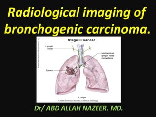
Radiological imaging of
bronchogenic carcinoma.
Dr/ ABD ALLAH NAZEER. MD.
Bronchogenic carcinoma, (or frequently known as lung
cancer) is the most common cause of cancer in men, and the 6th
most frequent cancer in women worldwide. It is the leading
cause of cancer mortality worldwide in both men and women
and accounts for approximately 20% of all cancer deaths.
Epidemiology.
The major risk factor is cigarette smoking which is implicated
in 90% of cases. Other risk factors include
radon, asbestos, uranium, arsenic, chromium, COPD and lung
fibrosis.
Nearly 90% of lung cancer are related to smoking.
226,000 are newly diagnosed: 160,000 die of lung cancer per
year.
The average 5 years is > 15% survival.
Introduction.
Squamous and adenocarcinoma have about the same 5-year
survival probability. It is worse for large cell carcinoma, and oat cell
carcinoma has almost no 5-year survival probability with a mean
lifespan of months from the time of diagnosis. Overall 8% of lung
cancer cases live past 5 years.
Squamous (epidermoid) carcinoma derived from reserve cells that
differentiate into squamous cells. Most commonly found centrally in
the lung, usually in the major lobar or first segmental bronchus.
Growth of the tumour occurs distally and proximally from site of
origin, destroying normal tissue and structures. Squamous
carcinomas tend to grow very large, and may kill by local growth.
These tumours often cavitate and resemble abscesses on
radiographic studies grossly. The tumour may be surgically resected
but cure rate depends on the clinical stage at the time of diagnosis.
Early diagnosis is possible by cytologic examination of a sputum
sample.
Adenocarcinoma:
Most adenocarcinoma are insidious and asymptomatic for a long
time. Adenocarcinoma tend to occur more peripherally in the lung.
Adenocarcinoma is the one cell type of primary lung tumour that
occurs more often in nonsmokers. Treatment is surgical and involves
removal of the entire lobe with associated lymph nodes..
Bronchiolo-alveolar carcinoma
A subtype of adenocarcinoma. The tumour grows on the underlying
supportive structure of the lung parenchyma without causing much
damage to that structure. A rapid ‘pneumonic’ spread occurs to
other areas of the lung. Thus, on CXR it resembles a pneumonia-like
infiltrate rather than a mass.
Small cell carcinoma (‘oat cell’ carcinoma)
A highly malignant form of ‘neuroendocrine’ tumour. Derived from
reserve cells, differentiate towards neuroendocrine (Kulchitsky)
cells. Occur almost exclusively in smokers.
Often associated with paraneoplastic syndromes e.g. hormonal
effects - Ectopic ACTH production - Inappropriate ADH secretion.
Small cell carcinomas arise centrally and tends to spread diffusely
along and into the bronchial wall. Extensive necrosis within the tumour
is common due to its rapid growth outgrowing the blood supply.
Small cell carcinomas are not generally considered resectable
cancers since dissemination is likely to have occured by the time they
are discovered. They are responsive to chemotherapy. Radiotherapy
is also useful, but they are rarely curable.
Large Cell Carcinoma
Carcinomas which are so poorly differentiated that by routine light
microscopy they cannot be placed into either the epidermoid or
glandular groups. Ultrastructurally, however, these tumours frequently
demonstrate features of either epidermoid cells or glandular cells or
both. Arise centrally in lung. Highly aggressive tumours.
Spread of lung carcinoma
Local.
Lymphogenic to regional lymph nodes: -
hilar, mediastinal, supraclavicular.
Hematogenous: Adrenal glands, brain, bones, liver.
Local infiltration
Bronchial obstruction ⇒ pneumonia, atelectasis, bronchiectasis distal
to obstruction.
Infiltration of lung parenchyma, pleura, pericardium, chest
wall, vertebrae.
Infiltration of superior vena cava ⇒ SVC syndrome (swelling of
face, fullness of neck veins).
Apical tumours may infiltrate:
-Cervical sympathetic nerves ⇒ Horner’s syndrome (ptosis, miosis,
anhydrosis on same side of lesion).
-Brachial plexus ⇒ Pancoast syndrome (neurological manifestations
e.g. pain in upper extremity).
New T-staging according to the 7th edition of the TNM-staging of lung cancer
Stage I Non-Small Cell Lung Cancer.
Cancer is found only in the
lung, usually less than 3 cm and
not invading the bronchi.
Surgical removal recommended.
Radiation therapy and/or
chemotherapy may also be used.
Stage II Non-Small Cell Lung Cancer.
The cancer has spread to lymph
nodes in the lung, more than 3
cm, reaching pleura or
bronchi, but away from carina
more 2 cm.
Treatment is surgery to remove
the tumor and nearby lymph
nodes.
Chemotherapy recommended;
radiation therapy sometimes
given after chemotherapy.
Stage III Non-Small Cell Lung Cancer.
The cancer has spread to the lymph nodes
located in the center of the chest, outside
the lung .
Stage IIIA cancer has spread to lymph
nodes in the chest, on the same side
where the cancer originated.
Stage IIIB cancer has spread to lymph
nodes on the opposite side of the
chest, under the collarbone, or the pleura
(lining of the chest cavity).
Surgery or radiation therapy with
chemotherapy recommended for stage
IIIA .
Chemotherapy and sometimes radiation
therapy recommended for stage IIIB.
Stage IV Non-Small Cell Lung Cancer.
The cancer has spread to
different lobes of the lung
or to other organs, such as
the brain, bones, and liver.
Stage IV non-small cell
lung cancer is treated with
chemotherapy.
Small Cell Lung Cancer–All Stages.
Patients with limited stage (confined to one area of the
chest) small cell lung cancer are treated with simultaneous
radiation therapy and chemotherapy.
Patients with extensive stage (not confined to one area of
the chest) small cell lung cancer are treated with
chemotherapy only.
Because small cell lung cancer can spread to the
brain, preventative radiation therapy to the brain is
routinely recommended to all patients whose tumors
disappear following chemotherapy and radiation therapy.
Left upper lobe bronchogenic carcinoma.
Peripheral bronchogenic carcinoma(T1 tumour).
T1 tumor.
T2 tumor
Adenocarcinoma in a 41-year-old man with right shoulder pain for several months. (a)
Apical lordotic chest radiograph demonstrates a right apical mass with poorly marginated
borders. (b) Chest CT scan (lung window) shows a homogeneous peripheral right upper
lobe mass with irregular borders. There is tumor involvement of a posterior rib (arrow).
Bronchioloalveolar carcinoma.
Squamous cell carcinoma
Adenosquamous carcinoma.
Undifferentiated large cell carcinoma.
Small cell carcinoma
Central lung cancer and atelectasis.
T3 tumor with invasion of the chest wall.
T4 tumor with invasion of the mediastinum.
T4 tumor constricting the right pulmonary artery (blue arrow)
Pancoast tumor in the right upper lobe with
displacement of the superior mediastinum and trachea.
T4N2-tumor
N3-stage disease.
Chest wall invasion.
MRI advantages.
Multiplanar imaging.
Can differentiate tumor tissue from chest wall muscles.
Cine MRI during breathing can assess the
tumor movement against the pleural surface.
In Pancoast’s tumors, MR can assess invasion of the
brachial plexus and subclavian vessels.
The accuracy of MRI versus CT in this domain in 94% for
MRI compared to 63% for CT.
Pancoast tumor
Peripheral bronchogenic carcinoma with positive
axillary and mediastinal nodes by PET Scan.
Pulmonary lymphoma refers to lung parenchymal involvement
with lymphoma. It can be broadly divided as primary or
secondary. primary pulmonary lymphoma - (rare) usually non-
Hodgkin lymphoma which is limited to the lung with or without
mediastinal lymph node involvement and with no evidence of
extrathoracic dissemination for at least 3 months after the initial
diagnosis. low-grade B-cell lymphoma (MALToma) – commonest
80% of primary pulmonary lymphoma high grade B-cell
lymphoma - most commonly B-cell, occasionally anaplastic and
peripheral T-cell types primary pulmonary plasmacytoma
(rare)pulmonary intravascular lymphoma (rare) angiocentric
immunoproliferative lesion (lymphomatoid granulomatosis)
secondary pulmonary lymphoma - relatively common Hodgkin
disease, non-Hodgkin lymphoma
Two additional categories have also been described which include
lymphoma in patients with post-transplant lymphoproliferative
disorders (PTLD) AIDS-related pulmonary lymphoma (ARL - ARPL).
Radiographic features
HRCT - chest
A number of features may be present on HRCT which include, a mass or
mass-like consolidation greater than 1 cm with or without cavitations or
bronchograms - most common finding according to one study masses of
pleural origin nodules less than 1 cm alveolar or interstitial infiltrates
peribronchial or perivascular thickening with or without atelectasis
pleural effusions, hilar or mediastinal lymphadenopathy
Pulmonary lymphoma in immune compromised patient.
Pulmonary metastases are common and the result of
metastatic spread to the lungs from a variety of tumours. Spread
can occur via blood, or lymphatics. The remainder of this article
concerns itself with hematogenous pulmonary metastases.
Lymphangitis carcinomatosis is discussed separately.
Pulmonary metastases from testicular cancer.
Presentation1.pptx. radiological imaging of bronchogenic carcinom.

Contenu connexe

Tendances

renal cell carcinoma radiology
renal cell carcinoma radiologyrenal cell carcinoma radiology
renal cell carcinoma radiologydocaashishgupt
 
Radiological imaging of pulmonary neoplasms
Radiological imaging of pulmonary neoplasmsRadiological imaging of pulmonary neoplasms
Radiological imaging of pulmonary neoplasmsPankaj Kaira
 
Diagnostic Imaging of Pulmonary infections
Diagnostic Imaging of Pulmonary infectionsDiagnostic Imaging of Pulmonary infections
Diagnostic Imaging of Pulmonary infectionsMohamed M.A. Zaitoun
 
Radiology Spots PPT- 2 by Dr Chandni Wadhwani
Radiology Spots PPT- 2 by Dr Chandni WadhwaniRadiology Spots PPT- 2 by Dr Chandni Wadhwani
Radiology Spots PPT- 2 by Dr Chandni WadhwaniChandni Wadhwani
 
Radiological approach to gastric ulcer disease
Radiological approach to gastric ulcer diseaseRadiological approach to gastric ulcer disease
Radiological approach to gastric ulcer diseaseNavneet Ranjan
 
Radiology Spots PPT- 3 by Dr Chandni Wadhwani
 Radiology Spots PPT- 3 by Dr Chandni Wadhwani Radiology Spots PPT- 3 by Dr Chandni Wadhwani
Radiology Spots PPT- 3 by Dr Chandni WadhwaniChandni Wadhwani
 
Mediastinal lesions final
Mediastinal lesions   finalMediastinal lesions   final
Mediastinal lesions finalGobardhan Thapa
 
Testicular tumor imaging
Testicular tumor imaging Testicular tumor imaging
Testicular tumor imaging Bom BC
 
INFLAMMATORY BOWEL DISEASE IMAGING(RADIOLOGY)
INFLAMMATORY BOWEL DISEASE IMAGING(RADIOLOGY)INFLAMMATORY BOWEL DISEASE IMAGING(RADIOLOGY)
INFLAMMATORY BOWEL DISEASE IMAGING(RADIOLOGY)Khyati Vadera
 
Diagnostic Imaging of Pulmonary Tumors
Diagnostic Imaging of Pulmonary TumorsDiagnostic Imaging of Pulmonary Tumors
Diagnostic Imaging of Pulmonary TumorsMohamed M.A. Zaitoun
 
radiology Spotters mixed bag
radiology Spotters mixed bagradiology Spotters mixed bag
radiology Spotters mixed bagAnish Choudhary
 
Radiological approach to lung neoplasms
Radiological approach to lung neoplasmsRadiological approach to lung neoplasms
Radiological approach to lung neoplasmsSnehaMandal5
 
Imaging of the Pancreas
Imaging of the PancreasImaging of the Pancreas
Imaging of the PancreasAtit Ghoda
 
Imaging of the large bowel
Imaging of the large bowelImaging of the large bowel
Imaging of the large bowelArchana Koshy
 
Benign bone disease spotters
Benign bone disease spottersBenign bone disease spotters
Benign bone disease spottersAGRAWAL14
 
Radiological imaging of mediastinal masses
Radiological imaging of mediastinal massesRadiological imaging of mediastinal masses
Radiological imaging of mediastinal massesPankaj Kaira
 

Tendances (20)

LUNG MASSES
LUNG MASSESLUNG MASSES
LUNG MASSES
 
renal cell carcinoma radiology
renal cell carcinoma radiologyrenal cell carcinoma radiology
renal cell carcinoma radiology
 
Radiological imaging of pulmonary neoplasms
Radiological imaging of pulmonary neoplasmsRadiological imaging of pulmonary neoplasms
Radiological imaging of pulmonary neoplasms
 
Diagnostic Imaging of Pulmonary infections
Diagnostic Imaging of Pulmonary infectionsDiagnostic Imaging of Pulmonary infections
Diagnostic Imaging of Pulmonary infections
 
Radiology Spots PPT- 2 by Dr Chandni Wadhwani
Radiology Spots PPT- 2 by Dr Chandni WadhwaniRadiology Spots PPT- 2 by Dr Chandni Wadhwani
Radiology Spots PPT- 2 by Dr Chandni Wadhwani
 
Radiological approach to gastric ulcer disease
Radiological approach to gastric ulcer diseaseRadiological approach to gastric ulcer disease
Radiological approach to gastric ulcer disease
 
Radiology Spots PPT- 3 by Dr Chandni Wadhwani
 Radiology Spots PPT- 3 by Dr Chandni Wadhwani Radiology Spots PPT- 3 by Dr Chandni Wadhwani
Radiology Spots PPT- 3 by Dr Chandni Wadhwani
 
Mediastinal lesions final
Mediastinal lesions   finalMediastinal lesions   final
Mediastinal lesions final
 
Testicular tumor imaging
Testicular tumor imaging Testicular tumor imaging
Testicular tumor imaging
 
INFLAMMATORY BOWEL DISEASE IMAGING(RADIOLOGY)
INFLAMMATORY BOWEL DISEASE IMAGING(RADIOLOGY)INFLAMMATORY BOWEL DISEASE IMAGING(RADIOLOGY)
INFLAMMATORY BOWEL DISEASE IMAGING(RADIOLOGY)
 
Diagnostic Imaging of Pulmonary Tumors
Diagnostic Imaging of Pulmonary TumorsDiagnostic Imaging of Pulmonary Tumors
Diagnostic Imaging of Pulmonary Tumors
 
Gastric carcinoma radiology ppt
Gastric carcinoma radiology  ppt Gastric carcinoma radiology  ppt
Gastric carcinoma radiology ppt
 
radiology Spotters mixed bag
radiology Spotters mixed bagradiology Spotters mixed bag
radiology Spotters mixed bag
 
Radiological approach to lung neoplasms
Radiological approach to lung neoplasmsRadiological approach to lung neoplasms
Radiological approach to lung neoplasms
 
Imaging of the Pancreas
Imaging of the PancreasImaging of the Pancreas
Imaging of the Pancreas
 
Radiology spotters
Radiology spottersRadiology spotters
Radiology spotters
 
Imaging of the large bowel
Imaging of the large bowelImaging of the large bowel
Imaging of the large bowel
 
Git signs
Git signsGit signs
Git signs
 
Benign bone disease spotters
Benign bone disease spottersBenign bone disease spotters
Benign bone disease spotters
 
Radiological imaging of mediastinal masses
Radiological imaging of mediastinal massesRadiological imaging of mediastinal masses
Radiological imaging of mediastinal masses
 

En vedette

Radiological features of Lung cancer Dr. Muhammad Bin Zulfiqar
Radiological features of Lung cancer Dr. Muhammad Bin ZulfiqarRadiological features of Lung cancer Dr. Muhammad Bin Zulfiqar
Radiological features of Lung cancer Dr. Muhammad Bin ZulfiqarDr. Muhammad Bin Zulfiqar
 
Diagnostic imaging of lung cancer
Diagnostic imaging of lung cancerDiagnostic imaging of lung cancer
Diagnostic imaging of lung cancerDouble M
 
Bronchogenic carcinoma
Bronchogenic carcinomaBronchogenic carcinoma
Bronchogenic carcinomamaimusirdan
 
Ppt lung carcinoma part1
Ppt lung carcinoma part1Ppt lung carcinoma part1
Ppt lung carcinoma part1Juned Khan
 
Lung Cancer: Diagnosis, Staging, and Treatment
Lung Cancer: Diagnosis, Staging, and TreatmentLung Cancer: Diagnosis, Staging, and Treatment
Lung Cancer: Diagnosis, Staging, and TreatmentDene W. Daugherty
 
Fundamentals of chest radiology
Fundamentals of chest radiologyFundamentals of chest radiology
Fundamentals of chest radiologyDr. Sreedhar Rao
 
Presentation1.pptx, radiological imaging of pulmonary infection.
Presentation1.pptx, radiological imaging of pulmonary infection.Presentation1.pptx, radiological imaging of pulmonary infection.
Presentation1.pptx, radiological imaging of pulmonary infection.Abdellah Nazeer
 
Ct chest pneumonias and neoplasms
Ct chest pneumonias and neoplasmsCt chest pneumonias and neoplasms
Ct chest pneumonias and neoplasmsRikin Hasnani
 
Lung cancer staging the noninvasive tools
Lung cancer staging the noninvasive toolsLung cancer staging the noninvasive tools
Lung cancer staging the noninvasive toolsAbdulsalam Taha
 
Ablation of HCC
Ablation of HCCAblation of HCC
Ablation of HCCPAIRS WEB
 
group 3
group 3group 3
group 3kilaye
 

En vedette (20)

Lung tumor radiology
Lung tumor radiologyLung tumor radiology
Lung tumor radiology
 
Radiological features of Lung cancer Dr. Muhammad Bin Zulfiqar
Radiological features of Lung cancer Dr. Muhammad Bin ZulfiqarRadiological features of Lung cancer Dr. Muhammad Bin Zulfiqar
Radiological features of Lung cancer Dr. Muhammad Bin Zulfiqar
 
Diagnostic imaging of lung cancer
Diagnostic imaging of lung cancerDiagnostic imaging of lung cancer
Diagnostic imaging of lung cancer
 
X-Ray Chest: Carcinoma Lung
X-Ray Chest: Carcinoma LungX-Ray Chest: Carcinoma Lung
X-Ray Chest: Carcinoma Lung
 
Bronchogenic carcinoma
Bronchogenic carcinomaBronchogenic carcinoma
Bronchogenic carcinoma
 
Lung Cancer
Lung CancerLung Cancer
Lung Cancer
 
Ppt lung carcinoma part1
Ppt lung carcinoma part1Ppt lung carcinoma part1
Ppt lung carcinoma part1
 
Lung Cancer: Diagnosis, Staging, and Treatment
Lung Cancer: Diagnosis, Staging, and TreatmentLung Cancer: Diagnosis, Staging, and Treatment
Lung Cancer: Diagnosis, Staging, and Treatment
 
Lung Cancer
Lung CancerLung Cancer
Lung Cancer
 
Fundamentals of chest radiology
Fundamentals of chest radiologyFundamentals of chest radiology
Fundamentals of chest radiology
 
Lung cancer, 3rd ed
Lung cancer, 3rd edLung cancer, 3rd ed
Lung cancer, 3rd ed
 
Presentation1.pptx, radiological imaging of pulmonary infection.
Presentation1.pptx, radiological imaging of pulmonary infection.Presentation1.pptx, radiological imaging of pulmonary infection.
Presentation1.pptx, radiological imaging of pulmonary infection.
 
Chest x ray pathology
Chest x ray pathologyChest x ray pathology
Chest x ray pathology
 
Lung Carcinoma
Lung CarcinomaLung Carcinoma
Lung Carcinoma
 
V/Q scan
V/Q scanV/Q scan
V/Q scan
 
Ct chest pneumonias and neoplasms
Ct chest pneumonias and neoplasmsCt chest pneumonias and neoplasms
Ct chest pneumonias and neoplasms
 
Lung cancer staging the noninvasive tools
Lung cancer staging the noninvasive toolsLung cancer staging the noninvasive tools
Lung cancer staging the noninvasive tools
 
Chest imaging
Chest imagingChest imaging
Chest imaging
 
Ablation of HCC
Ablation of HCCAblation of HCC
Ablation of HCC
 
group 3
group 3group 3
group 3
 

Similaire à Presentation1.pptx. radiological imaging of bronchogenic carcinom.

BENIGN AND MALIGNANT LUNG NEOPLASAM MASSES
BENIGN AND MALIGNANT LUNG NEOPLASAM MASSESBENIGN AND MALIGNANT LUNG NEOPLASAM MASSES
BENIGN AND MALIGNANT LUNG NEOPLASAM MASSESnishit viradia
 
Lungs Cancer etiology sign symtom causes.pptx
Lungs Cancer etiology sign symtom causes.pptxLungs Cancer etiology sign symtom causes.pptx
Lungs Cancer etiology sign symtom causes.pptxShaheerShakeel1
 
Non Small Cell Lung Cancer
Non Small Cell Lung CancerNon Small Cell Lung Cancer
Non Small Cell Lung Cancerfondas vakalis
 
Lung Cancer and bronchopulmonary neoplasia
Lung Cancer and bronchopulmonary neoplasiaLung Cancer and bronchopulmonary neoplasia
Lung Cancer and bronchopulmonary neoplasiaYohannes Bisewer
 
Lung Cancer Pathology & Clinical
Lung Cancer Pathology & Clinical Lung Cancer Pathology & Clinical
Lung Cancer Pathology & Clinical Shahd Al Ali
 
Lung cancer by Dr. Aabidullah Rahimee
Lung cancer by Dr. Aabidullah RahimeeLung cancer by Dr. Aabidullah Rahimee
Lung cancer by Dr. Aabidullah RahimeeAabidullah
 
Malignant epthelial tumors of the lung
Malignant epthelial tumors of the lungMalignant epthelial tumors of the lung
Malignant epthelial tumors of the lungJoy Mridha
 
Lung tumors dr. rishi
Lung tumors dr. rishiLung tumors dr. rishi
Lung tumors dr. rishi1drrishisaini
 
lung neoplasms
lung neoplasmslung neoplasms
lung neoplasmsbbxoxo
 

Similaire à Presentation1.pptx. radiological imaging of bronchogenic carcinom. (20)

Primary pulmonary neoplasm
Primary pulmonary neoplasmPrimary pulmonary neoplasm
Primary pulmonary neoplasm
 
CARCINOMA OF LUNG.pptx
CARCINOMA OF LUNG.pptxCARCINOMA OF LUNG.pptx
CARCINOMA OF LUNG.pptx
 
Lung cancer
Lung cancerLung cancer
Lung cancer
 
Lung cancer.
Lung cancer.Lung cancer.
Lung cancer.
 
Lung cancer
Lung cancer Lung cancer
Lung cancer
 
lung tumors.pptx
lung tumors.pptxlung tumors.pptx
lung tumors.pptx
 
BENIGN AND MALIGNANT LUNG NEOPLASAM MASSES
BENIGN AND MALIGNANT LUNG NEOPLASAM MASSESBENIGN AND MALIGNANT LUNG NEOPLASAM MASSES
BENIGN AND MALIGNANT LUNG NEOPLASAM MASSES
 
Lung cancer
Lung cancerLung cancer
Lung cancer
 
Lung ca
Lung caLung ca
Lung ca
 
Lungs Cancer etiology sign symtom causes.pptx
Lungs Cancer etiology sign symtom causes.pptxLungs Cancer etiology sign symtom causes.pptx
Lungs Cancer etiology sign symtom causes.pptx
 
Pulmonary nodule
Pulmonary nodulePulmonary nodule
Pulmonary nodule
 
Non Small Cell Lung Cancer
Non Small Cell Lung CancerNon Small Cell Lung Cancer
Non Small Cell Lung Cancer
 
Lung Cancer and bronchopulmonary neoplasia
Lung Cancer and bronchopulmonary neoplasiaLung Cancer and bronchopulmonary neoplasia
Lung Cancer and bronchopulmonary neoplasia
 
Lung Cancer Pathology & Clinical
Lung Cancer Pathology & Clinical Lung Cancer Pathology & Clinical
Lung Cancer Pathology & Clinical
 
Lung cancer by Dr. Aabidullah Rahimee
Lung cancer by Dr. Aabidullah RahimeeLung cancer by Dr. Aabidullah Rahimee
Lung cancer by Dr. Aabidullah Rahimee
 
Malignant epthelial tumors of the lung
Malignant epthelial tumors of the lungMalignant epthelial tumors of the lung
Malignant epthelial tumors of the lung
 
Tumurs of the lung
Tumurs of the lungTumurs of the lung
Tumurs of the lung
 
Lung tumors dr. rishi
Lung tumors dr. rishiLung tumors dr. rishi
Lung tumors dr. rishi
 
lung neoplasms
lung neoplasmslung neoplasms
lung neoplasms
 
Lung cancer
Lung cancerLung cancer
Lung cancer
 

Plus de Abdellah Nazeer

Muculoskeletal Pediatic Imaging..pptx
Muculoskeletal Pediatic Imaging..pptxMuculoskeletal Pediatic Imaging..pptx
Muculoskeletal Pediatic Imaging..pptxAbdellah Nazeer
 
Presentation1, Ultrasound of the bowel loops and the lymph nodes..pptx
Presentation1, Ultrasound of the bowel loops and the lymph nodes..pptxPresentation1, Ultrasound of the bowel loops and the lymph nodes..pptx
Presentation1, Ultrasound of the bowel loops and the lymph nodes..pptxAbdellah Nazeer
 
Presentation1 Short cases MD..pptx
Presentation1 Short cases MD..pptxPresentation1 Short cases MD..pptx
Presentation1 Short cases MD..pptxAbdellah Nazeer
 
Presentation1, MD MCQ Cases..pptx
Presentation1, MD MCQ Cases..pptxPresentation1, MD MCQ Cases..pptx
Presentation1, MD MCQ Cases..pptxAbdellah Nazeer
 
Presentation1, Short Cases Quiz..pptx
Presentation1, Short Cases Quiz..pptxPresentation1, Short Cases Quiz..pptx
Presentation1, Short Cases Quiz..pptxAbdellah Nazeer
 
Presentation1, radiological imaging of lateral hindfoot impingement.
Presentation1, radiological imaging of lateral hindfoot impingement.Presentation1, radiological imaging of lateral hindfoot impingement.
Presentation1, radiological imaging of lateral hindfoot impingement.Abdellah Nazeer
 
Presentation2, radiological anatomy of the liver and spleen.
Presentation2, radiological anatomy of the liver and spleen.Presentation2, radiological anatomy of the liver and spleen.
Presentation2, radiological anatomy of the liver and spleen.Abdellah Nazeer
 
Presentation1, artifacts and pitfalls of the wrist and elbow joints.
Presentation1, artifacts and pitfalls of the wrist and elbow joints.Presentation1, artifacts and pitfalls of the wrist and elbow joints.
Presentation1, artifacts and pitfalls of the wrist and elbow joints.Abdellah Nazeer
 
Presentation1, artifact and pitfalls of the knee, hip and ankle joints.
Presentation1, artifact and pitfalls of the knee, hip and ankle joints.Presentation1, artifact and pitfalls of the knee, hip and ankle joints.
Presentation1, artifact and pitfalls of the knee, hip and ankle joints.Abdellah Nazeer
 
Presentation1, radiological imaging of artifact and pitfalls in shoulder join...
Presentation1, radiological imaging of artifact and pitfalls in shoulder join...Presentation1, radiological imaging of artifact and pitfalls in shoulder join...
Presentation1, radiological imaging of artifact and pitfalls in shoulder join...Abdellah Nazeer
 
Presentation1, radiological imaging of internal abdominal hernia.
Presentation1, radiological imaging of internal abdominal hernia.Presentation1, radiological imaging of internal abdominal hernia.
Presentation1, radiological imaging of internal abdominal hernia.Abdellah Nazeer
 
Presentation11, radiological imaging of ovarian torsion.
Presentation11, radiological imaging of ovarian torsion.Presentation11, radiological imaging of ovarian torsion.
Presentation11, radiological imaging of ovarian torsion.Abdellah Nazeer
 
Presentation1, musculoskeletal anatomy.
Presentation1, musculoskeletal anatomy.Presentation1, musculoskeletal anatomy.
Presentation1, musculoskeletal anatomy.Abdellah Nazeer
 
Presentation1, new mri techniques in the diagnosis and monitoring of multiple...
Presentation1, new mri techniques in the diagnosis and monitoring of multiple...Presentation1, new mri techniques in the diagnosis and monitoring of multiple...
Presentation1, new mri techniques in the diagnosis and monitoring of multiple...Abdellah Nazeer
 
Presentation1, radiological application of diffusion weighted mri in neck mas...
Presentation1, radiological application of diffusion weighted mri in neck mas...Presentation1, radiological application of diffusion weighted mri in neck mas...
Presentation1, radiological application of diffusion weighted mri in neck mas...Abdellah Nazeer
 
Presentation1, radiological application of diffusion weighted images in breas...
Presentation1, radiological application of diffusion weighted images in breas...Presentation1, radiological application of diffusion weighted images in breas...
Presentation1, radiological application of diffusion weighted images in breas...Abdellah Nazeer
 
Presentation1, radiological application of diffusion weighted images in abdom...
Presentation1, radiological application of diffusion weighted images in abdom...Presentation1, radiological application of diffusion weighted images in abdom...
Presentation1, radiological application of diffusion weighted images in abdom...Abdellah Nazeer
 
Presentation1, radiological application of diffusion weighted imges in neuror...
Presentation1, radiological application of diffusion weighted imges in neuror...Presentation1, radiological application of diffusion weighted imges in neuror...
Presentation1, radiological application of diffusion weighted imges in neuror...Abdellah Nazeer
 
Presentation1, mr physics.
Presentation1, mr physics.Presentation1, mr physics.
Presentation1, mr physics.Abdellah Nazeer
 
Presentation1. ct physics.
Presentation1. ct physics.Presentation1. ct physics.
Presentation1. ct physics.Abdellah Nazeer
 

Plus de Abdellah Nazeer (20)

Muculoskeletal Pediatic Imaging..pptx
Muculoskeletal Pediatic Imaging..pptxMuculoskeletal Pediatic Imaging..pptx
Muculoskeletal Pediatic Imaging..pptx
 
Presentation1, Ultrasound of the bowel loops and the lymph nodes..pptx
Presentation1, Ultrasound of the bowel loops and the lymph nodes..pptxPresentation1, Ultrasound of the bowel loops and the lymph nodes..pptx
Presentation1, Ultrasound of the bowel loops and the lymph nodes..pptx
 
Presentation1 Short cases MD..pptx
Presentation1 Short cases MD..pptxPresentation1 Short cases MD..pptx
Presentation1 Short cases MD..pptx
 
Presentation1, MD MCQ Cases..pptx
Presentation1, MD MCQ Cases..pptxPresentation1, MD MCQ Cases..pptx
Presentation1, MD MCQ Cases..pptx
 
Presentation1, Short Cases Quiz..pptx
Presentation1, Short Cases Quiz..pptxPresentation1, Short Cases Quiz..pptx
Presentation1, Short Cases Quiz..pptx
 
Presentation1, radiological imaging of lateral hindfoot impingement.
Presentation1, radiological imaging of lateral hindfoot impingement.Presentation1, radiological imaging of lateral hindfoot impingement.
Presentation1, radiological imaging of lateral hindfoot impingement.
 
Presentation2, radiological anatomy of the liver and spleen.
Presentation2, radiological anatomy of the liver and spleen.Presentation2, radiological anatomy of the liver and spleen.
Presentation2, radiological anatomy of the liver and spleen.
 
Presentation1, artifacts and pitfalls of the wrist and elbow joints.
Presentation1, artifacts and pitfalls of the wrist and elbow joints.Presentation1, artifacts and pitfalls of the wrist and elbow joints.
Presentation1, artifacts and pitfalls of the wrist and elbow joints.
 
Presentation1, artifact and pitfalls of the knee, hip and ankle joints.
Presentation1, artifact and pitfalls of the knee, hip and ankle joints.Presentation1, artifact and pitfalls of the knee, hip and ankle joints.
Presentation1, artifact and pitfalls of the knee, hip and ankle joints.
 
Presentation1, radiological imaging of artifact and pitfalls in shoulder join...
Presentation1, radiological imaging of artifact and pitfalls in shoulder join...Presentation1, radiological imaging of artifact and pitfalls in shoulder join...
Presentation1, radiological imaging of artifact and pitfalls in shoulder join...
 
Presentation1, radiological imaging of internal abdominal hernia.
Presentation1, radiological imaging of internal abdominal hernia.Presentation1, radiological imaging of internal abdominal hernia.
Presentation1, radiological imaging of internal abdominal hernia.
 
Presentation11, radiological imaging of ovarian torsion.
Presentation11, radiological imaging of ovarian torsion.Presentation11, radiological imaging of ovarian torsion.
Presentation11, radiological imaging of ovarian torsion.
 
Presentation1, musculoskeletal anatomy.
Presentation1, musculoskeletal anatomy.Presentation1, musculoskeletal anatomy.
Presentation1, musculoskeletal anatomy.
 
Presentation1, new mri techniques in the diagnosis and monitoring of multiple...
Presentation1, new mri techniques in the diagnosis and monitoring of multiple...Presentation1, new mri techniques in the diagnosis and monitoring of multiple...
Presentation1, new mri techniques in the diagnosis and monitoring of multiple...
 
Presentation1, radiological application of diffusion weighted mri in neck mas...
Presentation1, radiological application of diffusion weighted mri in neck mas...Presentation1, radiological application of diffusion weighted mri in neck mas...
Presentation1, radiological application of diffusion weighted mri in neck mas...
 
Presentation1, radiological application of diffusion weighted images in breas...
Presentation1, radiological application of diffusion weighted images in breas...Presentation1, radiological application of diffusion weighted images in breas...
Presentation1, radiological application of diffusion weighted images in breas...
 
Presentation1, radiological application of diffusion weighted images in abdom...
Presentation1, radiological application of diffusion weighted images in abdom...Presentation1, radiological application of diffusion weighted images in abdom...
Presentation1, radiological application of diffusion weighted images in abdom...
 
Presentation1, radiological application of diffusion weighted imges in neuror...
Presentation1, radiological application of diffusion weighted imges in neuror...Presentation1, radiological application of diffusion weighted imges in neuror...
Presentation1, radiological application of diffusion weighted imges in neuror...
 
Presentation1, mr physics.
Presentation1, mr physics.Presentation1, mr physics.
Presentation1, mr physics.
 
Presentation1. ct physics.
Presentation1. ct physics.Presentation1. ct physics.
Presentation1. ct physics.
 

Dernier

Mumbai ] (Call Girls) in Mumbai 10k @ I'm VIP Independent Escorts Girls 98333...
Mumbai ] (Call Girls) in Mumbai 10k @ I'm VIP Independent Escorts Girls 98333...Mumbai ] (Call Girls) in Mumbai 10k @ I'm VIP Independent Escorts Girls 98333...
Mumbai ] (Call Girls) in Mumbai 10k @ I'm VIP Independent Escorts Girls 98333...Ishani Gupta
 
Coimbatore Call Girls in Thudiyalur : 7427069034 High Profile Model Escorts |...
Coimbatore Call Girls in Thudiyalur : 7427069034 High Profile Model Escorts |...Coimbatore Call Girls in Thudiyalur : 7427069034 High Profile Model Escorts |...
Coimbatore Call Girls in Thudiyalur : 7427069034 High Profile Model Escorts |...chennailover
 
💚Call Girls In Amritsar 💯Anvi 📲🔝8725944379🔝Amritsar Call Girl No💰Advance Cash...
💚Call Girls In Amritsar 💯Anvi 📲🔝8725944379🔝Amritsar Call Girl No💰Advance Cash...💚Call Girls In Amritsar 💯Anvi 📲🔝8725944379🔝Amritsar Call Girl No💰Advance Cash...
💚Call Girls In Amritsar 💯Anvi 📲🔝8725944379🔝Amritsar Call Girl No💰Advance Cash...Sheetaleventcompany
 
Call Girl in Indore 8827247818 {LowPrice} ❤️ (ahana) Indore Call Girls * UPA...
Call Girl in Indore 8827247818 {LowPrice} ❤️ (ahana) Indore Call Girls  * UPA...Call Girl in Indore 8827247818 {LowPrice} ❤️ (ahana) Indore Call Girls  * UPA...
Call Girl in Indore 8827247818 {LowPrice} ❤️ (ahana) Indore Call Girls * UPA...mahaiklolahd
 
Premium Bangalore Call Girls Jigani Dail 6378878445 Escort Service For Hot Ma...
Premium Bangalore Call Girls Jigani Dail 6378878445 Escort Service For Hot Ma...Premium Bangalore Call Girls Jigani Dail 6378878445 Escort Service For Hot Ma...
Premium Bangalore Call Girls Jigani Dail 6378878445 Escort Service For Hot Ma...tanya dube
 
Call Girls Amritsar Just Call 8250077686 Top Class Call Girl Service Available
Call Girls Amritsar Just Call 8250077686 Top Class Call Girl Service AvailableCall Girls Amritsar Just Call 8250077686 Top Class Call Girl Service Available
Call Girls Amritsar Just Call 8250077686 Top Class Call Girl Service AvailableDipal Arora
 
Call Girls Ahmedabad Just Call 9630942363 Top Class Call Girl Service Available
Call Girls Ahmedabad Just Call 9630942363 Top Class Call Girl Service AvailableCall Girls Ahmedabad Just Call 9630942363 Top Class Call Girl Service Available
Call Girls Ahmedabad Just Call 9630942363 Top Class Call Girl Service AvailableGENUINE ESCORT AGENCY
 
Call Girls Varanasi Just Call 8250077686 Top Class Call Girl Service Available
Call Girls Varanasi Just Call 8250077686 Top Class Call Girl Service AvailableCall Girls Varanasi Just Call 8250077686 Top Class Call Girl Service Available
Call Girls Varanasi Just Call 8250077686 Top Class Call Girl Service AvailableDipal Arora
 
Call Girls Hyderabad Just Call 8250077686 Top Class Call Girl Service Available
Call Girls Hyderabad Just Call 8250077686 Top Class Call Girl Service AvailableCall Girls Hyderabad Just Call 8250077686 Top Class Call Girl Service Available
Call Girls Hyderabad Just Call 8250077686 Top Class Call Girl Service AvailableDipal Arora
 
Top Rated Pune Call Girls (DIPAL) ⟟ 8250077686 ⟟ Call Me For Genuine Sex Serv...
Top Rated Pune Call Girls (DIPAL) ⟟ 8250077686 ⟟ Call Me For Genuine Sex Serv...Top Rated Pune Call Girls (DIPAL) ⟟ 8250077686 ⟟ Call Me For Genuine Sex Serv...
Top Rated Pune Call Girls (DIPAL) ⟟ 8250077686 ⟟ Call Me For Genuine Sex Serv...Dipal Arora
 
Call Girls Mysore Just Call 8250077686 Top Class Call Girl Service Available
Call Girls Mysore Just Call 8250077686 Top Class Call Girl Service AvailableCall Girls Mysore Just Call 8250077686 Top Class Call Girl Service Available
Call Girls Mysore Just Call 8250077686 Top Class Call Girl Service AvailableDipal Arora
 
9630942363 Genuine Call Girls In Ahmedabad Gujarat Call Girls Service
9630942363 Genuine Call Girls In Ahmedabad Gujarat Call Girls Service9630942363 Genuine Call Girls In Ahmedabad Gujarat Call Girls Service
9630942363 Genuine Call Girls In Ahmedabad Gujarat Call Girls ServiceGENUINE ESCORT AGENCY
 
(Low Rate RASHMI ) Rate Of Call Girls Jaipur ❣ 8445551418 ❣ Elite Models & Ce...
(Low Rate RASHMI ) Rate Of Call Girls Jaipur ❣ 8445551418 ❣ Elite Models & Ce...(Low Rate RASHMI ) Rate Of Call Girls Jaipur ❣ 8445551418 ❣ Elite Models & Ce...
(Low Rate RASHMI ) Rate Of Call Girls Jaipur ❣ 8445551418 ❣ Elite Models & Ce...parulsinha
 
Call Girls Service Jaipur {9521753030} ❤️VVIP RIDDHI Call Girl in Jaipur Raja...
Call Girls Service Jaipur {9521753030} ❤️VVIP RIDDHI Call Girl in Jaipur Raja...Call Girls Service Jaipur {9521753030} ❤️VVIP RIDDHI Call Girl in Jaipur Raja...
Call Girls Service Jaipur {9521753030} ❤️VVIP RIDDHI Call Girl in Jaipur Raja...Sheetaleventcompany
 
Independent Call Girls Service Mohali Sector 116 | 6367187148 | Call Girl Ser...
Independent Call Girls Service Mohali Sector 116 | 6367187148 | Call Girl Ser...Independent Call Girls Service Mohali Sector 116 | 6367187148 | Call Girl Ser...
Independent Call Girls Service Mohali Sector 116 | 6367187148 | Call Girl Ser...karishmasinghjnh
 
Call Girls Service Jaipur {8445551418} ❤️VVIP BHAWNA Call Girl in Jaipur Raja...
Call Girls Service Jaipur {8445551418} ❤️VVIP BHAWNA Call Girl in Jaipur Raja...Call Girls Service Jaipur {8445551418} ❤️VVIP BHAWNA Call Girl in Jaipur Raja...
Call Girls Service Jaipur {8445551418} ❤️VVIP BHAWNA Call Girl in Jaipur Raja...parulsinha
 
Call Girls Kolkata Kalikapur 💯Call Us 🔝 8005736733 🔝 💃 Top Class Call Girl Se...
Call Girls Kolkata Kalikapur 💯Call Us 🔝 8005736733 🔝 💃 Top Class Call Girl Se...Call Girls Kolkata Kalikapur 💯Call Us 🔝 8005736733 🔝 💃 Top Class Call Girl Se...
Call Girls Kolkata Kalikapur 💯Call Us 🔝 8005736733 🔝 💃 Top Class Call Girl Se...Namrata Singh
 
Call Girls Rishikesh Just Call 9667172968 Top Class Call Girl Service Available
Call Girls Rishikesh Just Call 9667172968 Top Class Call Girl Service AvailableCall Girls Rishikesh Just Call 9667172968 Top Class Call Girl Service Available
Call Girls Rishikesh Just Call 9667172968 Top Class Call Girl Service Availableperfect solution
 
VIP Hyderabad Call Girls Bahadurpally 7877925207 ₹5000 To 25K With AC Room 💚😋
VIP Hyderabad Call Girls Bahadurpally 7877925207 ₹5000 To 25K With AC Room 💚😋VIP Hyderabad Call Girls Bahadurpally 7877925207 ₹5000 To 25K With AC Room 💚😋
VIP Hyderabad Call Girls Bahadurpally 7877925207 ₹5000 To 25K With AC Room 💚😋TANUJA PANDEY
 

Dernier (20)

Mumbai ] (Call Girls) in Mumbai 10k @ I'm VIP Independent Escorts Girls 98333...
Mumbai ] (Call Girls) in Mumbai 10k @ I'm VIP Independent Escorts Girls 98333...Mumbai ] (Call Girls) in Mumbai 10k @ I'm VIP Independent Escorts Girls 98333...
Mumbai ] (Call Girls) in Mumbai 10k @ I'm VIP Independent Escorts Girls 98333...
 
Coimbatore Call Girls in Thudiyalur : 7427069034 High Profile Model Escorts |...
Coimbatore Call Girls in Thudiyalur : 7427069034 High Profile Model Escorts |...Coimbatore Call Girls in Thudiyalur : 7427069034 High Profile Model Escorts |...
Coimbatore Call Girls in Thudiyalur : 7427069034 High Profile Model Escorts |...
 
💚Call Girls In Amritsar 💯Anvi 📲🔝8725944379🔝Amritsar Call Girl No💰Advance Cash...
💚Call Girls In Amritsar 💯Anvi 📲🔝8725944379🔝Amritsar Call Girl No💰Advance Cash...💚Call Girls In Amritsar 💯Anvi 📲🔝8725944379🔝Amritsar Call Girl No💰Advance Cash...
💚Call Girls In Amritsar 💯Anvi 📲🔝8725944379🔝Amritsar Call Girl No💰Advance Cash...
 
🌹Attapur⬅️ Vip Call Girls Hyderabad 📱9352852248 Book Well Trand Call Girls In...
🌹Attapur⬅️ Vip Call Girls Hyderabad 📱9352852248 Book Well Trand Call Girls In...🌹Attapur⬅️ Vip Call Girls Hyderabad 📱9352852248 Book Well Trand Call Girls In...
🌹Attapur⬅️ Vip Call Girls Hyderabad 📱9352852248 Book Well Trand Call Girls In...
 
Call Girl in Indore 8827247818 {LowPrice} ❤️ (ahana) Indore Call Girls * UPA...
Call Girl in Indore 8827247818 {LowPrice} ❤️ (ahana) Indore Call Girls  * UPA...Call Girl in Indore 8827247818 {LowPrice} ❤️ (ahana) Indore Call Girls  * UPA...
Call Girl in Indore 8827247818 {LowPrice} ❤️ (ahana) Indore Call Girls * UPA...
 
Premium Bangalore Call Girls Jigani Dail 6378878445 Escort Service For Hot Ma...
Premium Bangalore Call Girls Jigani Dail 6378878445 Escort Service For Hot Ma...Premium Bangalore Call Girls Jigani Dail 6378878445 Escort Service For Hot Ma...
Premium Bangalore Call Girls Jigani Dail 6378878445 Escort Service For Hot Ma...
 
Call Girls Amritsar Just Call 8250077686 Top Class Call Girl Service Available
Call Girls Amritsar Just Call 8250077686 Top Class Call Girl Service AvailableCall Girls Amritsar Just Call 8250077686 Top Class Call Girl Service Available
Call Girls Amritsar Just Call 8250077686 Top Class Call Girl Service Available
 
Call Girls Ahmedabad Just Call 9630942363 Top Class Call Girl Service Available
Call Girls Ahmedabad Just Call 9630942363 Top Class Call Girl Service AvailableCall Girls Ahmedabad Just Call 9630942363 Top Class Call Girl Service Available
Call Girls Ahmedabad Just Call 9630942363 Top Class Call Girl Service Available
 
Call Girls Varanasi Just Call 8250077686 Top Class Call Girl Service Available
Call Girls Varanasi Just Call 8250077686 Top Class Call Girl Service AvailableCall Girls Varanasi Just Call 8250077686 Top Class Call Girl Service Available
Call Girls Varanasi Just Call 8250077686 Top Class Call Girl Service Available
 
Call Girls Hyderabad Just Call 8250077686 Top Class Call Girl Service Available
Call Girls Hyderabad Just Call 8250077686 Top Class Call Girl Service AvailableCall Girls Hyderabad Just Call 8250077686 Top Class Call Girl Service Available
Call Girls Hyderabad Just Call 8250077686 Top Class Call Girl Service Available
 
Top Rated Pune Call Girls (DIPAL) ⟟ 8250077686 ⟟ Call Me For Genuine Sex Serv...
Top Rated Pune Call Girls (DIPAL) ⟟ 8250077686 ⟟ Call Me For Genuine Sex Serv...Top Rated Pune Call Girls (DIPAL) ⟟ 8250077686 ⟟ Call Me For Genuine Sex Serv...
Top Rated Pune Call Girls (DIPAL) ⟟ 8250077686 ⟟ Call Me For Genuine Sex Serv...
 
Call Girls Mysore Just Call 8250077686 Top Class Call Girl Service Available
Call Girls Mysore Just Call 8250077686 Top Class Call Girl Service AvailableCall Girls Mysore Just Call 8250077686 Top Class Call Girl Service Available
Call Girls Mysore Just Call 8250077686 Top Class Call Girl Service Available
 
9630942363 Genuine Call Girls In Ahmedabad Gujarat Call Girls Service
9630942363 Genuine Call Girls In Ahmedabad Gujarat Call Girls Service9630942363 Genuine Call Girls In Ahmedabad Gujarat Call Girls Service
9630942363 Genuine Call Girls In Ahmedabad Gujarat Call Girls Service
 
(Low Rate RASHMI ) Rate Of Call Girls Jaipur ❣ 8445551418 ❣ Elite Models & Ce...
(Low Rate RASHMI ) Rate Of Call Girls Jaipur ❣ 8445551418 ❣ Elite Models & Ce...(Low Rate RASHMI ) Rate Of Call Girls Jaipur ❣ 8445551418 ❣ Elite Models & Ce...
(Low Rate RASHMI ) Rate Of Call Girls Jaipur ❣ 8445551418 ❣ Elite Models & Ce...
 
Call Girls Service Jaipur {9521753030} ❤️VVIP RIDDHI Call Girl in Jaipur Raja...
Call Girls Service Jaipur {9521753030} ❤️VVIP RIDDHI Call Girl in Jaipur Raja...Call Girls Service Jaipur {9521753030} ❤️VVIP RIDDHI Call Girl in Jaipur Raja...
Call Girls Service Jaipur {9521753030} ❤️VVIP RIDDHI Call Girl in Jaipur Raja...
 
Independent Call Girls Service Mohali Sector 116 | 6367187148 | Call Girl Ser...
Independent Call Girls Service Mohali Sector 116 | 6367187148 | Call Girl Ser...Independent Call Girls Service Mohali Sector 116 | 6367187148 | Call Girl Ser...
Independent Call Girls Service Mohali Sector 116 | 6367187148 | Call Girl Ser...
 
Call Girls Service Jaipur {8445551418} ❤️VVIP BHAWNA Call Girl in Jaipur Raja...
Call Girls Service Jaipur {8445551418} ❤️VVIP BHAWNA Call Girl in Jaipur Raja...Call Girls Service Jaipur {8445551418} ❤️VVIP BHAWNA Call Girl in Jaipur Raja...
Call Girls Service Jaipur {8445551418} ❤️VVIP BHAWNA Call Girl in Jaipur Raja...
 
Call Girls Kolkata Kalikapur 💯Call Us 🔝 8005736733 🔝 💃 Top Class Call Girl Se...
Call Girls Kolkata Kalikapur 💯Call Us 🔝 8005736733 🔝 💃 Top Class Call Girl Se...Call Girls Kolkata Kalikapur 💯Call Us 🔝 8005736733 🔝 💃 Top Class Call Girl Se...
Call Girls Kolkata Kalikapur 💯Call Us 🔝 8005736733 🔝 💃 Top Class Call Girl Se...
 
Call Girls Rishikesh Just Call 9667172968 Top Class Call Girl Service Available
Call Girls Rishikesh Just Call 9667172968 Top Class Call Girl Service AvailableCall Girls Rishikesh Just Call 9667172968 Top Class Call Girl Service Available
Call Girls Rishikesh Just Call 9667172968 Top Class Call Girl Service Available
 
VIP Hyderabad Call Girls Bahadurpally 7877925207 ₹5000 To 25K With AC Room 💚😋
VIP Hyderabad Call Girls Bahadurpally 7877925207 ₹5000 To 25K With AC Room 💚😋VIP Hyderabad Call Girls Bahadurpally 7877925207 ₹5000 To 25K With AC Room 💚😋
VIP Hyderabad Call Girls Bahadurpally 7877925207 ₹5000 To 25K With AC Room 💚😋
 

Presentation1.pptx. radiological imaging of bronchogenic carcinom.

  • 1. Radiological imaging of bronchogenic carcinoma. Dr/ ABD ALLAH NAZEER. MD.
  • 2. Bronchogenic carcinoma, (or frequently known as lung cancer) is the most common cause of cancer in men, and the 6th most frequent cancer in women worldwide. It is the leading cause of cancer mortality worldwide in both men and women and accounts for approximately 20% of all cancer deaths. Epidemiology. The major risk factor is cigarette smoking which is implicated in 90% of cases. Other risk factors include radon, asbestos, uranium, arsenic, chromium, COPD and lung fibrosis. Nearly 90% of lung cancer are related to smoking. 226,000 are newly diagnosed: 160,000 die of lung cancer per year. The average 5 years is > 15% survival. Introduction.
  • 3.
  • 4.
  • 5.
  • 6.
  • 7.
  • 8.
  • 9. Squamous and adenocarcinoma have about the same 5-year survival probability. It is worse for large cell carcinoma, and oat cell carcinoma has almost no 5-year survival probability with a mean lifespan of months from the time of diagnosis. Overall 8% of lung cancer cases live past 5 years. Squamous (epidermoid) carcinoma derived from reserve cells that differentiate into squamous cells. Most commonly found centrally in the lung, usually in the major lobar or first segmental bronchus. Growth of the tumour occurs distally and proximally from site of origin, destroying normal tissue and structures. Squamous carcinomas tend to grow very large, and may kill by local growth. These tumours often cavitate and resemble abscesses on radiographic studies grossly. The tumour may be surgically resected but cure rate depends on the clinical stage at the time of diagnosis. Early diagnosis is possible by cytologic examination of a sputum sample.
  • 10. Adenocarcinoma: Most adenocarcinoma are insidious and asymptomatic for a long time. Adenocarcinoma tend to occur more peripherally in the lung. Adenocarcinoma is the one cell type of primary lung tumour that occurs more often in nonsmokers. Treatment is surgical and involves removal of the entire lobe with associated lymph nodes.. Bronchiolo-alveolar carcinoma A subtype of adenocarcinoma. The tumour grows on the underlying supportive structure of the lung parenchyma without causing much damage to that structure. A rapid ‘pneumonic’ spread occurs to other areas of the lung. Thus, on CXR it resembles a pneumonia-like infiltrate rather than a mass. Small cell carcinoma (‘oat cell’ carcinoma) A highly malignant form of ‘neuroendocrine’ tumour. Derived from reserve cells, differentiate towards neuroendocrine (Kulchitsky) cells. Occur almost exclusively in smokers.
  • 11. Often associated with paraneoplastic syndromes e.g. hormonal effects - Ectopic ACTH production - Inappropriate ADH secretion. Small cell carcinomas arise centrally and tends to spread diffusely along and into the bronchial wall. Extensive necrosis within the tumour is common due to its rapid growth outgrowing the blood supply. Small cell carcinomas are not generally considered resectable cancers since dissemination is likely to have occured by the time they are discovered. They are responsive to chemotherapy. Radiotherapy is also useful, but they are rarely curable. Large Cell Carcinoma Carcinomas which are so poorly differentiated that by routine light microscopy they cannot be placed into either the epidermoid or glandular groups. Ultrastructurally, however, these tumours frequently demonstrate features of either epidermoid cells or glandular cells or both. Arise centrally in lung. Highly aggressive tumours.
  • 12. Spread of lung carcinoma Local. Lymphogenic to regional lymph nodes: - hilar, mediastinal, supraclavicular. Hematogenous: Adrenal glands, brain, bones, liver. Local infiltration Bronchial obstruction ⇒ pneumonia, atelectasis, bronchiectasis distal to obstruction. Infiltration of lung parenchyma, pleura, pericardium, chest wall, vertebrae. Infiltration of superior vena cava ⇒ SVC syndrome (swelling of face, fullness of neck veins). Apical tumours may infiltrate: -Cervical sympathetic nerves ⇒ Horner’s syndrome (ptosis, miosis, anhydrosis on same side of lesion). -Brachial plexus ⇒ Pancoast syndrome (neurological manifestations e.g. pain in upper extremity).
  • 13.
  • 14.
  • 15.
  • 16.
  • 17.
  • 18.
  • 19.
  • 20.
  • 21. New T-staging according to the 7th edition of the TNM-staging of lung cancer
  • 22.
  • 23.
  • 24.
  • 25.
  • 26. Stage I Non-Small Cell Lung Cancer. Cancer is found only in the lung, usually less than 3 cm and not invading the bronchi. Surgical removal recommended. Radiation therapy and/or chemotherapy may also be used.
  • 27. Stage II Non-Small Cell Lung Cancer. The cancer has spread to lymph nodes in the lung, more than 3 cm, reaching pleura or bronchi, but away from carina more 2 cm. Treatment is surgery to remove the tumor and nearby lymph nodes. Chemotherapy recommended; radiation therapy sometimes given after chemotherapy.
  • 28. Stage III Non-Small Cell Lung Cancer. The cancer has spread to the lymph nodes located in the center of the chest, outside the lung . Stage IIIA cancer has spread to lymph nodes in the chest, on the same side where the cancer originated. Stage IIIB cancer has spread to lymph nodes on the opposite side of the chest, under the collarbone, or the pleura (lining of the chest cavity). Surgery or radiation therapy with chemotherapy recommended for stage IIIA . Chemotherapy and sometimes radiation therapy recommended for stage IIIB.
  • 29. Stage IV Non-Small Cell Lung Cancer. The cancer has spread to different lobes of the lung or to other organs, such as the brain, bones, and liver. Stage IV non-small cell lung cancer is treated with chemotherapy.
  • 30. Small Cell Lung Cancer–All Stages. Patients with limited stage (confined to one area of the chest) small cell lung cancer are treated with simultaneous radiation therapy and chemotherapy. Patients with extensive stage (not confined to one area of the chest) small cell lung cancer are treated with chemotherapy only. Because small cell lung cancer can spread to the brain, preventative radiation therapy to the brain is routinely recommended to all patients whose tumors disappear following chemotherapy and radiation therapy.
  • 31.
  • 32.
  • 33.
  • 34.
  • 35.
  • 36.
  • 37.
  • 38. Left upper lobe bronchogenic carcinoma.
  • 42. Adenocarcinoma in a 41-year-old man with right shoulder pain for several months. (a) Apical lordotic chest radiograph demonstrates a right apical mass with poorly marginated borders. (b) Chest CT scan (lung window) shows a homogeneous peripheral right upper lobe mass with irregular borders. There is tumor involvement of a posterior rib (arrow).
  • 49. Central lung cancer and atelectasis.
  • 50. T3 tumor with invasion of the chest wall.
  • 51. T4 tumor with invasion of the mediastinum.
  • 52. T4 tumor constricting the right pulmonary artery (blue arrow)
  • 53. Pancoast tumor in the right upper lobe with displacement of the superior mediastinum and trachea.
  • 56. Chest wall invasion. MRI advantages. Multiplanar imaging. Can differentiate tumor tissue from chest wall muscles. Cine MRI during breathing can assess the tumor movement against the pleural surface. In Pancoast’s tumors, MR can assess invasion of the brachial plexus and subclavian vessels. The accuracy of MRI versus CT in this domain in 94% for MRI compared to 63% for CT.
  • 57.
  • 59.
  • 60. Peripheral bronchogenic carcinoma with positive axillary and mediastinal nodes by PET Scan.
  • 61.
  • 62. Pulmonary lymphoma refers to lung parenchymal involvement with lymphoma. It can be broadly divided as primary or secondary. primary pulmonary lymphoma - (rare) usually non- Hodgkin lymphoma which is limited to the lung with or without mediastinal lymph node involvement and with no evidence of extrathoracic dissemination for at least 3 months after the initial diagnosis. low-grade B-cell lymphoma (MALToma) – commonest 80% of primary pulmonary lymphoma high grade B-cell lymphoma - most commonly B-cell, occasionally anaplastic and peripheral T-cell types primary pulmonary plasmacytoma (rare)pulmonary intravascular lymphoma (rare) angiocentric immunoproliferative lesion (lymphomatoid granulomatosis) secondary pulmonary lymphoma - relatively common Hodgkin disease, non-Hodgkin lymphoma Two additional categories have also been described which include lymphoma in patients with post-transplant lymphoproliferative disorders (PTLD) AIDS-related pulmonary lymphoma (ARL - ARPL).
  • 63. Radiographic features HRCT - chest A number of features may be present on HRCT which include, a mass or mass-like consolidation greater than 1 cm with or without cavitations or bronchograms - most common finding according to one study masses of pleural origin nodules less than 1 cm alveolar or interstitial infiltrates peribronchial or perivascular thickening with or without atelectasis pleural effusions, hilar or mediastinal lymphadenopathy Pulmonary lymphoma in immune compromised patient.
  • 64. Pulmonary metastases are common and the result of metastatic spread to the lungs from a variety of tumours. Spread can occur via blood, or lymphatics. The remainder of this article concerns itself with hematogenous pulmonary metastases. Lymphangitis carcinomatosis is discussed separately. Pulmonary metastases from testicular cancer.