SlideShare une entreprise Scribd logo
1  sur  85
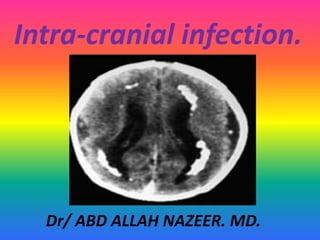
Intra-cranial infection.
Dr/ ABD ALLAH NAZEER. MD.
Intra-cranial infection.
. Congenital.
. Encephalitis.
. Meningitis.
. Cerebritis, ependymitis, abscess formation.
. Granulomatous infection.
. Parasitic and fungal infection.
. HIV infection.
Congenital Infections.
TORCH.
Toxoplasmosis.
Other (syphilis).
Rubella.
Cytomegalovirus (CMV).
Herpes simplex virus (HSV).
Varicella zoster (the chickenpox virus).
Entroviruses.
Hepatitis B.
Parvovirus.
HIV (human immune deficiency virus).
Chlamydia trachomatis.
Mycoplasma.
Group B streptococcus.
Malaria.
COMMON CLINICAL FEATURES.
Low birth weight
for gestational age.
Prematurity.
Seizures.
Chorio-retinitis.
Cataracts.
Purpura.
Cerebral calcification.
Micro-ophthalmia.
Jaundice.
Anemia.
Hepatosplenomegaly
Pneumonitis.
PATHOGENESIS.
Neonatal
1.Antenatal (in utero) - 80-96% of cases
Primary Maternal Infection
Recurrent Maternal Infection
2.Perinatal.
3.Postnatal.
Childhood
1. Horizontal Transmission
CMV excreted in saliva, urine, stool, tears
2. Organ Transplantation
kidney, marrow, heart, liver, blood (leukocytes)
CLINICAL FEATURES.
90% of infants with congenital CMV
infection are clinically silent.
CNS Manifestations
70% - microcephaly.
60% - intellectual impairment.
35% - sensorineural hearing loss
seizures.
22% - chorioretinitis.
CONGENITAL TOXOPLASMOSIS.
Caused by the protozoan Toxoplasma gondii
ocular, central nervous system (CNS)
incidence: 0.3-1/1000 live births.
Routes of Transmission.
Neonatal (in utero).
Primary Maternal Infection.
acquired by the ingestion of raw or undercooked meat
( cattle), or of infectious oocysts in feces (cats, birds).
1st trimester - 17% - spontaneous abortion.
2nd trimester - 25% - spontaneous abortion or
severe disease.
3rd trimester - 65% - subclinical disease.
Congenital toxoplasmosis show the typical multifocal
periventricular and cortical/subcortical punctuate calcifications as
well as a moderate ventriculomegaly as complication of the infection.
Congenital toxoplasmosis shows bilateral microophthalmia and chorionic
calcifications as well as deformity of the lenses (white arrows).
Toxoplasmosis of immune compromised
patient with multiple ring enhancing lesions.
CONGENITAL RUBELLA(German measles).
Caused by an RNA Togavirus.
Vaccine-preventable disease.
Routes of Transmission.
Antenatal (in utero).
1st trimester - 75-90%.
2nd trimester - 35-40%.
3rd trimester - 25-50%.
CLINICAL FEATURES:
Neonatal Manifestations
IUGR low birth weight -
prematurity.
stillbirth - spontaneous
abortion.
Early Manifestations
cloudy corneas.
Cataracts.
microcephaly.
Hepatomegally.
splenomegally .
Jaundice.
pulmonary valve stenosis.
patent ductus arteriosus.
thrombocytopenia purpura.
Congenital rubella infection show small calcifications within the basal ganglia,
along the intramedullary veins within both frontal lobes and within the deep
Layers of the overlying cerebral cortex and adjacent subcortical white matter
(white arrows on A). MRI shows ill-defined, multifocal regions of dysmyelination
within the periventricular white matter of both cerebral hemisphere.
Rubella. Unenhanced axial CT (a) in a 3-day-old with congenital
rubella demonstrates punctate calcifications of the basal ganglia
(arrow) and low attenuation of the white matter.
The classic clinical
picture of cytomegalic inclusion disease (CID) is characterized
by involvement of multiple organs, in particular the
reticuloendothelial and central nervous system, with or
without ocular and auditory damage. Obvious symptoms of
CID could be jaundice, Hepatosplenomegaly and petechiae in
a growth retarded often prematurely born baby. The
neurological involvement includes microcephaly, seizures,
hypotonia and lethargy. The most severely affected infants
have a mortality rate of about 30%. Deaths are usually due to
hepatic dysfunction, bleeding, disseminated intravascular
coagulation or secondary bacterial infections. Some 85- 90%
of children with a congenital CMV infection are asymptomatic
in the neonatal period.
Congenital cytomegalovirus infection.
Congenital CMV-infection show multifocal, predominantly periventricular
located calcifications within the supra- and infratentorial brain as well as a
thickened, smooth dysplastic cortex, moderate ventriculomegaly, a small
cerebellum and a CT-hypodense, T2-hyperintense dysmyelination of the
periventricular white matter. In addition, a mild microcephaly is noted.
Axial CT-images (A, D), T2-weighted images (B, E), and SWI images (C, F) of with congenital
cytomegalovirus infection presenting with moderate hydrocephalus and white matter
volume loss as well as subtle subependymal calcifications are seen on the CT study (white
arrow on D). The extent of calcium depositions is however much better appreciated on the
SWI images as SWI-hypointense signal abnormalities (C, F). In addition, cerebellar
hypoplasia (A, B) and high-grade loss of the hemispheric white matter (E), including a
more focal subcortical defect in the right parieto-occipital region are noted.
MENINGITIS & ENCEPHALITIS.
Definitions.
Meningitis – inflammation of
the meninges
Encephalitis – infection of the
brain parenchyma
Meningoencephalitis –
inflammation of brain +
meninges.
Aseptic meningitis –
inflammation of meninges
with sterile CSF
Symptoms of encephalitis.
.Many encephalitides are mild and
recovery occurs.
.In a minority, serious illness develops with
high fever, headache, mood change and
drowsiness over hours or days.
Focal signs, seizures and coma ensue.
Death, or brain injury follows.
Herpes simplex encephalitis.
Axial T2-weighted (A), coronal FLAIR (B), axial DWI (C) MR images, and axial
ADC map (D) with encephalitis . MRI shows symmetrical, ill defined T2- and
FLAIR-hyperintense signal abnormalities in both thalami with matching
regions of restricted diffusion (DWI-hyperintense, ADC-hypointense) on
diffusion weighted imaging (white arrows on A-D) encephalitis.
Rasmussen's encephalitis with hemispheric volume loss.
Acute disseminated encephalomyelitis.
Symptoms of meningitis.
Fever
.Altered consciousness, irritability, photophobia vomiting,
poor appetite.
. Seizures 20 - 30%
. Bulging fontanel 30%
. Stiff neck or nuchal rigidity
. Meningismus (stiff neck + Brudzinski + Kernig signs).
Meningitis:
• Infection of the meninges which may be pyogenic, viral or
granulomatous.
• Inflammation of the leptomeninges can be divided into acute
pyogenic (bacterial), lymphocytic (viral), and chronic (TB)
meningitis. The diagnosis is usually clinical.
• The role of neuroimaging is to exclude complication of
meningitis(e. g, abscess, ventriculitis, empyema).
CT and MRI of meningitis with dense basal cistern
which is seen enhanced at MRI images.
Acute pyogenic meningitis with basal cistern
exudates with enhancement and hydrocephalus.
Meningitis with predominant leptomeningeal enhancement.
Post-meningitis sequale with epidural and subdural empyema.
Cerebritis and abscess formation.
Ventriculitis with wall enhancement.
Tuberculosis meningitis Multiple enhancing tuberculomas.
T.B abscesses with left frontal subdural collection.
Left parietal tuberculous abscess.
Miliary tuberculosis of the brain.
Parasitic infection.
CT and MRI of
Cysticercosis
Hydatid cyst with marginal calcification.
Multiple coalescing thin walled hydatid cysts.
HIV Encephalitis with progressive encephalopathy.
HIV Encephalitis
Presentation3.pptx, intra cranial infection.

Contenu connexe

Tendances

Imaging of spinal dysraphism
Imaging of spinal dysraphismImaging of spinal dysraphism
Imaging of spinal dysraphismMuthu Magesh
 
Adult brain tumors imaging
Adult brain tumors imagingAdult brain tumors imaging
Adult brain tumors imagingrzgar hamed
 
Cervical Spine Trauma Imaging
Cervical Spine Trauma ImagingCervical Spine Trauma Imaging
Cervical Spine Trauma ImagingSCGH ED CME
 
Anatomy of sellar suprasellar region
Anatomy of sellar suprasellar regionAnatomy of sellar suprasellar region
Anatomy of sellar suprasellar regionPGINeurosurgery
 
Radiographic manifestations of pulmonary tuberculosis
Radiographic manifestations of pulmonary tuberculosisRadiographic manifestations of pulmonary tuberculosis
Radiographic manifestations of pulmonary tuberculosisDev Lakhera
 
Radiological anatomy & Techniques of the Ventricular system
Radiological anatomy & Techniques of the Ventricular systemRadiological anatomy & Techniques of the Ventricular system
Radiological anatomy & Techniques of the Ventricular systemMohamed M.A. Zaitoun
 
Dandy-Walker Malformation: Classification and Management
Dandy-Walker Malformation: Classification and ManagementDandy-Walker Malformation: Classification and Management
Dandy-Walker Malformation: Classification and ManagementDr. Shahnawaz Alam
 
Presentation1.pptx, radiological imaging of hydrocephalus.
Presentation1.pptx, radiological imaging of hydrocephalus.Presentation1.pptx, radiological imaging of hydrocephalus.
Presentation1.pptx, radiological imaging of hydrocephalus.Abdellah Nazeer
 
IMAGING IN CEREBRAL VENOUS THROMBOSIS
IMAGING IN CEREBRAL VENOUS THROMBOSIS IMAGING IN CEREBRAL VENOUS THROMBOSIS
IMAGING IN CEREBRAL VENOUS THROMBOSIS Shivshankar Badole
 
Spinal dysraphism
Spinal dysraphismSpinal dysraphism
Spinal dysraphismViral Patel
 
Tethered Cord Syndrome
Tethered Cord SyndromeTethered Cord Syndrome
Tethered Cord SyndromeAde Wijaya
 
BRAIN ARTERIOVENOUS MALFORMATION
BRAIN ARTERIOVENOUS MALFORMATIONBRAIN ARTERIOVENOUS MALFORMATION
BRAIN ARTERIOVENOUS MALFORMATIONsuresh Bishokarma
 
Presentation2.pptx pediatric brain tumour
Presentation2.pptx pediatric brain tumourPresentation2.pptx pediatric brain tumour
Presentation2.pptx pediatric brain tumourAbdellah Nazeer
 
Imaging in head trauma
Imaging in head traumaImaging in head trauma
Imaging in head traumaSCGH ED CME
 
CEREBRAL VENOUS THROMBOSIS
CEREBRAL VENOUS THROMBOSISCEREBRAL VENOUS THROMBOSIS
CEREBRAL VENOUS THROMBOSISNija Panchal
 
Supratentorial brain tumours
Supratentorial brain tumoursSupratentorial brain tumours
Supratentorial brain tumourstrial4neha
 

Tendances (20)

Imaging of spinal dysraphism
Imaging of spinal dysraphismImaging of spinal dysraphism
Imaging of spinal dysraphism
 
Adult brain tumors imaging
Adult brain tumors imagingAdult brain tumors imaging
Adult brain tumors imaging
 
Ring enhancing lesions
Ring enhancing lesionsRing enhancing lesions
Ring enhancing lesions
 
Cervical Spine Trauma Imaging
Cervical Spine Trauma ImagingCervical Spine Trauma Imaging
Cervical Spine Trauma Imaging
 
Anatomy of sellar suprasellar region
Anatomy of sellar suprasellar regionAnatomy of sellar suprasellar region
Anatomy of sellar suprasellar region
 
Radiographic manifestations of pulmonary tuberculosis
Radiographic manifestations of pulmonary tuberculosisRadiographic manifestations of pulmonary tuberculosis
Radiographic manifestations of pulmonary tuberculosis
 
Radiological anatomy & Techniques of the Ventricular system
Radiological anatomy & Techniques of the Ventricular systemRadiological anatomy & Techniques of the Ventricular system
Radiological anatomy & Techniques of the Ventricular system
 
Holoprosencephaly
HoloprosencephalyHoloprosencephaly
Holoprosencephaly
 
Dandy-Walker Malformation: Classification and Management
Dandy-Walker Malformation: Classification and ManagementDandy-Walker Malformation: Classification and Management
Dandy-Walker Malformation: Classification and Management
 
Cranial Ultrasound of neonate
Cranial Ultrasound of neonateCranial Ultrasound of neonate
Cranial Ultrasound of neonate
 
Presentation1.pptx, radiological imaging of hydrocephalus.
Presentation1.pptx, radiological imaging of hydrocephalus.Presentation1.pptx, radiological imaging of hydrocephalus.
Presentation1.pptx, radiological imaging of hydrocephalus.
 
IMAGING IN CEREBRAL VENOUS THROMBOSIS
IMAGING IN CEREBRAL VENOUS THROMBOSIS IMAGING IN CEREBRAL VENOUS THROMBOSIS
IMAGING IN CEREBRAL VENOUS THROMBOSIS
 
Spinal dysraphism
Spinal dysraphismSpinal dysraphism
Spinal dysraphism
 
Intra axial posterior fossa tumor
Intra axial posterior fossa tumorIntra axial posterior fossa tumor
Intra axial posterior fossa tumor
 
Tethered Cord Syndrome
Tethered Cord SyndromeTethered Cord Syndrome
Tethered Cord Syndrome
 
BRAIN ARTERIOVENOUS MALFORMATION
BRAIN ARTERIOVENOUS MALFORMATIONBRAIN ARTERIOVENOUS MALFORMATION
BRAIN ARTERIOVENOUS MALFORMATION
 
Presentation2.pptx pediatric brain tumour
Presentation2.pptx pediatric brain tumourPresentation2.pptx pediatric brain tumour
Presentation2.pptx pediatric brain tumour
 
Imaging in head trauma
Imaging in head traumaImaging in head trauma
Imaging in head trauma
 
CEREBRAL VENOUS THROMBOSIS
CEREBRAL VENOUS THROMBOSISCEREBRAL VENOUS THROMBOSIS
CEREBRAL VENOUS THROMBOSIS
 
Supratentorial brain tumours
Supratentorial brain tumoursSupratentorial brain tumours
Supratentorial brain tumours
 

En vedette

Pestana knight diagnosisepilepsy-5.8.2014
Pestana knight diagnosisepilepsy-5.8.2014Pestana knight diagnosisepilepsy-5.8.2014
Pestana knight diagnosisepilepsy-5.8.2014Cleveland Clinic
 
171&Sch ch130 Dorsal root entry zone lesion (DREZ)
171&Sch ch130 Dorsal root entry zone lesion (DREZ)171&Sch ch130 Dorsal root entry zone lesion (DREZ)
171&Sch ch130 Dorsal root entry zone lesion (DREZ)Neurosurgery Vajira
 
331 Clinical pathophhysiology of traumatic brain injury
331 Clinical pathophhysiology of traumatic brain injury331 Clinical pathophhysiology of traumatic brain injury
331 Clinical pathophhysiology of traumatic brain injuryNeurosurgery Vajira
 
338 Indications and technique for cranial decompression after traumatic brain...
338 Indications and technique for cranial decompression after traumatic brain...338 Indications and technique for cranial decompression after traumatic brain...
338 Indications and technique for cranial decompression after traumatic brain...Neurosurgery Vajira
 
034 Clinical evaluation of adult hydrocephalus
034 Clinical evaluation of adult hydrocephalus034 Clinical evaluation of adult hydrocephalus
034 Clinical evaluation of adult hydrocephalusNeurosurgery Vajira
 
Mr imaging findings in spinal ligamentous injury
Mr imaging findings in spinal ligamentous injuryMr imaging findings in spinal ligamentous injury
Mr imaging findings in spinal ligamentous injuryBattulga Munkhtsetseg
 
Imaging of cns viral infection
Imaging of cns viral infectionImaging of cns viral infection
Imaging of cns viral infectionsks200166
 
Torch infections by Dr. Dilip
Torch infections by Dr. DilipTorch infections by Dr. Dilip
Torch infections by Dr. DilipDrDilip86
 
Trigeminal neuralgia
Trigeminal neuralgiaTrigeminal neuralgia
Trigeminal neuralgiayury
 

En vedette (20)

CNS INFECTION
CNS INFECTIONCNS INFECTION
CNS INFECTION
 
CNS infections
CNS infectionsCNS infections
CNS infections
 
Cns infections
Cns infections Cns infections
Cns infections
 
Modernidade
ModernidadeModernidade
Modernidade
 
Pestana knight diagnosisepilepsy-5.8.2014
Pestana knight diagnosisepilepsy-5.8.2014Pestana knight diagnosisepilepsy-5.8.2014
Pestana knight diagnosisepilepsy-5.8.2014
 
171&Sch ch130 Dorsal root entry zone lesion (DREZ)
171&Sch ch130 Dorsal root entry zone lesion (DREZ)171&Sch ch130 Dorsal root entry zone lesion (DREZ)
171&Sch ch130 Dorsal root entry zone lesion (DREZ)
 
Awake Craniotomy
Awake CraniotomyAwake Craniotomy
Awake Craniotomy
 
13 atlantoaxial subluxation
13 atlantoaxial subluxation13 atlantoaxial subluxation
13 atlantoaxial subluxation
 
331 Clinical pathophhysiology of traumatic brain injury
331 Clinical pathophhysiology of traumatic brain injury331 Clinical pathophhysiology of traumatic brain injury
331 Clinical pathophhysiology of traumatic brain injury
 
338 Indications and technique for cranial decompression after traumatic brain...
338 Indications and technique for cranial decompression after traumatic brain...338 Indications and technique for cranial decompression after traumatic brain...
338 Indications and technique for cranial decompression after traumatic brain...
 
Lumbar Injuries
Lumbar InjuriesLumbar Injuries
Lumbar Injuries
 
034 Clinical evaluation of adult hydrocephalus
034 Clinical evaluation of adult hydrocephalus034 Clinical evaluation of adult hydrocephalus
034 Clinical evaluation of adult hydrocephalus
 
Syllabus exercise
Syllabus exerciseSyllabus exercise
Syllabus exercise
 
Sepsis
SepsisSepsis
Sepsis
 
212 Birth head trauma
212 Birth head trauma212 Birth head trauma
212 Birth head trauma
 
Mr imaging findings in spinal ligamentous injury
Mr imaging findings in spinal ligamentous injuryMr imaging findings in spinal ligamentous injury
Mr imaging findings in spinal ligamentous injury
 
029 Incision and closure
029 Incision and closure 029 Incision and closure
029 Incision and closure
 
Imaging of cns viral infection
Imaging of cns viral infectionImaging of cns viral infection
Imaging of cns viral infection
 
Torch infections by Dr. Dilip
Torch infections by Dr. DilipTorch infections by Dr. Dilip
Torch infections by Dr. Dilip
 
Trigeminal neuralgia
Trigeminal neuralgiaTrigeminal neuralgia
Trigeminal neuralgia
 

Similaire à Presentation3.pptx, intra cranial infection.

Imaging of common viral brain infection in india PPT
Imaging of common  viral brain infection in india PPTImaging of common  viral brain infection in india PPT
Imaging of common viral brain infection in india PPTNaba Kumar Barman
 
MENINGITIS & ENCEPHALITIS - Ayushi.pptx pdf
MENINGITIS & ENCEPHALITIS - Ayushi.pptx pdfMENINGITIS & ENCEPHALITIS - Ayushi.pptx pdf
MENINGITIS & ENCEPHALITIS - Ayushi.pptx pdfAditiShah380128
 
Imaging of cns viral infection
Imaging of cns viral infectionImaging of cns viral infection
Imaging of cns viral infectionNeurologyKota
 
Presentation1.pptx, radiological imaging of intra cranial calcification.
Presentation1.pptx, radiological imaging of intra cranial calcification.Presentation1.pptx, radiological imaging of intra cranial calcification.
Presentation1.pptx, radiological imaging of intra cranial calcification.Abdellah Nazeer
 
Radiology of demyelinating diseases
Radiology of demyelinating diseases Radiology of demyelinating diseases
Radiology of demyelinating diseases NeurologyKota
 
CONGENITAL,PYOGENIC AND VIRAL INFECTIONS OF CNS @22-02-23.pptx
CONGENITAL,PYOGENIC AND VIRAL INFECTIONS OF CNS @22-02-23.pptxCONGENITAL,PYOGENIC AND VIRAL INFECTIONS OF CNS @22-02-23.pptx
CONGENITAL,PYOGENIC AND VIRAL INFECTIONS OF CNS @22-02-23.pptxaasrithakotha2
 
CONGENITAL,PYOGENIC AND VIRAL INFECTIONS OF CNS @22-02-23.pptx
CONGENITAL,PYOGENIC AND VIRAL INFECTIONS OF CNS @22-02-23.pptxCONGENITAL,PYOGENIC AND VIRAL INFECTIONS OF CNS @22-02-23.pptx
CONGENITAL,PYOGENIC AND VIRAL INFECTIONS OF CNS @22-02-23.pptxaasrithakotha2
 
CNS Radiography for helminth infections.pptx
 CNS Radiography for helminth infections.pptx CNS Radiography for helminth infections.pptx
CNS Radiography for helminth infections.pptxIbrahimAboAlasaad
 
Congenital, Acquired Pyogenic, and Acquired Viral Infection
Congenital, Acquired Pyogenic, and Acquired Viral InfectionCongenital, Acquired Pyogenic, and Acquired Viral Infection
Congenital, Acquired Pyogenic, and Acquired Viral Infectionpriyanka rana
 
Central Nervous System (CNS) TBacterialM
Central Nervous System (CNS) TBacterialMCentral Nervous System (CNS) TBacterialM
Central Nervous System (CNS) TBacterialMDr. Ajit Surya Singh
 
Intracranial infections
Intracranial infectionsIntracranial infections
Intracranial infectionsMilan Silwal
 
imagingofintracranialinfectionsincludingcovid19pk2-200605190339 (1).pptx
imagingofintracranialinfectionsincludingcovid19pk2-200605190339 (1).pptximagingofintracranialinfectionsincludingcovid19pk2-200605190339 (1).pptx
imagingofintracranialinfectionsincludingcovid19pk2-200605190339 (1).pptxRose635887
 
Approach to a patient of neurological tuberculosis
Approach to a patient of neurological tuberculosisApproach to a patient of neurological tuberculosis
Approach to a patient of neurological tuberculosisRahul Arya
 
Imaging in multiple ring enhancing brain lesions
Imaging in multiple ring enhancing brain lesionsImaging in multiple ring enhancing brain lesions
Imaging in multiple ring enhancing brain lesionsSumiya Arshad
 
Brain abscess
Brain abscessBrain abscess
Brain abscessjoemdas
 
04 Neurologic
04 Neurologic04 Neurologic
04 NeurologicDeep Deep
 
04 Neurologic
04 Neurologic04 Neurologic
04 NeurologicDeep Deep
 
Short presentation version cns infections Lecture
Short presentation version cns infections LectureShort presentation version cns infections Lecture
Short presentation version cns infections Lecturetest
 

Similaire à Presentation3.pptx, intra cranial infection. (20)

Imaging of common viral brain infection in india PPT
Imaging of common  viral brain infection in india PPTImaging of common  viral brain infection in india PPT
Imaging of common viral brain infection in india PPT
 
MENINGITIS & ENCEPHALITIS - Ayushi.pptx pdf
MENINGITIS & ENCEPHALITIS - Ayushi.pptx pdfMENINGITIS & ENCEPHALITIS - Ayushi.pptx pdf
MENINGITIS & ENCEPHALITIS - Ayushi.pptx pdf
 
Imaging of cns viral infection
Imaging of cns viral infectionImaging of cns viral infection
Imaging of cns viral infection
 
Presentation1.pptx, radiological imaging of intra cranial calcification.
Presentation1.pptx, radiological imaging of intra cranial calcification.Presentation1.pptx, radiological imaging of intra cranial calcification.
Presentation1.pptx, radiological imaging of intra cranial calcification.
 
Radiology of demyelinating diseases
Radiology of demyelinating diseases Radiology of demyelinating diseases
Radiology of demyelinating diseases
 
CONGENITAL,PYOGENIC AND VIRAL INFECTIONS OF CNS @22-02-23.pptx
CONGENITAL,PYOGENIC AND VIRAL INFECTIONS OF CNS @22-02-23.pptxCONGENITAL,PYOGENIC AND VIRAL INFECTIONS OF CNS @22-02-23.pptx
CONGENITAL,PYOGENIC AND VIRAL INFECTIONS OF CNS @22-02-23.pptx
 
CONGENITAL,PYOGENIC AND VIRAL INFECTIONS OF CNS @22-02-23.pptx
CONGENITAL,PYOGENIC AND VIRAL INFECTIONS OF CNS @22-02-23.pptxCONGENITAL,PYOGENIC AND VIRAL INFECTIONS OF CNS @22-02-23.pptx
CONGENITAL,PYOGENIC AND VIRAL INFECTIONS OF CNS @22-02-23.pptx
 
CNS Radiography for helminth infections.pptx
 CNS Radiography for helminth infections.pptx CNS Radiography for helminth infections.pptx
CNS Radiography for helminth infections.pptx
 
Congenital, Acquired Pyogenic, and Acquired Viral Infection
Congenital, Acquired Pyogenic, and Acquired Viral InfectionCongenital, Acquired Pyogenic, and Acquired Viral Infection
Congenital, Acquired Pyogenic, and Acquired Viral Infection
 
Central Nervous System (CNS) TBacterialM
Central Nervous System (CNS) TBacterialMCentral Nervous System (CNS) TBacterialM
Central Nervous System (CNS) TBacterialM
 
Intracranial infections
Intracranial infectionsIntracranial infections
Intracranial infections
 
imagingofintracranialinfectionsincludingcovid19pk2-200605190339 (1).pptx
imagingofintracranialinfectionsincludingcovid19pk2-200605190339 (1).pptximagingofintracranialinfectionsincludingcovid19pk2-200605190339 (1).pptx
imagingofintracranialinfectionsincludingcovid19pk2-200605190339 (1).pptx
 
Cns TB
Cns TBCns TB
Cns TB
 
CNS Infections
CNS InfectionsCNS Infections
CNS Infections
 
Approach to a patient of neurological tuberculosis
Approach to a patient of neurological tuberculosisApproach to a patient of neurological tuberculosis
Approach to a patient of neurological tuberculosis
 
Imaging in multiple ring enhancing brain lesions
Imaging in multiple ring enhancing brain lesionsImaging in multiple ring enhancing brain lesions
Imaging in multiple ring enhancing brain lesions
 
Brain abscess
Brain abscessBrain abscess
Brain abscess
 
04 Neurologic
04 Neurologic04 Neurologic
04 Neurologic
 
04 Neurologic
04 Neurologic04 Neurologic
04 Neurologic
 
Short presentation version cns infections Lecture
Short presentation version cns infections LectureShort presentation version cns infections Lecture
Short presentation version cns infections Lecture
 

Plus de Abdellah Nazeer

Muculoskeletal Pediatic Imaging..pptx
Muculoskeletal Pediatic Imaging..pptxMuculoskeletal Pediatic Imaging..pptx
Muculoskeletal Pediatic Imaging..pptxAbdellah Nazeer
 
Presentation1, Ultrasound of the bowel loops and the lymph nodes..pptx
Presentation1, Ultrasound of the bowel loops and the lymph nodes..pptxPresentation1, Ultrasound of the bowel loops and the lymph nodes..pptx
Presentation1, Ultrasound of the bowel loops and the lymph nodes..pptxAbdellah Nazeer
 
Presentation1 Short cases MD..pptx
Presentation1 Short cases MD..pptxPresentation1 Short cases MD..pptx
Presentation1 Short cases MD..pptxAbdellah Nazeer
 
Presentation1, MD MCQ Cases..pptx
Presentation1, MD MCQ Cases..pptxPresentation1, MD MCQ Cases..pptx
Presentation1, MD MCQ Cases..pptxAbdellah Nazeer
 
Presentation1, Short Cases Quiz..pptx
Presentation1, Short Cases Quiz..pptxPresentation1, Short Cases Quiz..pptx
Presentation1, Short Cases Quiz..pptxAbdellah Nazeer
 
Presentation1, radiological imaging of lateral hindfoot impingement.
Presentation1, radiological imaging of lateral hindfoot impingement.Presentation1, radiological imaging of lateral hindfoot impingement.
Presentation1, radiological imaging of lateral hindfoot impingement.Abdellah Nazeer
 
Presentation2, radiological anatomy of the liver and spleen.
Presentation2, radiological anatomy of the liver and spleen.Presentation2, radiological anatomy of the liver and spleen.
Presentation2, radiological anatomy of the liver and spleen.Abdellah Nazeer
 
Presentation1, artifacts and pitfalls of the wrist and elbow joints.
Presentation1, artifacts and pitfalls of the wrist and elbow joints.Presentation1, artifacts and pitfalls of the wrist and elbow joints.
Presentation1, artifacts and pitfalls of the wrist and elbow joints.Abdellah Nazeer
 
Presentation1, artifact and pitfalls of the knee, hip and ankle joints.
Presentation1, artifact and pitfalls of the knee, hip and ankle joints.Presentation1, artifact and pitfalls of the knee, hip and ankle joints.
Presentation1, artifact and pitfalls of the knee, hip and ankle joints.Abdellah Nazeer
 
Presentation1, radiological imaging of artifact and pitfalls in shoulder join...
Presentation1, radiological imaging of artifact and pitfalls in shoulder join...Presentation1, radiological imaging of artifact and pitfalls in shoulder join...
Presentation1, radiological imaging of artifact and pitfalls in shoulder join...Abdellah Nazeer
 
Presentation1, radiological imaging of internal abdominal hernia.
Presentation1, radiological imaging of internal abdominal hernia.Presentation1, radiological imaging of internal abdominal hernia.
Presentation1, radiological imaging of internal abdominal hernia.Abdellah Nazeer
 
Presentation11, radiological imaging of ovarian torsion.
Presentation11, radiological imaging of ovarian torsion.Presentation11, radiological imaging of ovarian torsion.
Presentation11, radiological imaging of ovarian torsion.Abdellah Nazeer
 
Presentation1, musculoskeletal anatomy.
Presentation1, musculoskeletal anatomy.Presentation1, musculoskeletal anatomy.
Presentation1, musculoskeletal anatomy.Abdellah Nazeer
 
Presentation1, new mri techniques in the diagnosis and monitoring of multiple...
Presentation1, new mri techniques in the diagnosis and monitoring of multiple...Presentation1, new mri techniques in the diagnosis and monitoring of multiple...
Presentation1, new mri techniques in the diagnosis and monitoring of multiple...Abdellah Nazeer
 
Presentation1, radiological application of diffusion weighted mri in neck mas...
Presentation1, radiological application of diffusion weighted mri in neck mas...Presentation1, radiological application of diffusion weighted mri in neck mas...
Presentation1, radiological application of diffusion weighted mri in neck mas...Abdellah Nazeer
 
Presentation1, radiological application of diffusion weighted images in breas...
Presentation1, radiological application of diffusion weighted images in breas...Presentation1, radiological application of diffusion weighted images in breas...
Presentation1, radiological application of diffusion weighted images in breas...Abdellah Nazeer
 
Presentation1, radiological application of diffusion weighted images in abdom...
Presentation1, radiological application of diffusion weighted images in abdom...Presentation1, radiological application of diffusion weighted images in abdom...
Presentation1, radiological application of diffusion weighted images in abdom...Abdellah Nazeer
 
Presentation1, radiological application of diffusion weighted imges in neuror...
Presentation1, radiological application of diffusion weighted imges in neuror...Presentation1, radiological application of diffusion weighted imges in neuror...
Presentation1, radiological application of diffusion weighted imges in neuror...Abdellah Nazeer
 
Presentation1, mr physics.
Presentation1, mr physics.Presentation1, mr physics.
Presentation1, mr physics.Abdellah Nazeer
 
Presentation1. ct physics.
Presentation1. ct physics.Presentation1. ct physics.
Presentation1. ct physics.Abdellah Nazeer
 

Plus de Abdellah Nazeer (20)

Muculoskeletal Pediatic Imaging..pptx
Muculoskeletal Pediatic Imaging..pptxMuculoskeletal Pediatic Imaging..pptx
Muculoskeletal Pediatic Imaging..pptx
 
Presentation1, Ultrasound of the bowel loops and the lymph nodes..pptx
Presentation1, Ultrasound of the bowel loops and the lymph nodes..pptxPresentation1, Ultrasound of the bowel loops and the lymph nodes..pptx
Presentation1, Ultrasound of the bowel loops and the lymph nodes..pptx
 
Presentation1 Short cases MD..pptx
Presentation1 Short cases MD..pptxPresentation1 Short cases MD..pptx
Presentation1 Short cases MD..pptx
 
Presentation1, MD MCQ Cases..pptx
Presentation1, MD MCQ Cases..pptxPresentation1, MD MCQ Cases..pptx
Presentation1, MD MCQ Cases..pptx
 
Presentation1, Short Cases Quiz..pptx
Presentation1, Short Cases Quiz..pptxPresentation1, Short Cases Quiz..pptx
Presentation1, Short Cases Quiz..pptx
 
Presentation1, radiological imaging of lateral hindfoot impingement.
Presentation1, radiological imaging of lateral hindfoot impingement.Presentation1, radiological imaging of lateral hindfoot impingement.
Presentation1, radiological imaging of lateral hindfoot impingement.
 
Presentation2, radiological anatomy of the liver and spleen.
Presentation2, radiological anatomy of the liver and spleen.Presentation2, radiological anatomy of the liver and spleen.
Presentation2, radiological anatomy of the liver and spleen.
 
Presentation1, artifacts and pitfalls of the wrist and elbow joints.
Presentation1, artifacts and pitfalls of the wrist and elbow joints.Presentation1, artifacts and pitfalls of the wrist and elbow joints.
Presentation1, artifacts and pitfalls of the wrist and elbow joints.
 
Presentation1, artifact and pitfalls of the knee, hip and ankle joints.
Presentation1, artifact and pitfalls of the knee, hip and ankle joints.Presentation1, artifact and pitfalls of the knee, hip and ankle joints.
Presentation1, artifact and pitfalls of the knee, hip and ankle joints.
 
Presentation1, radiological imaging of artifact and pitfalls in shoulder join...
Presentation1, radiological imaging of artifact and pitfalls in shoulder join...Presentation1, radiological imaging of artifact and pitfalls in shoulder join...
Presentation1, radiological imaging of artifact and pitfalls in shoulder join...
 
Presentation1, radiological imaging of internal abdominal hernia.
Presentation1, radiological imaging of internal abdominal hernia.Presentation1, radiological imaging of internal abdominal hernia.
Presentation1, radiological imaging of internal abdominal hernia.
 
Presentation11, radiological imaging of ovarian torsion.
Presentation11, radiological imaging of ovarian torsion.Presentation11, radiological imaging of ovarian torsion.
Presentation11, radiological imaging of ovarian torsion.
 
Presentation1, musculoskeletal anatomy.
Presentation1, musculoskeletal anatomy.Presentation1, musculoskeletal anatomy.
Presentation1, musculoskeletal anatomy.
 
Presentation1, new mri techniques in the diagnosis and monitoring of multiple...
Presentation1, new mri techniques in the diagnosis and monitoring of multiple...Presentation1, new mri techniques in the diagnosis and monitoring of multiple...
Presentation1, new mri techniques in the diagnosis and monitoring of multiple...
 
Presentation1, radiological application of diffusion weighted mri in neck mas...
Presentation1, radiological application of diffusion weighted mri in neck mas...Presentation1, radiological application of diffusion weighted mri in neck mas...
Presentation1, radiological application of diffusion weighted mri in neck mas...
 
Presentation1, radiological application of diffusion weighted images in breas...
Presentation1, radiological application of diffusion weighted images in breas...Presentation1, radiological application of diffusion weighted images in breas...
Presentation1, radiological application of diffusion weighted images in breas...
 
Presentation1, radiological application of diffusion weighted images in abdom...
Presentation1, radiological application of diffusion weighted images in abdom...Presentation1, radiological application of diffusion weighted images in abdom...
Presentation1, radiological application of diffusion weighted images in abdom...
 
Presentation1, radiological application of diffusion weighted imges in neuror...
Presentation1, radiological application of diffusion weighted imges in neuror...Presentation1, radiological application of diffusion weighted imges in neuror...
Presentation1, radiological application of diffusion weighted imges in neuror...
 
Presentation1, mr physics.
Presentation1, mr physics.Presentation1, mr physics.
Presentation1, mr physics.
 
Presentation1. ct physics.
Presentation1. ct physics.Presentation1. ct physics.
Presentation1. ct physics.
 

Dernier

call girls in paharganj DELHI 🔝 >༒9540349809 🔝 genuine Escort Service 🔝✔️✔️
call girls in paharganj DELHI 🔝 >༒9540349809 🔝 genuine Escort Service 🔝✔️✔️call girls in paharganj DELHI 🔝 >༒9540349809 🔝 genuine Escort Service 🔝✔️✔️
call girls in paharganj DELHI 🔝 >༒9540349809 🔝 genuine Escort Service 🔝✔️✔️saminamagar
 
See the 2,456 pharmacies on the National E-Pharmacy Platform
See the 2,456 pharmacies on the National E-Pharmacy PlatformSee the 2,456 pharmacies on the National E-Pharmacy Platform
See the 2,456 pharmacies on the National E-Pharmacy PlatformKweku Zurek
 
Measurement of Radiation and Dosimetric Procedure.pptx
Measurement of Radiation and Dosimetric Procedure.pptxMeasurement of Radiation and Dosimetric Procedure.pptx
Measurement of Radiation and Dosimetric Procedure.pptxDr. Dheeraj Kumar
 
epilepsy and status epilepticus for undergraduate.pptx
epilepsy and status epilepticus  for undergraduate.pptxepilepsy and status epilepticus  for undergraduate.pptx
epilepsy and status epilepticus for undergraduate.pptxMohamed Rizk Khodair
 
VarSeq 2.6.0: Advancing Pharmacogenomics and Genomic Analysis
VarSeq 2.6.0: Advancing Pharmacogenomics and Genomic AnalysisVarSeq 2.6.0: Advancing Pharmacogenomics and Genomic Analysis
VarSeq 2.6.0: Advancing Pharmacogenomics and Genomic AnalysisGolden Helix
 
Biomechanics- Shoulder Joint!!!!!!!!!!!!
Biomechanics- Shoulder Joint!!!!!!!!!!!!Biomechanics- Shoulder Joint!!!!!!!!!!!!
Biomechanics- Shoulder Joint!!!!!!!!!!!!ibtesaam huma
 
History and Development of Pharmacovigilence.pdf
History and Development of Pharmacovigilence.pdfHistory and Development of Pharmacovigilence.pdf
History and Development of Pharmacovigilence.pdfSasikiranMarri
 
97111 47426 Call Girls In Delhi MUNIRKAA
97111 47426 Call Girls In Delhi MUNIRKAA97111 47426 Call Girls In Delhi MUNIRKAA
97111 47426 Call Girls In Delhi MUNIRKAAjennyeacort
 
Presentation on Parasympathetic Nervous System
Presentation on Parasympathetic Nervous SystemPresentation on Parasympathetic Nervous System
Presentation on Parasympathetic Nervous SystemPrerana Jadhav
 
LUNG TUMORS AND ITS CLASSIFICATIONS.pdf
LUNG TUMORS AND ITS  CLASSIFICATIONS.pdfLUNG TUMORS AND ITS  CLASSIFICATIONS.pdf
LUNG TUMORS AND ITS CLASSIFICATIONS.pdfDolisha Warbi
 
call girls in Dwarka Sector 21 Metro DELHI 🔝 >༒9540349809 🔝 genuine Escort Se...
call girls in Dwarka Sector 21 Metro DELHI 🔝 >༒9540349809 🔝 genuine Escort Se...call girls in Dwarka Sector 21 Metro DELHI 🔝 >༒9540349809 🔝 genuine Escort Se...
call girls in Dwarka Sector 21 Metro DELHI 🔝 >༒9540349809 🔝 genuine Escort Se...saminamagar
 
Glomerular Filtration and determinants of glomerular filtration .pptx
Glomerular Filtration and  determinants of glomerular filtration .pptxGlomerular Filtration and  determinants of glomerular filtration .pptx
Glomerular Filtration and determinants of glomerular filtration .pptxDr.Nusrat Tariq
 
Big Data Analysis Suggests COVID Vaccination Increases Excess Mortality Of ...
Big Data Analysis Suggests COVID  Vaccination Increases Excess Mortality Of  ...Big Data Analysis Suggests COVID  Vaccination Increases Excess Mortality Of  ...
Big Data Analysis Suggests COVID Vaccination Increases Excess Mortality Of ...sdateam0
 
Wessex Health Partners Wessex Integrated Care, Population Health, Research & ...
Wessex Health Partners Wessex Integrated Care, Population Health, Research & ...Wessex Health Partners Wessex Integrated Care, Population Health, Research & ...
Wessex Health Partners Wessex Integrated Care, Population Health, Research & ...Wessex Health Partners
 
Presentació "Real-Life VR Integration for Mild Cognitive Impairment Rehabilit...
Presentació "Real-Life VR Integration for Mild Cognitive Impairment Rehabilit...Presentació "Real-Life VR Integration for Mild Cognitive Impairment Rehabilit...
Presentació "Real-Life VR Integration for Mild Cognitive Impairment Rehabilit...Badalona Serveis Assistencials
 
call girls in munirka DELHI 🔝 >༒9540349809 🔝 genuine Escort Service 🔝✔️✔️
call girls in munirka  DELHI 🔝 >༒9540349809 🔝 genuine Escort Service 🔝✔️✔️call girls in munirka  DELHI 🔝 >༒9540349809 🔝 genuine Escort Service 🔝✔️✔️
call girls in munirka DELHI 🔝 >༒9540349809 🔝 genuine Escort Service 🔝✔️✔️saminamagar
 
POST NATAL EXERCISES AND ITS IMPACT.pptx
POST NATAL EXERCISES AND ITS IMPACT.pptxPOST NATAL EXERCISES AND ITS IMPACT.pptx
POST NATAL EXERCISES AND ITS IMPACT.pptxvirengeeta
 
call girls in Connaught Place DELHI 🔝 >༒9540349809 🔝 genuine Escort Service ...
call girls in Connaught Place  DELHI 🔝 >༒9540349809 🔝 genuine Escort Service ...call girls in Connaught Place  DELHI 🔝 >༒9540349809 🔝 genuine Escort Service ...
call girls in Connaught Place DELHI 🔝 >༒9540349809 🔝 genuine Escort Service ...saminamagar
 
Introduction to Sports Injuries by- Dr. Anjali Rai
Introduction to Sports Injuries by- Dr. Anjali RaiIntroduction to Sports Injuries by- Dr. Anjali Rai
Introduction to Sports Injuries by- Dr. Anjali RaiGoogle
 
PULMONARY EDEMA AND ITS MANAGEMENT.pdf
PULMONARY EDEMA AND  ITS  MANAGEMENT.pdfPULMONARY EDEMA AND  ITS  MANAGEMENT.pdf
PULMONARY EDEMA AND ITS MANAGEMENT.pdfDolisha Warbi
 

Dernier (20)

call girls in paharganj DELHI 🔝 >༒9540349809 🔝 genuine Escort Service 🔝✔️✔️
call girls in paharganj DELHI 🔝 >༒9540349809 🔝 genuine Escort Service 🔝✔️✔️call girls in paharganj DELHI 🔝 >༒9540349809 🔝 genuine Escort Service 🔝✔️✔️
call girls in paharganj DELHI 🔝 >༒9540349809 🔝 genuine Escort Service 🔝✔️✔️
 
See the 2,456 pharmacies on the National E-Pharmacy Platform
See the 2,456 pharmacies on the National E-Pharmacy PlatformSee the 2,456 pharmacies on the National E-Pharmacy Platform
See the 2,456 pharmacies on the National E-Pharmacy Platform
 
Measurement of Radiation and Dosimetric Procedure.pptx
Measurement of Radiation and Dosimetric Procedure.pptxMeasurement of Radiation and Dosimetric Procedure.pptx
Measurement of Radiation and Dosimetric Procedure.pptx
 
epilepsy and status epilepticus for undergraduate.pptx
epilepsy and status epilepticus  for undergraduate.pptxepilepsy and status epilepticus  for undergraduate.pptx
epilepsy and status epilepticus for undergraduate.pptx
 
VarSeq 2.6.0: Advancing Pharmacogenomics and Genomic Analysis
VarSeq 2.6.0: Advancing Pharmacogenomics and Genomic AnalysisVarSeq 2.6.0: Advancing Pharmacogenomics and Genomic Analysis
VarSeq 2.6.0: Advancing Pharmacogenomics and Genomic Analysis
 
Biomechanics- Shoulder Joint!!!!!!!!!!!!
Biomechanics- Shoulder Joint!!!!!!!!!!!!Biomechanics- Shoulder Joint!!!!!!!!!!!!
Biomechanics- Shoulder Joint!!!!!!!!!!!!
 
History and Development of Pharmacovigilence.pdf
History and Development of Pharmacovigilence.pdfHistory and Development of Pharmacovigilence.pdf
History and Development of Pharmacovigilence.pdf
 
97111 47426 Call Girls In Delhi MUNIRKAA
97111 47426 Call Girls In Delhi MUNIRKAA97111 47426 Call Girls In Delhi MUNIRKAA
97111 47426 Call Girls In Delhi MUNIRKAA
 
Presentation on Parasympathetic Nervous System
Presentation on Parasympathetic Nervous SystemPresentation on Parasympathetic Nervous System
Presentation on Parasympathetic Nervous System
 
LUNG TUMORS AND ITS CLASSIFICATIONS.pdf
LUNG TUMORS AND ITS  CLASSIFICATIONS.pdfLUNG TUMORS AND ITS  CLASSIFICATIONS.pdf
LUNG TUMORS AND ITS CLASSIFICATIONS.pdf
 
call girls in Dwarka Sector 21 Metro DELHI 🔝 >༒9540349809 🔝 genuine Escort Se...
call girls in Dwarka Sector 21 Metro DELHI 🔝 >༒9540349809 🔝 genuine Escort Se...call girls in Dwarka Sector 21 Metro DELHI 🔝 >༒9540349809 🔝 genuine Escort Se...
call girls in Dwarka Sector 21 Metro DELHI 🔝 >༒9540349809 🔝 genuine Escort Se...
 
Glomerular Filtration and determinants of glomerular filtration .pptx
Glomerular Filtration and  determinants of glomerular filtration .pptxGlomerular Filtration and  determinants of glomerular filtration .pptx
Glomerular Filtration and determinants of glomerular filtration .pptx
 
Big Data Analysis Suggests COVID Vaccination Increases Excess Mortality Of ...
Big Data Analysis Suggests COVID  Vaccination Increases Excess Mortality Of  ...Big Data Analysis Suggests COVID  Vaccination Increases Excess Mortality Of  ...
Big Data Analysis Suggests COVID Vaccination Increases Excess Mortality Of ...
 
Wessex Health Partners Wessex Integrated Care, Population Health, Research & ...
Wessex Health Partners Wessex Integrated Care, Population Health, Research & ...Wessex Health Partners Wessex Integrated Care, Population Health, Research & ...
Wessex Health Partners Wessex Integrated Care, Population Health, Research & ...
 
Presentació "Real-Life VR Integration for Mild Cognitive Impairment Rehabilit...
Presentació "Real-Life VR Integration for Mild Cognitive Impairment Rehabilit...Presentació "Real-Life VR Integration for Mild Cognitive Impairment Rehabilit...
Presentació "Real-Life VR Integration for Mild Cognitive Impairment Rehabilit...
 
call girls in munirka DELHI 🔝 >༒9540349809 🔝 genuine Escort Service 🔝✔️✔️
call girls in munirka  DELHI 🔝 >༒9540349809 🔝 genuine Escort Service 🔝✔️✔️call girls in munirka  DELHI 🔝 >༒9540349809 🔝 genuine Escort Service 🔝✔️✔️
call girls in munirka DELHI 🔝 >༒9540349809 🔝 genuine Escort Service 🔝✔️✔️
 
POST NATAL EXERCISES AND ITS IMPACT.pptx
POST NATAL EXERCISES AND ITS IMPACT.pptxPOST NATAL EXERCISES AND ITS IMPACT.pptx
POST NATAL EXERCISES AND ITS IMPACT.pptx
 
call girls in Connaught Place DELHI 🔝 >༒9540349809 🔝 genuine Escort Service ...
call girls in Connaught Place  DELHI 🔝 >༒9540349809 🔝 genuine Escort Service ...call girls in Connaught Place  DELHI 🔝 >༒9540349809 🔝 genuine Escort Service ...
call girls in Connaught Place DELHI 🔝 >༒9540349809 🔝 genuine Escort Service ...
 
Introduction to Sports Injuries by- Dr. Anjali Rai
Introduction to Sports Injuries by- Dr. Anjali RaiIntroduction to Sports Injuries by- Dr. Anjali Rai
Introduction to Sports Injuries by- Dr. Anjali Rai
 
PULMONARY EDEMA AND ITS MANAGEMENT.pdf
PULMONARY EDEMA AND  ITS  MANAGEMENT.pdfPULMONARY EDEMA AND  ITS  MANAGEMENT.pdf
PULMONARY EDEMA AND ITS MANAGEMENT.pdf
 

Presentation3.pptx, intra cranial infection.

  • 2. Intra-cranial infection. . Congenital. . Encephalitis. . Meningitis. . Cerebritis, ependymitis, abscess formation. . Granulomatous infection. . Parasitic and fungal infection. . HIV infection.
  • 4. Varicella zoster (the chickenpox virus). Entroviruses. Hepatitis B. Parvovirus. HIV (human immune deficiency virus). Chlamydia trachomatis. Mycoplasma. Group B streptococcus. Malaria.
  • 5. COMMON CLINICAL FEATURES. Low birth weight for gestational age. Prematurity. Seizures. Chorio-retinitis. Cataracts. Purpura. Cerebral calcification. Micro-ophthalmia. Jaundice. Anemia. Hepatosplenomegaly Pneumonitis.
  • 6. PATHOGENESIS. Neonatal 1.Antenatal (in utero) - 80-96% of cases Primary Maternal Infection Recurrent Maternal Infection 2.Perinatal. 3.Postnatal. Childhood 1. Horizontal Transmission CMV excreted in saliva, urine, stool, tears 2. Organ Transplantation kidney, marrow, heart, liver, blood (leukocytes)
  • 7. CLINICAL FEATURES. 90% of infants with congenital CMV infection are clinically silent. CNS Manifestations 70% - microcephaly. 60% - intellectual impairment. 35% - sensorineural hearing loss seizures. 22% - chorioretinitis.
  • 8. CONGENITAL TOXOPLASMOSIS. Caused by the protozoan Toxoplasma gondii ocular, central nervous system (CNS) incidence: 0.3-1/1000 live births. Routes of Transmission. Neonatal (in utero). Primary Maternal Infection. acquired by the ingestion of raw or undercooked meat ( cattle), or of infectious oocysts in feces (cats, birds). 1st trimester - 17% - spontaneous abortion. 2nd trimester - 25% - spontaneous abortion or severe disease. 3rd trimester - 65% - subclinical disease.
  • 9. Congenital toxoplasmosis show the typical multifocal periventricular and cortical/subcortical punctuate calcifications as well as a moderate ventriculomegaly as complication of the infection.
  • 10. Congenital toxoplasmosis shows bilateral microophthalmia and chorionic calcifications as well as deformity of the lenses (white arrows).
  • 11. Toxoplasmosis of immune compromised patient with multiple ring enhancing lesions.
  • 12. CONGENITAL RUBELLA(German measles). Caused by an RNA Togavirus. Vaccine-preventable disease. Routes of Transmission. Antenatal (in utero). 1st trimester - 75-90%. 2nd trimester - 35-40%. 3rd trimester - 25-50%.
  • 13. CLINICAL FEATURES: Neonatal Manifestations IUGR low birth weight - prematurity. stillbirth - spontaneous abortion. Early Manifestations cloudy corneas. Cataracts. microcephaly. Hepatomegally. splenomegally . Jaundice. pulmonary valve stenosis. patent ductus arteriosus. thrombocytopenia purpura.
  • 14. Congenital rubella infection show small calcifications within the basal ganglia, along the intramedullary veins within both frontal lobes and within the deep Layers of the overlying cerebral cortex and adjacent subcortical white matter (white arrows on A). MRI shows ill-defined, multifocal regions of dysmyelination within the periventricular white matter of both cerebral hemisphere.
  • 15. Rubella. Unenhanced axial CT (a) in a 3-day-old with congenital rubella demonstrates punctate calcifications of the basal ganglia (arrow) and low attenuation of the white matter.
  • 16. The classic clinical picture of cytomegalic inclusion disease (CID) is characterized by involvement of multiple organs, in particular the reticuloendothelial and central nervous system, with or without ocular and auditory damage. Obvious symptoms of CID could be jaundice, Hepatosplenomegaly and petechiae in a growth retarded often prematurely born baby. The neurological involvement includes microcephaly, seizures, hypotonia and lethargy. The most severely affected infants have a mortality rate of about 30%. Deaths are usually due to hepatic dysfunction, bleeding, disseminated intravascular coagulation or secondary bacterial infections. Some 85- 90% of children with a congenital CMV infection are asymptomatic in the neonatal period. Congenital cytomegalovirus infection.
  • 17. Congenital CMV-infection show multifocal, predominantly periventricular located calcifications within the supra- and infratentorial brain as well as a thickened, smooth dysplastic cortex, moderate ventriculomegaly, a small cerebellum and a CT-hypodense, T2-hyperintense dysmyelination of the periventricular white matter. In addition, a mild microcephaly is noted.
  • 18.
  • 19. Axial CT-images (A, D), T2-weighted images (B, E), and SWI images (C, F) of with congenital cytomegalovirus infection presenting with moderate hydrocephalus and white matter volume loss as well as subtle subependymal calcifications are seen on the CT study (white arrow on D). The extent of calcium depositions is however much better appreciated on the SWI images as SWI-hypointense signal abnormalities (C, F). In addition, cerebellar hypoplasia (A, B) and high-grade loss of the hemispheric white matter (E), including a more focal subcortical defect in the right parieto-occipital region are noted.
  • 20. MENINGITIS & ENCEPHALITIS. Definitions. Meningitis – inflammation of the meninges Encephalitis – infection of the brain parenchyma Meningoencephalitis – inflammation of brain + meninges. Aseptic meningitis – inflammation of meninges with sterile CSF
  • 21.
  • 22. Symptoms of encephalitis. .Many encephalitides are mild and recovery occurs. .In a minority, serious illness develops with high fever, headache, mood change and drowsiness over hours or days. Focal signs, seizures and coma ensue. Death, or brain injury follows.
  • 23.
  • 24.
  • 25.
  • 26.
  • 27.
  • 28.
  • 30.
  • 31. Axial T2-weighted (A), coronal FLAIR (B), axial DWI (C) MR images, and axial ADC map (D) with encephalitis . MRI shows symmetrical, ill defined T2- and FLAIR-hyperintense signal abnormalities in both thalami with matching regions of restricted diffusion (DWI-hyperintense, ADC-hypointense) on diffusion weighted imaging (white arrows on A-D) encephalitis.
  • 32.
  • 33.
  • 34.
  • 35.
  • 36. Rasmussen's encephalitis with hemispheric volume loss.
  • 37.
  • 38.
  • 39.
  • 40.
  • 41.
  • 43.
  • 44.
  • 45.
  • 46. Symptoms of meningitis. Fever .Altered consciousness, irritability, photophobia vomiting, poor appetite. . Seizures 20 - 30% . Bulging fontanel 30% . Stiff neck or nuchal rigidity . Meningismus (stiff neck + Brudzinski + Kernig signs). Meningitis: • Infection of the meninges which may be pyogenic, viral or granulomatous. • Inflammation of the leptomeninges can be divided into acute pyogenic (bacterial), lymphocytic (viral), and chronic (TB) meningitis. The diagnosis is usually clinical. • The role of neuroimaging is to exclude complication of meningitis(e. g, abscess, ventriculitis, empyema).
  • 47.
  • 48. CT and MRI of meningitis with dense basal cistern which is seen enhanced at MRI images.
  • 49. Acute pyogenic meningitis with basal cistern exudates with enhancement and hydrocephalus.
  • 50.
  • 51. Meningitis with predominant leptomeningeal enhancement.
  • 52.
  • 53.
  • 54. Post-meningitis sequale with epidural and subdural empyema.
  • 56.
  • 57.
  • 58.
  • 59.
  • 60.
  • 61.
  • 62. Ventriculitis with wall enhancement.
  • 63.
  • 64.
  • 65. Tuberculosis meningitis Multiple enhancing tuberculomas.
  • 66.
  • 67. T.B abscesses with left frontal subdural collection.
  • 68.
  • 72.
  • 73.
  • 74.
  • 75. CT and MRI of Cysticercosis
  • 76.
  • 77.
  • 78.
  • 79. Hydatid cyst with marginal calcification.
  • 80. Multiple coalescing thin walled hydatid cysts.
  • 81.
  • 82.
  • 83. HIV Encephalitis with progressive encephalopathy.