The gut microbiota — masters of host development and physiology
•
1 like•4,498 views


Recommended
Recommended
More Related Content
What's hot
What's hot (20)
Similar to The gut microbiota — masters of host development and physiology
Similar to The gut microbiota — masters of host development and physiology (20)
More from Alfonso Enrique Islas Rodríguez
More from Alfonso Enrique Islas Rodríguez (20)
The gut microbiota — masters of host development and physiology
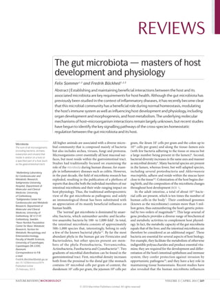
- 1. REVIEWS The gut microbiota — masters of host development and physiology Felix Sommer1,2 and Fredrik Bäckhed1,2,3 Abstract | Establishing and maintaining beneficial interactions between the host and its associated microbiota are key requirements for host health. Although the gut microbiota has previously been studied in the context of inflammatory diseases, it has recently become clear that this microbial community has a beneficial role during normal homeostasis, modulating the host’s immune system as well as influencing host development and physiology, including organ development and morphogenesis, and host metabolism. The underlying molecular mechanisms of host–microorganism interactions remain largely unknown, but recent studies have begun to identify the key signalling pathways of the cross-species homeostatic regulation between the gut microbiota and its host. Microbiota All higher animals are associated with a diverse micro- gram, the ileum 107 cells per gram and the colon up to The sum of all microorganisms bial community that is composed mainly of bacteria 1012 cells per gram) and along the tissue–lumen axis (including bacteria, archaea, but also includes archea, viruses, fungi and protozoa. (with few bacteria adhering to the tissue or mucus but eukaryotes and viruses) that Microorganisms cover essentially all host mucosal sur- a large number being present in the lumen)4. Second, reside in and/or on a host or a specified part of a host (such faces, but most reside within the gastrointestinal tract. bacterial diversity increases in the same axes and manner as the gastrointestinal tract). Studies had traditionally focused on examining the as microbial density 4. Many bacterial species are present role of the microbiota during human disease, for exam- in the lumen, whereas fewer, but well-adapted species, 1 Wallenberg Laboratory ple in inflammatory diseases such as colitis. However, including several proteobacteria and Akkermansia for Cardiovascular and in the past decade, the field of microbiota research has muciniphila, adhere and reside within the mucus layer Metabolic Research, exploded, resulting in the publication of a plethora of close to the tissue5,6. Colonization of the host begins dur- Sahlgrenska University reports that describe both the individual members of our ing birth, and the composition of the microbiota changes Hospital, Department of Molecular and Clinical intestinal microbiota and their wide-ranging impact on throughout host development (BOX 1). Medicine, University host physiology. Thus, the traditional anthropocentric In the adult intestine, a total of about 10 14 bacte- of Gothenburg. view of the gut microbiota as pathogenic and solely rial cells are present, which is ten times the number of 2 Sahlgrenska Center for an immunological threat has been substituted with human cells in the body 7. Their combined genomes Cardiovascular and Metabolic an appreciation of its mainly beneficial influence on (known as the microbiome) contain more than 5 mil- Research, Department of Molecular and Clinical human health. lion genes, thus outnumbering the host’s genetic poten- Medicine, University of The ‘normal’ gut microbiota is dominated by anaer- tial by two orders of magnitude2,8. This large arsenal of Gothenburg, SE‑413 45 obic bacteria, which outnumber aerobic and faculta- gene products provides a diverse range of biochemical Gothenburg, Sweden. tive anaerobic bacteria by 100- to 1,000‑fold1. In total, and metabolic activities to complement host physiol- 3 Novo Nordisk Foundation Center for Basic Metabolic the intestinal microbiota consists of approximately ogy. In fact, the metabolic capacity of the gut microbiota Research, Section for 500–1,000 species that, interestingly, belong to only equals that of the liver, and the intestinal microbiota can Metabolic Receptology and a few of the known bacterial phyla2,3. By far the most therefore be considered as an additional organ9. These Enteroendocrinology, abundant phyla in the human gut are Firmicutes and bacteria are essential for several aspects of host biology. Faculty of Health Sciences, Bacteriodetes, but other species present are mem- For example, they facilitate the metabolism of otherwise University of Copenhagen, Copenhagen DK‑2200, bers of the phyla Proteobacteria, Verrumicrobia, indigestible polysaccharides and produce essential vita- Denmark. Actinobacteria, Fusobacteria and Cyanobacteria2,3. Two mins; they are required for the development and differ Correspondence to F.B. gradients of microbial distribution can be found in the entiation of the host’s intestinal epithelium and immune e‑mail: gastrointestinal tract. First, microbial density increases system; they confer protection against invasion by Fredrik.Backhed@wlab.gu.se doi:10.1038/nrmicro2974 both from the proximal to the distal gut (the stomach opportunistic pathogens10; and they have a key role in Published online contains 101 microbial cells per gram of content, the maintaining tissue homeostasis. Recent studies have 25 February 2013 duodenum 103 cells per gram, the jejunum 104 cells per also revealed that the human microbiota influences NATURE REVIEWS | MICROBIOLOGY VOLUME 11 | APRIL 2013 | 227 © 2013 Macmillan Publishers Limited. All rights reserved
- 2. REVIEWS Box 1 | Colonization of the host are pathogens and thus the cause of infectious diseases. The realization that we live in a microbially dominated Human babies are colonized during passage through the birth canal by environmental world and in fact benefit greatly from our microbiota microorganisms (for example, from the mother’s vagina or skin) and during breast has led to a paradigm shift in immunology. Thus, the feeding by microorganisms present in the milk137. Owing to the highly oxidative definition of self in the superorganism theory has been environment in the gastrointestinal tract of the newborn, primary colonizers are extended to incorporate the constituents of both our facultative anaerobic bacteria such as proteobacteria, which are thought to adjust the environmental conditions by decreasing the oxygen concentration to allow successive own body and our microbiota12. It is also now widely colonization by anaerobic microorganisms such as members of the genus Bacteroides accepted that the host’s mucosal immune system is char- and members of the phyla Actinobacteria and Firmicutes. During the first year of life, acterized by tolerance to microorganisms rather than the intestinal microbiota composition is simple and fluctuates widely between responsiveness13. Furthermore, it has even been specu- individuals and over time. Microbial signatures stabilize and start to resemble the ‘adult lated that the highly sophisticated adaptive immune state’ when the infant reaches 1–2 years of age4. system of jawed vertebrates evolved to keep control of Interestingly, conflicting evidence has been published concerning the driving force the mutualistic or beneficial symbiosis with our complex for microbial transmission. In early studies of twins, the faecal microbial compositions in microbial ecosystem14. the mother and her children were similar, indicating a mainly maternal transmission108,111. The intestine, one essential organ in which the However, in a more recent and extensive study, the same research group found that the mucosal immune system operates, has to accomplish faecal microbiota of children was no more similar to that of their mothers than to that of their biological fathers, and genetically unrelated but co‑habiting mothers and fathers two apparently confounding tasks. First, it needs to were significantly more microbially similar to one another than to members of different facilitate nutrient absorption; thus, the total surface area families138. This indicates that, as well as genetics and kinship, environmental factors of the gastrointestinal tract amounts to about 200 m2 in have a considerable effect on the microbial composition of the infant. humans15. Second, it needs to be resistant to infection and inhibit microbial translocation across the tissue bar- rier. Bacterial densities in the gut are the highest known the development and homeostasis of other host tissues, in any habitat to date and reach up to 1012 cells per including the bone11. gram in the lower intestine16. This highly dense micro- The microbiota also benefits from this mutualistic bial community and the host intestinal epithelial cell association, as the mammalian intestine is a nutrient-rich (IEC) lining are separated by only a few micro etres of m environment that is maintained at a constant tempera- mucus in the small intestine and up to several hundred ture. However, it is also a dynamic habitat that undergoes micrometers in the colon, depending on the location17. constant and rapid changes in its physiological para Because of this unique nature of the intestinal tract, its Mutualistic meters owing to variations in, for example, host diet, mucosal immune system needs to fulfil several special Pertaining to a relationship lifestyle, hygiene or use of antibiotics, all of which affect requirements. It has to be non-responsive to or tolerant between two organisms: beneficial to both organisms. gut microbial composition (FIG. 1). Thus, unlike the host towards the huge number of mutualistic microorganisms The term originates from the genome, the microbiome can change rapidly as a result of that reside in the intestinal lumen. At the same time, it is Latin word mutuus (lent, modifications in either the composition of the microbial thought that the mucosal immune system has to assure borrowed or mutual). community or individual microbial genomes, resulting a beneficial microbiota composition by keeping patho- in modified transcriptomic, proteomic and metabolic bionts in check, restricting microbial overgrowth and Superorganism A term that extends the profiles. Accordingly, the establishment and preserva- reacting to penetrating microorganisms that breach the classical biological definition tion of beneficial interactions between the host and its intestinal chemical and physical barriers (such as secreted of an organism (a living system associated intestinal microbiota are key requirements soluble immunoglobulin A (IgA), antimicrobial peptides capable of autonomous for health. (AMPs), the mucus layer and the tightly interconnected metabolism and reproduction) by including the many The dynamic fluctuations in the microbiota com- IEC lining). In turn, the intestinal microbiota has a key microorganisms that live in and bined with the vast numbers of bacterial cells and their role in directing several aspects in the development and on that host organism, thus close proximity to the epithelial tissue represent a mas- regulation of the host’s immune tissues, immune cell yielding a superior degree of sive challenge to host immunity, as microbial growth populations and immune mediators. complexity. The term originates has to be restricted to ensure a beneficial homeostasis. from the Latin supra (above) and the Greek organon (organ, Furthermore, activation of the host immune system Mucus layer properties depend on intestinal bacteria. instrument, tool). has to be controlled to circumvent the detrimental The intestinal mucus layer covers the epithelial cell effects of chronic inflammation, so the interaction of lining and functions as a lubricant, facilitating gastro- Symbiosis the gut microbiota with the host has to be tightly regu- intestinal transport, and as a protective layer against Any close physical association between two organisms, lated. In this Review, we discuss recent insights into the bacterial invasion, owing to its physical properties 18. usually from different species. impact of the normal microbiota on the development The colonic mucus layer is in fact composed of two This includes mutualism, and homeostasis of the immune system and other tis- layers17. Both the inner and outer mucus layers are commensalism and parasitism. sues and organs, as well as on host physiology. We also secreted by goblet cells and are mainly made up of gel- The term originates from the highlight recent advances in deciphering the under forming highly glyco ylated proteins termed mucins18. s Greek words syn (together) and bio (life). lying molecular mechanisms of host–microorganism Mucin 2 (MUC2) is the main mucin in the small and interactions. large intestines of both mice and humans18. The entire Pathobionts mucus layer represents a selective microbial habitat Normally harmless Tailoring immune development owing to microbial adhesion via lectins and glycosidases microorganism that can become pathogens under Immunology was originally based on the concept of that are expressed by only specific bacteria, and it also certain environmental ‘self ’ versus ‘non-self ’ discrimination, with the assump- serves as nutrient source19,20. However, bacteria are found conditions. tion that, because they are non-self, all microorganisms only in the outer layer 17, probably owing to the specific 228 | APRIL 2013 | VOLUME 11 www.nature.com/reviews/micro © 2013 Macmillan Publishers Limited. All rights reserved
- 3. REVIEWS Antibiotics Lifestyle Diet Hygiene Hyperimmunity Immunodeficiency IL-6 Altered intestinal microbiota NOD2 IL-12 IL-10 TNF Chronic Metabolic inflammation dysfunction Figure 1 | Factors shaping intestinal microbial composition and effects of dysbiosis on host health. The composition of the gut microbiota is influenced by various environmental factors, including the use of antibiotics, lifestyle, diet and hygiene preferences. The host’s genetic disposition also has a role: hyperimmunity (owing Nature Reviews | Microbiology to over-representation of pro-inflammatory mediators such as interleukin‑6 (IL‑6), IL‑12 or tumour necrosis factor (TNF)) or immunodeficiency (owing to mutations in regulatory immune proteins such as NOD2 (nucleotide-binding oligomerization domain protein 2) or IL‑10) can influence the gut microbiota composition. In turn, dysbiosis affects levels of immune mediators and induces both chronic inflammation and metabolic dysfunction. structure of the mucus layer as a whole, which is formed mislocalization of the same microbiota or merely on of interconnected sheets that create pores smaller than a increased bacterial load. bacterial cell and thus inhibit penetration21. Comparisons of germ-free and conventionally raised Microorganisms induce the development of lymphoid animals revealed that microorganisms have major effects structures. The lymphatic system consists of a network on mucus thickness and composition; compared with of lymphatic vessels connecting the primary and second- conventionally raised animals, germ-free animals have ary lymphoid organs. The main functions of this sys- fewer goblet cells, a thinner mucus layer and also a tem are the recirculation of interstitial fluid and blood higher percentage of neutral mucins in the colon 22. as well as the transport of lymphocytes (such as B cells Stimulation with bacterial products such as lipopolysac- and T cells) (BOX 2) and antigen-presenting cells to lymph charide (LPS) and peptidoglycan is sufficient to estab- nodes. Lymphoid tissue is classified as primary (thy- lish conventional mucus properties in germ-free mice23, mus and bone marrow) and secondary (lymph nodes, but the underlying mechanisms for how the gut micro Peyer’s patches, tonsils, spleen and lymphoid follicles). biota modulates goblet cells and mucus layer properties Lymphocytes are generated in primary lymphoid tissues remain largely elusive. and are then transported to secondary lymphoid tissues, Notably, Muc2‑deficient mice or those with aberrant where the mature lymphocytes are exposed to antigens mucin glycosylation profiles (owing to a lack of specific by antigen-presenting cells and are thus activated to glycosyl transferases) show bacterial overgrowth and initiate an adaptive immune response. The cellular either develop spontaneous colitis or are more suscep- interactions that occur during lymphoid tissue develop- tible to chemical induction of colitis, an effect that can ment and maturation are similar for both primary and be ameliorated by treatment with antibiotics23–25. This secondary lymphoid organs, although the molecular demonstrates the importance of the mucus layer for frameworks differ a little (for details see REF. 26). homeostasis in the gut and also highlights the recip- In addition to host genetics, several environmen- rocal interaction between the mucus layer and the gut tal factors, including contact with microorganisms, microbiota. It remains to be clarified whether disease influence both the development and maturation of the onset in these mouse strains depends on a selectively immune system. The development of secondary gut- altered and thus more colitogenic microbiota, on associated lymphoid tissue (GALT), such as Peyer’s NATURE REVIEWS | MICROBIOLOGY VOLUME 11 | APRIL 2013 | 229 © 2013 Macmillan Publishers Limited. All rights reserved
- 4. REVIEWS Box 2 | Lymphocyte subtypes induce the differentiation of stromal organizer cells to express several cytokines and adhesion molecules that All lymphocytes differentiate and mature in primary lymphoid organs (the thymus and attract further immune cells, causing GALT forma- bone marrow). Mature naive lymphocytes migrate to secondary lymphoid tissues, tion26. Maturation of these tissues, including an increase where they become activated by antigen-presenting cells such as dendritic cells. in tissue size and the development of germinal centres Gut-associated secondary lymphoid tissues include Peyer’s patches, mesenteric lymph (sites of B cell proliferation, differentiation and somatic nodes and lymphoid follicles139. Here, we list and describe the lymphocytes that are hypermutation in lymph nodes), depends on postnatal known to be modulated by the gut microbiota43,140,141. microbial colonization28 (FIG. 2). Consequently, Peyer’s Lymphoid tissue inducer cells patches, mesenteric lymph nodes and splenic white pulp (LTi cells). A unique T cell subpopulation that is characterized by the expression of RORγt, CD4 and interleukin‑7 receptor‑α and the absence of CD3, B220 (an isoform are underdeveloped in germ-free mice29. of CD45) and CD11c (also known as integrin αX). Their function is to recruit B cells and Furthermore, in parallel with microbial coloniza- T cells and thereby promote the formation of secondary lymphoid tissues. tion, clusters of LTi-like cells termed cryptopatches Natural killer cells form at birth in the connective tissue between intestinal (NK cells). Lymphocytes that recognize the abnormal antigen signatures of infected or crypts, known as the lamina propria30. Cryptopatches tumour cells, which NK cells kill by lysis or apoptosis. NK cells resemble cytotoxic T cells recruit B cells and develop into isolated lymphoid fol- in function but belong to the innate immune system. They express various NK cell licles (ILFs), a type of lymphoid tissue that is structurally receptors, including NKp46 (in mice; NKp44 in humans) and NKG2D. They can activate similar to Peyer’s patches and serves as an inductive site B cells and T cells and thereby stimulate an adaptive immune response. for intestinal immune reactions31,32. This process also Natural killer T cells depends on the gut microbiota, as ILFs fail to develop (NKT cells). These cells have properties of both T cells and NK cells, as they co-express fully in germ-free mice33. ILF formation can be induced NK cell markers with a T cell receptor. NKT cells mainly recognize lipids and glycolipids by exposing germ-free mice to purified peptidoglycan presented by antigen-presenting cells via CD1d. Following activation, NKT cells from Gram-negative bacteria, indicating that this pro- produce pro-inflammatory cytokines such as tumour necrosis factor (TNF) and cess is driven solely by a specific microbial pattern34. interleukin‑17 (IL‑17). Invariant NKT (iNKT) cells are a specific subpopulation expressing Stromal and epithelial cells recognize the peptido an invariant T cell receptor. glycan of resident microorganisms mainly via signalling T helper 1 cells through the pattern recognition receptor (PRR) NOD1 (TH1 cells). A subset of TH lymphocytes that is characterized by the expression of (nucleotide-binding oligomerization domain contain- interferon‑γ and transforming growth factor-β (TGFβ). TH1 cell differentiation is induced by contact with activated macrophages or NK cells. ing 1) but also partially through another family of PRRs, the Toll-like receptors (TLRs). Activation of NOD1 T helper 2 cells by the gut microbiota causes increased expression of (TH2 cells). A subset of TH lymphocytes that is characterized by the expression of the cytokines IL‑4, IL‑5 and IL‑13. TH2 cell differentiation is induced in response to, for CC-chemokine ligand 20 (CCL20) and presumably also example, allergens and extracellular microorganisms. of β‑defensin 3, both of which activate ILF formation by binding to CC-chemokine receptor 6 (CCR6) on LTi T helper 17 cells (TH17 cells). A subset of TH cells that is characterized by the expression of IL‑17, which cells34. stimulates stromal cells to express the pro-inflammatory cytokines IL‑6 and IL‑8, thereby attracting neutrophils and promoting inflammation to clear out invading The gut microbiota modulates immune cell differentia- microorganisms. tion. In addition to regulating the development of lym- Regulatory T cells phoid structures, the gut microbiota has been shown (TReg cells). A T cell subpopulation that is characterized by the expression of CD4, CD25 to modulate the differentiation of immune cell subsets and FOXP3 and the production of the anti-inflammatory cytokines TGFβ and IL‑10. (BOX 2) and, therefore, maintain homeostatic interactions These cells can be subdivided into natural TReg cells, which differentiate from CD4+ between the host and the gut microbiota. T cells in the thymus, and inducible TReg cells, which arise from naive T cells in secondary After birth, LTi-like cells that express nuclear RORγt lymphoid tissues. Both cell types function to suppress immune activation and prevent but lack NKp46 (in mice; also known as NCR1) or self-reactivity, thereby reducing the risk of autoimmune disease. NKp44 (in humans; also known as NCR2) markers accu- Type 1 regulatory T cells mulate in both the mouse and human GALT and lamina (TR1 cells). These CD4+CD25+FOXP3− T cells are functionally equivalent to the propria35–37. Interestingly, the RORγt+NKp46− LTi-like IL‑10‑producing TReg cells. They respond to microorganisms and regulate intestinal cells can differentiate into RORγt+NKp46+ natural killer tolerance through the secretion of IL‑10. (NK)‑like cells, which differ from regular NK cells (BOX 2) B cells in that they have intermediate expression of NK1.1 (also Lymphocytes that are activated when the unique B cell receptor binds its specific known as KLRB1C) and do not produce interleukin‑1β antigen and that then mediate humoral immunity through the production of antibodies. (IL‑1β) or kill tumour cells36,37. This differentiation B cells are also involved in lymphoid tissue organization. requires both IL‑23, which is produced by activated myeloid cells and epithelial or endothelial cells, and the presence of the intestinal microbiota, as germ-free mice patches and mesenteric lymph nodes, is initiated pre- have fewer RORγt+NKp46+ NK‑like cells than conven- natally in the sterile environment of the fetus through tionally raised mice37. These cells produce IL‑22, which in Somatic hypermutation induction by lymphoid tissue inducer (LTi) cells 27. mice promotes the integrity of the intestinal barrier and A programmed process of Briefly, mesenchymal cells are induced by retinoic acid reduces bacterial infiltration by inducing epithelial repair mutation affecting the variable regions of immunoglobulin to produce CXC-chemokine ligand 13 (CXCL13), which via signal transducer and activator of transcription 3 genes during affinity recruits LTi precursor cells and stimulates their cluster- (STAT3) signalling and the production of antimicrobial maturation of B cell receptors. ing, leading to their maturation into LTi cells. These then proteins38. Thus, the normal gut microbiota promotes 230 | APRIL 2013 | VOLUME 11 www.nature.com/reviews/micro © 2013 Macmillan Publishers Limited. All rights reserved
- 5. REVIEWS a Germ-free mice Conventionally raised mice ↓ Mucus thickness Altered mucus properties Microbiota Mucus Intestinal epithelial cell Goblet cell Villus IgA ↓ Vessel density AMP Intestinal crypt Blood vessel b c PSA TReg cell B. fragilis Peyer’s patch Immature Peyer’s patch Immature SFB TH17 cell MLN ↓ AMP and MLN IgA production T cell B cell IgA-producing Dendritic cell plasma cell Figure 2 | Microbiota-induced maturation of the gastrointestinal tract. The microbiota promotes substantial changes in gut morphology, including villus architecture, crypt depth, stem cell proliferation, bloodReviews | Microbiology Nature vessel density, mucus layer properties and maturation of mucosa-associated lymphoid tissues. a | In germ-free mice, the villi in the distal small intestine are longer and thinner and have a less complex vascular network than the villi of conventionally raised animals. In the absence of bacteria, intestinal crypts are less deep and contain fewer proliferating stem cells. Furthermore, germ-free animals show reduced mucus thickness and altered mucus properties. b | Moreover, very few isolated lymphoid follicles, immature Peyer’s patches and immature mesenteric lymph nodes (MLNs) are present under germ-free conditions, and levels of both immunoglobulin A (IgA) and antimicrobial peptides (AMPs) are lower than in conventionally raised animals. c | In conventionally raised mice, polysaccharide A (PSA) of Bacteroides fragilis is known to induce the expansion of CD4+CD25+FOXP3+ regulatory T (TReg) cells, which have an anti-inflammatory effect and dampen immune responses. By contrast, segmented filamentous bacteria (SFB) have been shown to induce the expansion of T helper 17 (TH17) cells, which are pro-inflammatory. NATURE REVIEWS | MICROBIOLOGY VOLUME 11 | APRIL 2013 | 231 © 2013 Macmillan Publishers Limited. All rights reserved
- 6. REVIEWS intestinal barrier function by modulating mucosal increased levels of the immunosuppressive cytokine homeostasis, in part by promoting the differentiation of IL‑10 (REF. 47). Notably, compared with non-colonized RORγt+NKp46+ NK‑like cells. germ-free mice, the Clostridia species-colonized mice The intestinal microbiota also modulates the abun- were more resistant to chemically induced disruption dance of invariant NK T cells (iNKT cells), a unique of the gut epithelium and displayed attenuated levels of T cell subset that expresses an invariant T cell receptor antigen-induced serum IgE. Furthermore, a similar TReg α-chain. These cells promote inflammation, as follow- cell response was observed when germ-free mice were ing activation they secrete pro-inflammatory T helper 1 colonized with altered Schaedler flora, a cocktail of eight (TH1)- and TH2‑type chemokines and cytokines, including defined bacteria including three Clostridia species48. interferon-γ, IL‑2, IL‑4, IL‑13, IL‑17A, IL‑21 and tumour However, the specific species in this community that are necrosis factor (TNF)39,40. In contrast to the NK‑like cells functionally responsible and the underlying molecular described above, there are more iNKT cells in the colon mechanisms of this effect are so far unknown. of germ-free mice than the colon of conventionally raised In contrast to the bacteria mentioned above, mice41, which suggests that the gut microbiota promotes segmented filamentous bacteria (SFB) elicit a pro- homeostasis by decreasing the number of these pro- inflammatory immune response by promoting the dif- inflammatory cells. Importantly, a recent study elegantly ferentiation of TH17 cells and, to a lesser extent, TH1 demonstrated an age dependency of the microbial effects cells49,50. SFB reside in the small intestine of mice and on the iNKT population and revealed that colonization of are in direct contact with epithelial cells, which these neonatal but not adult germ-free mice with conventional bacteria seem to readily invade. Invasion leads to local gut microbiota normalized iNKT cell numbers and pro- actin polymerization in the epithelium at the interac- tected against oxazolone-induced colitis as well as against tion site, and this presumably initiates a signalling event ovalbumin-induced allergic lung inflammation41. that activates the differentiation of TH17 cells. Notably, It has become evident that the gut microbiota shapes mono-association of germ-free mice with SFB is suffi- the T cell landscape not only in the lamina propria but cient to restore susceptibility to TH17 cell-mediated arthri- also systemically and therefore modulates the homeo- tis and experimental autoimmune encephalomyelitis51,52. So stasis of the superorganism42 (FIG. 2). Intestinal mucosal far, however, it is not known whether TH17 cell differ- T cells are important ‘legislators’ of intestinal homeo- entiation is induced by IEC-produced mediators, by stasis because they not only defend against intestinal direct interaction with antigen-presenting cells in the pathogens, but also promote wound healing, barrier lamina propria (dendritic cells or macrophages) or by repair and regeneration as they rapidly accumulate at bacterially secreted signalling molecules (for example, sites of injury and infection43. T cells can be assigned metabolites)15. to subpopulations that drive either a pro-inflammatory immune response (TH1, TH2 and TH17 cells) or an anti- Gut microorganisms tweak the production of immune inflammatory immune response (CD4+CD25+FOXP3+ mediators. It is clear that the gut microbiota regulates regulatory T (TReg) cells or CD4+CD25+FOXP3− type 1 the production of cytokines and chemokines to influ- regulatory T (TR1) cells), depending on the cytokines ence the T cell repertoire of the intestine and surround- that they produce44 (BOX 2). The balance between both ing tissue, but there is evidence that these bacteria also pro- and anti-inflammatory T cell subpopulations deter- modulate the production of other soluble immune mines the overall immune equilibrium. mediators (FIG. 2). IgA is produced by plasma cells (dif- Interestingly, individual members of the gut micro- ferentiated B cells) in the lamina propria and is trans biota have been found to drive specific T cell responses cytosed through the intestinal epithelium into the (FIG. 2c). The Gram-negative bacterium Bacteroides fragilis lumen, where it binds microbial antigens and thereby elicits an anti-inflammatory response by inducing the prevents bacterial translocation and infection53. The dif- differentiation of CD4+ T cells into TReg cells locally in ferentiation of B cells into IgA-producing plasma cells is the intestinal lamina propria but also in the circula- induced by sensing of gut microbiota-derived flagellin Experimental autoimmune encephalomyelitis tion45. TReg cells produce IL‑10 and thereby suppress the via TLR5 on lamina propria dendritic cells54. IgA has a An animal model of pro-inflammatory TH17 response46. This skewing event is key role in barrier homeostasis, as IgA-deficient mice T cell-mediated autoimmune mediated by polysaccharide A (PSA) on the outer mem- produce gut microbiota-specific serum IgG antibodies, disease in general and in brane of the bacterium, which is recognized by TLR2 on indicating that there is a breach in the mucosal barrier particular of demyelinating diseases of the central nervous CD4+ T cells and activates a signalling cascade involving of these mice and subsequent induction of the systemic system, such as multiple myeloid differentiation 88 (MYD88) to induce TReg cell immune system55. Furthermore, a recent study showed sclerosis. differentiation45. Indeed, a mutant strain of B. fragilis that microbial modulation of IgA homeostasis is in part lacking PSA fails to initiate differentiation of TReg cells, dependent on the host protein programmed cell death 1 T follicular helper cells whereas purified PSA has the same effect as the wild-type (PD1), which is expressed by T follicular helper cells in A T cell subtype that resides in the B cell follicles of secondary bacterium29. the germinal centre56. PD1‑deficient mice harbour an lymphoid organs and Bacteria from the Gram-positive class Clostridia have altered IgA repertoire owing to changes in B cell matura- expresses the B cell homing similar effects on the host immune system. A mixture of tion, leading to modified IgA specificity. This altered IgA receptor CXC-chemokine 46 Clostridia spp. belonging to clusters IV and XIVa was repertoire then shifts the normal gut microbiota com- receptor 5. These T cells mediate B cell activation and isolated from mouse faeces, and colonization of germ- position by reducing the numbers of bacteria from the trigger the formation of the free mice with this mixture induced the expansion of genera Bifidobacterium and Bacteroides and increasing germinal centre. TReg cells in the mucosal lamina propria and thereby the number from the family Enterobacteriaceae56. 232 | APRIL 2013 | VOLUME 11 www.nature.com/reviews/micro © 2013 Macmillan Publishers Limited. All rights reserved
- 7. REVIEWS In addition to IgA, the gut microbiota regulates the D. melanogaster insulin signalling involving phospho- production of AMPs. These molecules are produced by inositide 3‑kinase and the forkhead transcription factor IECs as a consequence of their tight contact with a dense FOXO by an as-yet-unknown mechanism and thereby and highly diverse microbial community and include tunes the homeostatic programmes in the fly. The squid defensins, C‑type lectins (such as REG3β and REG3γ), Euprymna scolopes has developed a close symbiosis with ribonucleases (for example, angiopoietin 4 (ANG4)) the bacterium Vibrio fischeri, in which the bacterium and S100 proteins (for example, psoriasin (also known releases a tetrapeptide peptidoglycan monomer that, as S100A7)), which rapidly kill or inactivate microorgan- together with the lipid A component of LPS, is sufficient isms (see REF. 57 for detailed information). Some AMPs, to drive the development of a light-emitting ‘organ’ in the such as α‑defensins and β‑defensin 1, are expressed squid68. Peptidoglycan signalling through a nuclear pep- constitutively 58, whereas others, such as ANG4 and tidoglycan recogniton protein induces apoptosis, which REG3γ, are induced following a microbial encounter 59,60, is an integral part of light organ morphogenesis69. This either via PRR signalling (through TLRs and NOD-like organ camouflages the squid at night, as it resembles a receptors (NLRs)) or in a PRR-independent manner star to predators below; remarkably, the organ is reas- (for example, by microbially fermented butyrate)61,62. sembled every night, as the bacteria are expelled every Furthermore, intestinal lymphocyte-derived IL‑17 and morning 70. IL‑22, which are bacterially modulated (see above), In humans and other mammals, studies have shown induce the production of AMPs by IECs and Paneth that the intestinal microbiota has a considerable effect cells59,63. Induction of AMPs in epithelial cells is likely to on the development of the gastrointestinal tract (FIG. 3). Crypts of Lieberkühn be one important mechanism for preventing breaches In newborns, the gastrointestinal tract is structurally Tubular invaginations of the of the mucosal barrier and, in particular, protecting the and functionally immature, and maturation is induced intestinal epithelium around stem cell niche in the crypts of Lieberkühn. Furthermore, by many factors, one them being the gut microbiota71 the villi. The crypt base contains Paneth cells, which AMPs not only help to sustain host–microorganism (BOX 1). Notably, the changes in microbiota composi- secrete mainly antimicrobial segregation, but also affect the microbiota composition. tion during weaning in both mice and humans coincide peptides as well as other Mice that are deficient in MYD88, NOD2 or matrilysin with gut maturation, indicating that specific bacteria in immune factors, and (MMP7; a protease involved in the regulation of defen- the pre- and post-weaning microbiota have differen- continually dividing stem sin activity), as well as mice that are transgenic for tial effects72. The most prominent feature of germ-free cells that are the source of all intestinal epithelial cells. α‑defensin 5, have an altered microbiota owing to shifted animals is a greatly enlarged caecum73. Furthermore, AMP production64–66. the overall intestinal surface area in germ-free mice is Xenobiotic metabolism reduced compared with that of conventional mice74. The metabolism of foreign Regulation of host physiology Germ-free mice are impaired in brush border differ- compounds that are neither produced by nor naturally As mentioned above, the microbiome contains >5 million entiation75 and have reduced villus thickness76 owing to found in the host, such as genes, many of which encode biosynthetic enzymes, pro- reduced cell regeneration77 and a longer cell cycle time78. drugs. teases and glycosidases, thereby greatly expanding the The number of serotonin-producing enterochromaffin host’s own biochemical and metabolic capability 2,3. The cells is also higher in the gut of germ-free compared Enterochromaffin cells effects of the gut microbiota on host metabolism — for with conventional mice16, but interestingly, germ-free A subtype of enteroendocrine cells in the intestinal or example, through the microbiota metabolizing other- mice have lower levels of serotonin79, and this is thought respiratory epithelium. wise indigestible polysaccharides, producing essential to correlate with decreased intestinal peristaltic activ- Enterochromaffin cells are the vitamins and carrying out xenobiotic metabolism — have ity and a prolonged gastrointestinal transit time80,81. main source of serotonin in the long been appreciated10. However, the effects on host Last, microorganisms modulate epithelial permeability body and are thereby involved in the regulation of intestinal physiology exceed these purely biochemical properties. in the gastrointestinal tract. For example, in mice the peristalsis and nausea. In fact, the microbiota also influences a wide range of host Gram-negative bacterium Bacteroides thetaiotaomicron processes and characteristics that were thought to depend increases the resistance of the gut to injury by inducing Desmosome solely on the genetic programme of the host, including the expression of SPRR2A, which is involved in desmo- A type of junctional complex organ development and morphogenesis, cell proliferation, some maintenance82. Moreover, several Lactobacillus that is mainly found in epithelia (specifically, in the lateral bone mass, adiposity and even behaviour (FIG. 3). Below, strains rigidify tight junctions between epithelial cells, plasma membrane of the we discuss recent advances in our understanding of the resulting in reduced epithelial permeability 83. PRR sig- epithelial cell) and mediates microbial modulation of these physiological properties nalling was found to be important in this process, as pep- cell‑to‑cell adhesion to allow of the superorganism. tidoglycan-induced TLR2 signalling in epithelial cells cells to withstand shearing forces. improves tight junction function and reduces apoptosis Development and morphogenesis. Microorganisms rates, thus enhancing barrier integrity and facilitating Tight junctions affect not only the development of immune tissues, wound repair after injury 84,85. Junctional complexes that are but also the development and morphogenesis of other Interestingly, recent work has shown that, in addition present only in vertebrates (the organs and body structures in a range of species. For to promoting gastrointestinal tract morphogenesis, the invertebrate equivalents are the septate junctions) and are example, the symbiotic interaction between the fruit gut microbiota influences the remodelling of the vascu- located at the transition of the fly Drosophila melanogaster and one of its gut bacteria, lar system76,86 (FIG. 3). Colonization of the gut in germ- apical and lateral membrane, Acetobacter pomorum, affects several host physiologi- free mice causes restructuring of intestinal villi, which closely connecting two cal properties, including developmental rate, body size, shorten and widen to prevent microbial infiltration. This epithelial cells and thereby making the epithelium wing area, metabolism and stem cell activity 67. Acetic restructuring increases the demand for oxygen in the gut impermeable to water and acid produced by the pyrroloquinoline quinone-depend- epithelium and leads to increased amounts of sprout- solutes. ent alcohol dehydrogenase of A. pomorum triggers ing endothelial cells and, consequently, angiogenesis76. NATURE REVIEWS | MICROBIOLOGY VOLUME 11 | APRIL 2013 | 233 © 2013 Macmillan Publishers Limited. All rights reserved
- 8. REVIEWS Behaviour microbiota are required for regaining tissue homeostasis Synaptic connectivity ↓ following injury in the intestine in mice85. Importantly, Anxiety ↑ the gut microbiota also has direct effects on tissue Pain perception ↑ homeo tasis, as germ-free mice have reduced epithelial s cell turnover in the small intestine owing to reduced IEC proliferation, reduced crypt-to-tip cellular migra- Metabolism tion and reduced apoptosis75,88,89 (FIG. 3). As the crypt Energy expenditure ↓ contains proliferative IECs and the villus contains differ- Nutrient accessibility ↑ Intestinal function entiated IECs that are in contact with the gut microbiota, Short-chain fatty acids ↑ GALT maturation ↑ these observations suggest that epithelial cells along the Adiposity ↑ Tissue regeneration ↑ crypt–tip axis differ in their responses to microbial Gut motility ↑ contact. Permeability ↓ Imbalanced cellular homeostasis can result in Intestinal vessel the development of cancer, and inflammation has formation a crucial role in cancer initiation and progression90. TF glycosylation Bone homeostasis Microorganisms modulate inflammation and thus could Thrombin cleavage TH17 cells ↑ TNF in colon and bone ↑ influence carcinogenesis91. Indeed, an increased bacterial PAR1 activation Osteoclastogenesis ↑ load was detected in colonic biopsies from patients with TF phosphorylation ANG1 expression Bone mass ↓ colorectal cancer or colonic adenomas92. In contrast to Vascularization the decreased microbial diversity that is associated with obesity and inflammatory bowel disease3,93, microbial diversity is increased in patients with colorectal adeno- mas94. In two mouse models of carcinogenesis, germ- free animals were protected from or showed reduced cancer development compared with conventionally Figure 3 | Microbial impact on host physiology. The gut microbiota has been shown Nature Reviews | Microbiology raised mice95,96. Notably, bacteria are also required for to affect several aspects of host physiology; arrows represent either stimulatory or inhibitory effects of the gut microbiota on host physiological processes. The microbiota the production of secondary bile acids, which have has been shown to influence intestinal function in the host, promoting gut-associated carcinogenic effects97. Some microbial species, such as lymphoid tissue (GALT) maturation, tissue regeneration (in particular of the villi) and gut B. fragilis, Streptococcus gallolyticus or Fusobacterium motility, and reducing the permeability of epithelial cells lining the gut, thus promoting nucleatum, have been associated with cancer develop- barrier integrity. Similarly, the gut microbiota influences the morphogenesis of the ment 98–100. This suggests that certain groups of bacteria vascular system surrounding the gut. This is associated with increased glycosylation of promote, whereas others protect against, colon cancer. tissue factor (TF), which leads to cleavage of thrombin, in turn activating proteinase- Therefore, selective manipulation of the gut microbiota activated receptor 1 (PAR1). This then phosphorylates TF to promote epithelial might provide new avenues to prevent carcinogenesis90. expression of angiopoietin 1 (ANG1), which promotes increased vascularization. In addition to its effect on immune system and gut Changes in the microbiota composition or a complete lack of a gut microbiota has been homeostasis, the gut microbiota affects homeostasis in shown to affect metabolism, behaviour and tissue homeostasis, suggesting that the microbiota also regulates these processes. Specifically, the gut microbiota can influence other tissues, for example by altering bone mineral den- the host’s nervous system, decreasing synaptic connectivity and promoting anxiety-like sity in mice (FIG. 3). Bone remodelling occurs through behaviour and pain perception. In the case of host metabolism, the gut microbiota has the antagonistic activity of bone-forming osteoblasts been shown to facilitate energy harvest from the diet, to modulate host metabolism (for and bone-resorbing osteoclasts101. Bone cells express example, by decreasing energy expenditure) and to promote host adiposity. Finally, the receptors for serotonin, which, as mentioned above, is gut microbiota can influence tissue homeostasis, for example decreasing bone mass by reduced in germ-free mice, and serotonin signalling promoting the function of osteoclasts (which cause bone resorption) and increasing inhibits bone formation102,103. Furthermore, bone loss is the numbers of pro-inflammatory T helper 17 (TH17) cells. associated with inflammation, and T cells are responsible for bone resorption in autoinflammatory diseases104,105. TH17 cells and the pro-inflammatory cytokines TNF and This process is associated with increased glycosylation IL‑1β all promote bone resorption by inducing osteo- and surface translocation of tissue factor (TF), leading clastogenesis105–107. Consistent with this, germ-free mice to increased activation of thrombin. In turn, thrombin have been shown to have a higher bone mineral den- activates proteinase-activated receptor 1 (PAR1), which sity than conventional mice, highlighting the fact that phosphorylates TF and promotes epithelial expression of microbial modulation of T cell function (see above), angiopoietin 1, a protein that is required for increased serotonin levels and cytokine profiles might contribute vascularization76. to microbial modulation of bone homeostasis11. Taken together, these findings suggest that the gut microbiota Tissue and organ homeostasis. Tissue homeostasis can be considered an environmental factor that might requires a balance between cell renewal and death, and contribute to osteoporosis. thus a tightly regulated cell cycle, which is also modified by microorganisms. For example, in D. melanogaster, Metabolism and adiposity. The microbiome encodes infection with the pathogenic bacterium Erwinia caroto a more versatile metabolome than the host 9. Although vora induces stem cell proliferation and epithelial cell gut microbial composition differs significantly between renewal87. Similarly, TLR signals derived from the gut individuals, a core microbiome can be identified, 234 | APRIL 2013 | VOLUME 11 www.nature.com/reviews/micro © 2013 Macmillan Publishers Limited. All rights reserved
- 9. REVIEWS indicating the requirement for stable functional meta- modulates mate choice in D. melanogaster 123; larval bolic interactions with the host 108. Studies have sug- settlement of the marine tubeworm Hydroides elegans is gested that there are differences in the gut microbiota regulated by the biofilm bacterium Pseudoalteromonas composition between obese and non-obese individuals, luteoviolacea 124; and the composition of the human although results about the gut microbiota composition skin microbiota influences attraction for mosquitoes125, in obese individuals have been conflicting 109. Compared with potential consequences for disease spread. In mice, with the gut microbiome of non-obese mice, that of the gut microbiota modulates the levels of several obese mice is enriched in genes encoding carbohydrate signalling molecules, such as brain-derived neuro- metabolism enzymes and was demonstrated to have trophic factor and noradrenaline, in different areas of a greater capacity to extract energy from the diet and the brain126. Germ-free mice display an altered stress to generate short-chain fatty acids110 (FIG. 3). Moreover, response, dysregulation of the hypothalamus–pituitary– obese humans harbour an altered microbiota with adrenal gland axis and decreased inflammatory pain reduced diversity 93,108,111, but the functional impact of perception127,128. this reduced diversity on the development of obesity is To date, the best studied microbial effects are the not yet clear. A recent report demonstrated that the gut effects on anxiety-like behaviour. Dysbiosis, as a result microbiome is altered in Chinese individuals with type 2 of either pathogenic infection or antibiotic treatment, diabetes112. Strikingly, the composition of the gut micro- increases anxiety-like behaviour in conventionally biota was able to predict type 2 diabetes in a second, raised mice129,130, whereas germ-free mice show little smaller cohort. anxiety-like behaviour 131,132. The neurological defects in A link between the gut microbiota and metabolism germ-free mice can be resolved only by colonization of has also been demonstrated in studies using germ-free neonates, indicating that there is a critical time window mice. These mice have reduced adiposity and require in which microbially induced maturation of the nervous a higher caloric intake to achieve the same weight as system occurs128,131. Notably, compared with the striatum conventionally raised mice113. This has in part been attrib- of conventionally raised mice, the striatum of germ-free uted to reduced energy extraction from a carbohydrate- mice has higher levels of the synaptic proteins synap- rich diet in germ-free mice113. However, these mice are tophysin and PSD95 (also known as DLG4), which are also resistant to diet-induced obesity when fed a fat- and both involved in synaptogenesis, indicating that the gut sucrose-rich ‘Western’ diet containing almost no com- microbiota might affect synaptic connectivity 131,133. plex carbohydrates114. Thus, the gut microbiota is likely to For further discussion of microbial effects on the directly modulate host metabolism (FIG. 3). For example, development of the nervous system and behaviour, compared with in conventionally raised mice, the small readers are referred to a recent review 134. intestine of germ-free mice has a higher expression of angiopoietin-like protein 4 (ANGPTL4; also known as Conclusion fasting-induced adipose factor), which promotes fatty Research over the past decade has accumulated a large acid oxidation in skeletal muscle114. Furthermore, the body of evidence linking alterations in the gut microbial gut microbiota might also contribute to increased adi- composition to several diseases, such as inflammatory posity and impaired glucose metabolism by stimulating bowel disease, asthma, arthritis, obesity and cardiovascu- inflammation and macrophage accumulation in adipose lar disease. Furthermore, it is now clear that the normal tissue115. Indeed, LPS from Gram-negative bacteria intestinal microbiota also influences numerous physio- promotes hepatic insulin resistance116. logical aspects in the healthy host, including organ mor- In addition to obesity, an altered gut microbiome phogenesis, immune system and gastrointestinal tract was recently associated with symptomatic atheroscle- development and maturation, intestinal vascularization, rosis117. The microbiomes of patients who have had a tissue regeneration, carcinogenesis, bone homeostasis, stroke were shown to have lower levels of carotene- metabolism and behaviour. and lycopene-producing enzymes and higher levels of An important insight that has come from these stud- peptidoglycan-producing enzymes than the microbiomes ies it that the timing of colonization of germ-free mice of individuals who have not had a stroke, suggesting seems to be crucial if these mice are to recapitulate the that patients who have had a stroke have a more inflam- phenotypes of conventionally raised mice. Whereas col- matory gut milieu. Furthermore, cardiovascular disease onization of adult germ-free mice restores adiposity to in humans is associated with altered microbial metabo- normal levels113, colonization before weaning is required lism of dietary choline118. For further details about how to normalize behaviour and protect against the iNKT cell host–microorganism interactions can programme host accumulation that is associated with asthma and inflam- metabolism, readers are referred to a recent review 119. matory bowel disease41,131. Such early microbial coloniza- tion might have an epigenetic effect on the host through Effects on the brain and behaviour. Animal behaviour and early-life imprinting, but it remains to be demonstrated social context have been shown to shape the microbiota how the gut microbiota achieves this. It is possible that Dysbiosis composition in several species, including bumblebees120, in Tlr2‑deficient mice, the altered microbiota contrib- An imbalance in the structural the squid E. scolopes 121 and chimpanzees122. In turn, the utes to altered DNA methylation patterns in cells of the and/or functional configuration of the microbiota, leading to a impact of the microbiota reaches far outside the gastro colonic mucosa135. The impact of the gut microbiota as disruption of host– intestinal tract, also affecting behaviour (FIG. 3). For a modulator of methylation in other organs remains to microorganism homeostasis. example, the gut bacterium Lactobacillus plantarum be identified. NATURE REVIEWS | MICROBIOLOGY VOLUME 11 | APRIL 2013 | 235 © 2013 Macmillan Publishers Limited. All rights reserved
- 10. REVIEWS Gnotobiotic Analyses of biopsy samples obtained from gnoto challenge when developing diagnostic markers based Pertaining to an organism: biotic mouse models or from mice treated with antibiot- on the gut microbiota. Although comparisons of gnoto associated with a defined ics have been extensively used to elucidate how the gut biotic and conventionally raised animals are useful for microbiota. For example, microbiota modulates metabolic interactions and gene identifying important physiological functions that are laboratory mice can be reared under sterile (germ-free) expression in different tissues. However, further studies modulated by the gut microbiota, such comparisons conditions or colonized with a are required to expand our currently limited knowledge cannot be automatically extrapolated to humans, and it specific collection of and to establish how the gut microbiota regulates the remains unclear whether an altered microbiota associ- microorganisms. From the functions of distinct cell populations in the gut. ated with a disease in humans is causing, contributing Greek gnosis (known or It is important to stress that findings obtained from to or merely a consequence of the disease state. knowledge) and bios (life), the study of animal models remain to be translated to Immense progress has been made not only in identi- diagnostic, prophylactic or therapeutic treatments for fying, isolating and culturing members of the gut micro- humans. One potential caveat is that microbiota mem- biota, but also in the development of genetic tools, such bers differ not only among host species but also between as whole-genome sequencing, and in the availability of individual host organisms136. For example, the potent novel genetic models to dissect the interplay between immune system-modulating SFB are found only in mice the microbiome, host genetics and host physiology. and have not yet been detected in humans. Diet is also Combining these tools for further studies in the upcom- one important factor modulating the composition of the ing years will greatly deepen our understanding of the gut microbial ecosystem136. Thus, variation in dietary molecular targets in the homeostatic interaction between habits among humans might contribute to the large the gut microbiota and the host, and thereby promises to inter-individual differences in the relative abundances reveal new ways to treat chronic inflammatory diseases of given microorganisms. This might constitute a major and maintain health. 1. Clemente, J. C., Ursell, L. K., Parfrey, L. W. & bacteria. Proc. Natl Acad. Sci. USA 105, 34. Bouskra, D. et al. Lymphoid tissue genesis induced by Knight, R. The impact of the gut microbiota on human 15064–15069 (2008). commensals through NOD1 regulates intestinal health: an integrative view. Cell 148, 1258–1270 An article showing that the inner mucus layer homeostasis. Nature 456, 507–510 (2008). (2012). shields the intestinal epithelium from bacterial A study which reveals that microbial induction 2. Human Microbiome Project Consortium. Structure, contact. of ileal lymphoid follicles is mediated via function and diversity of the healthy human 18. Johansson, M. E., Larsson, J. M. & Hansson, G. C. peptidoglycans that are recognized mainly by microbiome. Nature 486, 207–214 (2012). The two mucus layers of colon are organized by the the intracellular NOD1 receptor. A detailed catalogue of the human gut microbiome. MUC2 mucin, whereas the outer layer is a legislator of 35. Cupedo, T. et al. Human fetal lymphoid tissue-inducer 3. Qin, J. et al. A human gut microbial gene catalogue host–microbial interactions. Proc. Natl Acad. Sci. USA cells are interleukin 17‑producing precursors to established by metagenomic sequencing. Nature 464, 108 (Suppl. 1), 4659–4665 (2011). RORC+ CD127+ natural killer-like cells. Nature 59–65 (2010). 19. Juge, N. Microbial adhesins to gastrointestinal mucus. Immunol. 10, 66–74 (2009). The first catalogue of the human microbiome. Trends Microbiol. 20, 30–39 (2012). 36. Luci, C. et al. Influence of the transcription factor RORγt 4. Sekirov, I., Russell, S. L., Antunes, L. C. & Finlay, B. B. 20. Derrien, M. et al. Mucin-bacterial interactions in the on the development of NKp46+ cell populations in gut Gut microbiota in health and disease. Physiol. Rev. 90, human oral cavity and digestive tract. Gut Microbes 1, and skin. Nature Immunol. 10, 75–82 (2009). 859–904 (2010). 254–268 (2010). 37. Sanos, S. L. et al. RORγt and commensal microflora 5. Sina, C. et al. Extracellular cathepsin K exerts 21. Ambort, D. et al. Calcium and pH‑dependent packing are required for the differentiation of mucosal antimicrobial activity and is protective against chronic and release of the gel-forming MUC2 mucin. Proc. interleukin 22‑producing NKp46+ cells. Nature intestinal inflammation in mice. Gut 22 Mar 2012 Natl Acad. Sci. USA 109, 5645–5650 (2012). Immunol. 10, 83–91 (2009). (doi:10.1136/gutjnl-2011-300076). 22. Sharma, R., Schumacher, U., Ronaasen, V. & Coates, M. 38. Zheng, Y. et al. Interleukin‑22 mediates early host 6. Swidsinski, A., Loening-Baucke, V., Lochs, H. & Rat intestinal mucosal responses to a microbial flora defense against attaching and effacing bacterial Hale, L. P. Spatial organization of bacterial flora in and different diets. Gut 36, 209–214 (1995). pathogens. Nature Med. 14, 282–289 (2008). normal and inflamed intestine: a fluorescence in situ 23. Petersson, J. et al. Importance and regulation of the 39. Cohen, N. R., Garg, S. & Brenner, M. B. Antigen hybridization study in mice. World J. Gastroenterol. colonic mucus barrier in a mouse model of colitis. presentation by CD1 lipids, T cells, and NKT cells in 11, 1131–1140 (2005). Am. J. Physiol. Gastrointest. Liver Physiol. 300, microbial immunity. Adv. Immunol. 102, 1–94 (2009). 7. Xu, J. & Gordon, J. I. Honor thy symbionts. Proc. Natl G327–G333 (2011). 40. Van Kaer, L., Parekh, V. V. & Wu, L. Invariant natural Acad. Sci. USA 100, 10452–10459 (2003). 24. An, G. et al. Increased susceptibility to colitis and killer T cells: bridging innate and adaptive immunity. 8. Human Microbiome Project Consortium. A framework colorectal tumors in mice lacking core 3‑derived Cell Tissue Res. 343, 43–55 (2011). for human microbiome research. Nature 486, O‑glycans. J. Exp. Med. 204, 1417–1429 (2007). 41. Olszak, T. et al. Microbial exposure during early life 215–221 (2012). 25. Fu, J. et al. Loss of intestinal core 1‑derived O‑glycans has persistent effects on natural killer T cell function. 9. Gill, S. R. et al. Metagenomic analysis of the human causes spontaneous colitis in mice. J. Clin. Invest. 121, Science 336, 489–493 (2012). distal gut microbiome. Science 312, 1355–1359 1657–1666 (2011). An elegant study demonstrating that the gut (2006). 26. van de Pavert, S. A. & Mebius, R. E. New insights into microbiota is required for normal development of 10. Smith, K., McCoy, K. D. & Macpherson, A. J. Use of the development of lymphoid tissues. Nature Rev. iNKT cells in neonates and thereby protects from axenic animals in studying the adaptation of mammals Immunol. 10, 664–674 (2010). inflammatory diseases, thus confirming the hygiene to their commensal intestinal microbiota. Semin. 27. Mebius, R. E. Organogenesis of lymphoid tissues. hypothesis. Immunol. 19, 59–69 (2007). Nature Rev. Immunol. 3, 292–303 (2003). 42. Kieper, W. C. et al. Recent immune status determines 11. Sjogren, K. et al. The gut microbiota regulates bone 28. Renz, H., Brandtzaeg, P. & Hornef, M. The impact the source of antigens that drive homeostatic T cell mass in mice. J. Bone Miner. Res. 27, 1357–1367 of perinatal immune development on mucosal expansion. J. Immunol. 174, 3158–3163 (2005). (2012). homeostasis and chronic inflammation. Nature Rev. 43. Smith, P. M. & Garrett, W. S. The gut microbiota and A study demonstrating that the gut microbiota Immunol. 12, 9–23 (2012). mucosal T cells. Front. Microbiol. 2, 111 (2011). affects bone mass, possibly by inhibiting 29. Round, J. L. & Mazmanian, S. K. The gut microbiota 44. Hooper, L. V. & Macpherson, A. J. Immune osteoclastogenesis through modulation of the T cell shapes intestinal immune responses during health and adaptations that maintain homeostasis with the profile. disease. Nature Rev. Immunol. 9, 313–323 (2009). intestinal microbiota. Nature Rev. Immunol. 10, 12. Lederberg, J. Infectious history. Science 288, 287–293 30. Kanamori, Y. et al. Identification of novel lymphoid 159–169 (2010). (2000). tissues in murine intestinal mucosa where clusters of 45. Round, J. L. et al. The Toll-like receptor 2 pathway 13. Arrieta, M. C. & Finlay, B. B. The commensal microbiota c‑kit+ IL‑7R+ Thy1+ lympho-hemopoietic progenitors establishes colonization by a commensal of the human drives immune homeostasis. Front. Immunol. 3, 33 develop. J. Exp. Med. 184, 1449–1459 (1996). microbiota. Science 332, 974–977 (2011). (2012). 31. Eberl, G. Inducible lymphoid tissues in the adult gut: 46. Maynard, C. L. et al. Regulatory T cells expressing 14. McFall-Ngai, M. Adaptive immunity: care for the recapitulation of a fetal developmental pathway? interleukin 10 develop from Foxp3+ and Foxp3- community. Nature 445, 153 (2007). Nature Rev. Immunol. 5, 413–420 (2005). precursor cells in the absence of interleukin 10. 15. Hooper, L. V., Littman, D. R. & Macpherson, A. J. 32. Eberl, G. & Littman, D. R. Thymic origin of intestinal Nature Immunol. 8, 931–941 (2007). Interactions between the microbiota and the immune αβ T cells revealed by fate mapping of RORγt+ cells. 47. Atarashi, K. et al. Induction of colonic regulatory system. Science 336, 1268–1273 (2012). Science 305, 248–251 (2004). T cells by indigenous Clostridium species. Science 16. O’Hara, A. M. & Shanahan, F. The gut flora as a 33. Hamada, H. et al. Identification of multiple isolated 331, 337–341 (2011). forgotten organ. EMBO Rep. 7, 688–693 (2006). lymphoid follicles on the antimesenteric wall of the 48. Geuking, M. B. et al. Intestinal bacterial colonization 17. Johansson, M. E. et al. The inner of the two Muc2 mouse small intestine. J. Immunol. 168, 57–64 induces mutualistic regulatory T cell responses. mucin-dependent mucus layers in colon is devoid of (2002). Immunity 34, 794–806 (2011). 236 | APRIL 2013 | VOLUME 11 www.nature.com/reviews/micro © 2013 Macmillan Publishers Limited. All rights reserved