5. Fundic Glands: * Occupy most of mucosal thickness. * Perpendicular to the surface, reaching to muscularis mucosa (MM). MM *Very Crowded Minimal C.T. .
6. * Simple Branched Tubular Glands. * Open into lumen by short narrow ducts (Gastric Pits) * Pit: Secretory Portion : 1:3
8. Surface Mucous Cells: * Simple Columnar cells with basal oval Nuclei. * Line the surface epithelium and extend to line the pits . Function: Secrete “Neutral Mucus” which forms a “thick Blanket” for lubrication & protection.
10. Mucous Neck Cells * Located at the neck of the glands. * Low Columnar Cells with Vacuolated foamy cytoplasm. * Function: Secrete “Acidic Mucus” which protect gastric mucosa.
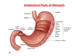
21. Musculosa Of Pylorus 2 Layers Inner Circular (IC) Outer Longitudinal (OL) Circular muscle is Thickened to form “ Pyloric Sphincter ” (PS ) IC OL (PS)
24. Identify the organ (be specific). Describe the characters of the glands. (2)
25. This is a section in the fundic glands Identify the cell indicated by the arrow. (3)
26. (4) a) Identify the cell indicated by the black arrow. b) Identify the layer indicated by the red arrow. This is a section in the lower part of the fundic glands:
27. (a) (b) (5) (a) & (b) are sections in the fundus of the stomach. Identify the layer indicated by the arrow in (a) & (b).
28. (a) (b) (6) i) In slides a & b , identify the organs. ii) Identify 1, 2 & 3 in each slide.
29.
30. 13- Identify the organ. 14- Identify the structure indicated by the arrow. 15- Identify the tissue indicated by the arrow. 16- identify the structure indicated by the arrow. 16 (9)
31. (10) a) Identify the type of junction. b) Name the abbreviations marked by the letters: S – D – PS – G- CM - LM