Ferster & Spruston propose that there are approximately one trillion neurons that are responsible for controlling our behavior
Although there are a variety of functions that the neurons are responsible for the basic structure is the same
The dendrites contain receptor sites (along with the soma) that receive messages from other neurons. In short, incoming messages to the neuron arrive at the dendrites and soma
The axon may be described as the information highway of the neuron. Electrochemical messages travel one way down these long slim tube like extensions that may travel for several millimeters (and in some cases) 3 feet in length from the soma to the terminal button.
The myelin sheath acts as insulation for most axons in order to provide insulation (to avoid short circuiting one another) and speed up the transmission of the message. They are a series of specialized cells that are composed of fat and protein which wrap themselves around the axon. The old name “fat head” takes a different connotation when you think of how such cells improve the functioning of the brain.
In such diseases as Multiple Sclerosis (MS) the myelin deteriorates leaving the axon without insulation and prone to short circuiting. As a result, disturbances occur between the brain and muscles creating such symptoms as an inability to walk, problems with vision, and general muscle impairment.
Significant to note that some substances (e.g. chemical substances needed for nourishment) travel the opposite direction down the axon {terminal button to soma}that may be disrupted by such diseases as amyotrophic lateral sclerosis (ALS) also known as Lou Gehrig’s disease.
“To fire or not to fire, that is the question”, the all-or-none law outlines the basic functioning of the neuron - it is either “on” or “off”. If messages from other neurons trigger an individual neuron beyond a specific point it will fire. However, if other neurons inhibit an individual neuron it will not fire. Basic to this premise is that the neuron always fires at the same speed.
At a resting state the neuron is at approximately -70 millivotes (1 thousandth of a volt). This indicates that the inside of the axon is more negatively charged than the outside of the neuron - (somewhat similar to a car battery with the inside of the axon being the negative pole and the outside of the axon being the positive pole). This is a result of negatively charged and positively charged ions both inside and outside of the axon. Once the neuron is being triggered to fire the axon reaches approximately -50 millivolts (threshold) and eventually reaches an action potential.
Similar to a series of dominos falling, the axon opens and closes ion channels that allow the influx of positive ions to travel down the axon to the terminal buttons. This is known as the “action potential”. After the action potential has occurred, positive ions are pumped outside of the axon and the resting potential is restored. However, the neuron cannot fire until the neuron has recovered. Although the action potential is always at the same speed as the all-or-none law indicates (speed determined by the presence of myelin and the diameter of the axon - small diameter axons average speed about 2 mph and larger diameter can average speeds of 225 mph), neurons vary concerning the rate of firing. In other words, some neurons have the potential to fire as many times as 1,000 times per second while others have a rate that is much slower. The intensity of the stimulus that provokes the neuron depends on how much of that neuron’s rate is actually reached.
Unlike computer chips and other electrical wiring, neurotransmitters do not
make direct contact with one another. Instead, a gap exists between the terminal
button and the dendrite/soma of the adjacent neuron. This gap is known as the
synapse where a chemical substance is released and travels across the gap in
order to communicate to the next neuron.
A neurotransmitter is a chemical courier that is released from the terminal button
and travels across the synapse in order to bind to the receptor site located on the
dendrite or soma.
Neurotransmitters act as a chemical key that fit into a specific lock known as the
receptor site. If you have ever grabbed the wrong key for a lock and experienced
the everyday frustration when it does not work, you have experienced a similar
process to the functioning of the neurotransmitter and receptor site (minus the
frustration). Basically, if the chemical key does not fit it does not make it to the
receptor site of the receiving neuron. When is does fit, the neurotransmitter will
communicate one of two basic types of messages. Excitatory messages which
increases the likelihood that the receiving neuron will fire and inhibitory
messages which decreases the likelihood that the receiving neuron will fire.
(ACh) is the most common neurotransmitter that is found throughout the nervous
system. It is related to our every “movement” in that it is involved in transmitting
messages to our skeletal muscles. Feldman reports that it is also related to the
drug Curare used on the tips of poisonous darts that paralyze skeletal muscles
(as it keeps ACh from reaching receptor cells) and can cause eventual death by
suffocation because the victim can no longer breathe.
It is also reported that some scientist identify a deficiency in ACh production in
patients suffering from Alzheimer’s disease. Therefore, implicating ACh as being
related to memory capabilities.
Glutamate plays an important role along with other neurotransmitters in
producing specific biochemical changes at specific synapses in the formation of
memories.
GABBA is found in both the brain and spinal cord and appears to be the nervous
system’s primary inhibitory neurotransmitter. It is the moderator of a host of
behaviors ranging from eating to aggression. As reported in the chapter,
strychnine disrupts the transmission of GABBA across synapses thereby
preventing its inhibitory role and resulting in neurons firing wildly and producing
convulsions.
The destruction of certain neurons in Parkinson’s disease reduce the production
of dopamine that results in muscular rigidity and tremors. Techniques that
increase production of dopamine has proved helpful to Parkinson’s patients.
Serotonin has been associated with the regulation of sleep, eating, mood, and
pain. Higley, Suomi, & Linnoila indicate that a growing body of research
implicate this neurotransmitter with such conditions as alcoholism, depression,
suicide, impulsivity, aggression, and coping with stress in their 1996 study.
Endorphins are our bodies’ natural “opiates” that are produced by the brain in
order to deal with pain. They can also provide a euphoric experience after
physical exertion commonly referred to as a “runners high”(Kremer & Scully,
1994; Dishman, 1997). This has also led researchers to hypothesize that the
endorphins are responsible for the “placebo effect” and the positive impact of
acupuncture in dealing with pain.
It is helpful to emphasize that the nervous system contains all of the neurons
contained in our body and truly works together as an interactive and dynamic
system that controls every aspect of our body. Divisions are made in order to
outline general functioning for various aspects of this complex system.
The peripheral nervous system are all those neuronal pathways that lead to and
from the central nervous system. It is further divided into the somatic and
autonomic nervous systems.
The somatic nervous system is responsible for all voluntary motor movements
and the communication of information to and from the sense organs. It is
helpful to demonstrate this by having students close their eyes and identify where
their body parts (e g feet, hands) are located and what direction they are pointed.
The autonomic nervous system controls all of the involuntary movements of the
body, e. g. heart, breathing, and other organs. This is subdivided into the sympathetic
(part of the autonomic nervous system that prepares the body to deal with stressful
situations and evokes the fight/flight response) and the parasympathetic
system (part of the autonomic system that acts to calm the body). It is helpful to
have the students to mimic the movement of the heart by squeezing and relaxing
their fist in order to sample how life would be without the autonomic system.
The central nervous system includes the brain and spinal cord.
As the axon is the information highway for the neuron, the spinal cord is the
primary highway for the brain. It is helpful to point out the impact of spinal cord
injuries - (the chapter discusses the injury sustained by Christopher Reeve that
resulted in quadriplegia), as well as the usage of such procedures as an epidural
or spinal block during pregnancy or surgeries.
The spinal cord also can initiate a behavior via a reflex arc - the process where an
interneuron acts as a “breaker switch” when we are experiencing pain. The basic
process involves a painful stimuli that travels to the spinal cord by an afferent
passageway that’s sufficient to trip the interneuron which stimulates an efferent
passageway in order to contract the muscle.
As mentioned above, sensory (afferent) neurons carry information from the body
to the central nervous system and motor (efferent) neurons communicate information
from the central nervous system to muscles and glands of the body. Interneurons
communicate messages between sensory and motor neurons.
Evolutionary psychology examines the progression of development for our
nervous system through the lens of Darwinian Evolutionary Theory. From the
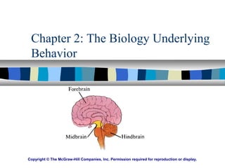
common elements of the primitive brain - (3 main parts that were dedicated to close
stimuli such as smell, distant stimuli such as sight and sound, and the ability to
maintain balance and coordination) to the complexity of our cerebral cortex, this
area of psychology examines the progression of our human brain. Our nervous
system is viewed as being hierarchically organized that suggests newer evolutionary
aspects of our brain control and regulate more primitive parts of our nervous
system. As we move up the spinal cord more complex and sophisticated behaviors
are controlled by the brain.
The area of behavioral genetics examines the effects of heredity on behavior.
Behavioral research explores the heart of the nature-nurture issue and examines
the impact of genetics on cognitive abilities, personality traits, sexual orientation,
and psychological disorders.
A variety of methods have been utilized in order to explore the various functioning and structures of the brain. Collectively they have provided the fund of information that we now possess.
EEG - Provides an electrical recording of the brain that is produced by the firing of the neurons. Recent techniques allow visual representation of the brain to aid in the diagnosis of epilepsy and learning disabilities.
CAT - A computerized enhancement of thousands of x-ray images taken at slightly different angels provides a structural view of the brain.
fMRI - A powerful magnetic field is utilized in order to show structure and functioning of the brain.
SQUID - Highly sensitive to minute changes in magnetic fields, the SQUID can pinpoint neuronal activity.
PET - With the use of radioactive water into the bloodstream, the PET scan can identify active areas of the brain and examine the brain at work.
Case Studies - Phineus Gauge presents the first case study for the behavioral changes associated with brain injury.
The patient is typically awake during neuro-surgical procedures which has allowed various areas and their corresponding functioning to have been mapped.
The central core is sometimes referred to as the “old brain” as its “evolutionary
underpinnings” can be traced back some 500 million years to primitive structures
found in nonhuman species.
The medulla controls primary life functioning such as heart beat and breathing.
The pons is involved in transmitting motor information, coordination muscles, and
integrating movement between right and left halves of the body. It is also involved
in arousal and sleep.
The reticular formation is responsible for screening in important information and
screening out non important information. With its association to attention and
concentration the ARAS (ascending reticular activating system) has been implicated
with ADD/ADHD. It is also responsible for scanning the environment while we
sleep in order to awaken us in case there is an unexpected noise.
The cerebellum is well known for its role in coordination and balance of movement.
It constantly monitors muscles and coordinates their placement, movement, and
tension. As noted in chapter 3, alcohol depresses the functioning of the cerebellum
and results in unsteady gait and lack of coordination associated with drunkenness.
Chapter 3 also reports of research conducted by Raymond, Lisberger, & Mauk, 1996;
Barinaga, 1996, and Gao et al. 1996 that implicates the cerebellum as having a
role in intellectual functioning ranging from the analysis of sensory information to
solving problems.
The thalamus is known for being the primary relay station of the brain. There are
numerous thalamic relays that extend to various parts of the cerebral cortex in
order to transmit information. It also is used as a pathway of communication from
higher parts of the brain to the cerebellum and medulla.
The relatively tiny hypothalamus (about the size of our fingertip) controls key aspects
of our functioning such as maintaining homeostasis and was identified by
Kupfermann, 1991 as critical in basic survival of the species by regulating the four
F’s: fight, flight, feeding, and fornication.
The limbic system consists of such structures as the amygdala, hippocampus, fornix,
septal area, cingulate cortex, and hypothalamus. It is sometimes referred to as our
“animal brain” as its structure and function is similar to that of other mammals.
Together, the aforementioned structures integrate their efforts and control such
behaviors as emotions and self-preservation such as eating, aggression, and
reproduction. It also has a significant role in learning and memory. Chapter 3
cites such research as Fanelli, Burright, & Donovick, 1983 and Bedard &
Persinger, 1995 that indicate the effects of lesions made within the limbic system as
turning docile animals hostile and hostile animals docile. Also, Routtenberg &
Lindy, 1965 demonstrated that some rats would bar press in order to stimulate the
pleasure centers within the limbic system until they collapsed with fatigue. The
chapter also reports of human studies that stimulated the limbic system which reported
the experience being somewhat similar to an orgasm. It is also notable to mention
the research conducted by Milner, 1966 that reports of memory impairments that
a patient experienced after having neurosurgery on the limbic system.
The cerebral cortex, also known as the “new brain”, enables us to think, evaluate, and make complex judgements. As pointed out in chapter 3, it is a mass of deeply folded, rippled, convoluted tissue (known as coritcalization) that allows for maximum cortical space in a limited area. If it were smooth and compact we would lose precious space occupied by neurons thereby limiting the sophistication and complexity of our behavior. This convoluted tissue also allows for a high degree of neuronal integration to allow for sophisticated processing of information.
The cerebral cortex is divided into four major sections by their general function that are called lobes. As seen in the side view of the brain, the frontal lobes lie at the front center of the cortex and the parietal lobes lie behind them. The temporal lobes are found in the lower center of the cortex and the occipital lobes lie behind them. The lobes are physically separated by deep grooves that are referred to as sulci.
The association areas are responsible for higher order thinking such as thinking, language, and speech. Damage to such areas may result in apraxia and the
individual may be unable to integrate activities in a rational or logical manner. The text identifies such cases as reported by Lechtenberg, 1982 of a patient being unable to follow simple directions to unlock a door with a key, but later able to perform such behavior when left alone in a locked room.
The temporal lobes are the primary site where auditory information is registered. Language typically is deciphered on the left temporal lobe - Wernicke’s area and music is typically registered on the right temporal lobe.
The occipital lobe is the region where visual input is registered. It is significant to discriminate the difference between sensation and perception. The occipital lobe stores our visual memories in the association areas and keeps our world constant by maintaining a fluid visual experience from the raw sensory material provided by our eyes.
It is significant to point out that memory and “thinking” are conducted within the association areas of the cerebral cortex. Also, areas of the brain are specified by the function that they perform. The more neurons dedicated to a specific function, the more complex and sophisticated that function may be.
The frontal lobe is dedicated to “executive functioning” - planning and organization ability, as well as, coordinating and initiating complex body movements. It is helpful to point out the devastating effect early frontal lobotomies had on patients - specifically it did not reduce aggressive tendencies as predicted and impaired the patient’s executive functions. On the side of the frontal lobe and beside the central sulci lies the primary motor strip. This area is dedicated to the voluntary movements for particular parts of our body. It is important to point out that some parts of our body (e. g. hands) have a larger area dedicated to their movement as compared to other parts (e. g. wrist). As pointed out before, the more brain real estate dedicated to a function, the more neurons occupy that space and the more complex and sophisticated the behavior.
Broca’s area, (named from French physician Paul Broca who discovered this area in 1861), is located within the frontal lobe (on the left hemisphere for the majority of individuals) and controls speech production. Damage to this area results in Broca’s aphasia and results in the individual struggling to produce words.
Wernicke’s area, (named for Carl Wernicke who discovered this area in the 1870’s), is located within the temporal lobe. Damage to this area results in Wernicke’s aphasia and impairs language usage.
The somatosensory strip is located beside the central sulci on the side of the parietal lobe. It has specific areas that are responsible for receiving bodily sensations for specific areas of the body. Again, it is significant to note that more brain real estate is distributed for some areas of the body as compared to others (e. g. lips/face vs. leg). The more real estate, the more neurons that increase the sensitivity of that area. A good experiment is to have students close their eyes and identify objects on their desk - while integrating the use of afferent passages from the somatic nervous system traveling through the thalamus to the somatosensory strip.
The cerebral cortex is divided into two halves known as the right and left hemispheres.
The hemispheres operate contralaterally meaning that the right hemisphere controls
the left side of the body and the left hemisphere controls the right side of the body. It
is helpful to point out how impairments in functioning on the left side of the body
reflect right hemispheric damage and vice versa. Also, that speech aphasia typically
indicates damage on the left hemisphere.
The notion of lateralization (the dominance of one hemisphere of the brain in relation
to a specific function) had been apparent from individuals experiencing speech aphasia
as having damage to the left hemisphere (Bradshaw & Rogers, 1993; Zaidel et al.
1995; Grossi et al., 1996). Other researchers (Zaidel, 1994; Davidson & Hugdahl,
1995, Siegal, Carrington, & Radel, 1996; LaMendola & Bever, 1997) have identified
such general differences in functioning as the left hemisphere being more concentrated
on tasks that require verbal abilities such as speaking, reading, thinking and reasoning.
The right hemisphere being more adept at non verbal tasks such as visual-spatial
integration, recognition of patterns and drawings, music, and emotional expression.
As pointed out in chapter 3, Roger Sperry earned the Nobel Prize for his work in
examining patients whom had their corpus callosum cut in order to eliminate intense
and frequent seizures that were non responsive to medication. Such individuals
are referred to as “split brain” patients. It is important to note that the corpus callosum
is the primary band of fibers that connects the right and left hemispheres together - thus
allowing them to communicate with one another. Also interesting to add that women tend
to have larger a corpus collosum than men.
The classic experiment involved split brain patients and the use of their somatosensory
system to identify and name objects out of their visual field. Basically, the subjects
were blind folded and then asked to touch particular objects. When using their right
hand (which communicates to the left hemisphere) they were able to provide a verbal
report of the object they had touched. When using their left hand (which communicates
to the right hemisphere) they were unable to verbalize what they had touched in
their hand. Note the breakdown of the right hemisphere to alert the left since the
corpus callosum had been severed. Also, it is significant to note that the subjects
were able to identify the object once the blindfold was removed with their left hand.
It is also interesting to report Gazzaniga’s observations in 1970 where some split brain
patients would exhibit unusual behavior. The text gives the example of the patient
who pulled his pants up with one hand while pulling them down with the other. This
can help to discuss the independent functioning of each hemisphere.
The endocrine system is a chemical communications network that is controlled and
monitored by the hypothalamus via the pituitary gland. Basically, hormones are chemicals
that are produced by the endocrine system and secreted into our bloodstream. The
chemicals affect the functioning or growth of our body – e. g. growth hormones and
the role of adrenaline during the fight flight response. It is helpful to tie in the connection
between the hypothalamus - pituitary gland - endocrine system. Also, pointing out
the role of the suprachiasmatic nuclei within the hypothalamus that utilizes the
pituitary gland to regulate our melatonin levels in order to set our sleep-wake cycle.
The text provides a wonderful metaphor for hormones as being similar to radio waves
that are distributed throughout the body and “tuned in” by specific areas that the hormone
has an effect.