2. Traumatic spinal cord injury 179
Imaging Evaluation
The initial computed tomography (CT) scan obtained within
2 hours of the fall showed findings of early diffuse cerebral
edema, SDH, and SAH, including a prominent left parietal
focus of SAH and a focal right frontal SDH (Fig 1). A much
smaller SAH/SDH were also present along the convexities,
falx (ie, interhemispheric), and tentorium. There was no ev-
idence of brain hemorrhage. No scalp or skull abnormalities
were identified, although 1 observer could not rule out a
“healed” skull fracture along the sutures in the parieto-occip-
ital region. A CT scan performed 5 hours later showed pro-
gression of the cerebral edema and no change in the SAH or
SDH (Fig 2). A CT scan of the cervical spine was negative
(Fig 3). Magnetic resonance imaging was recommended but
never obtained. A skeletal survey showed anterior wedge-like
vertebral body deformities from T5 through T12 and inferior
L2. Some widening of the cranial sutures was present, but no
fractures were confirmed on the plain radiographs. The re-
mainder of the survey was negative. A postmortem CT scan of
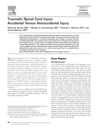
the entire spine (Fig 4), confirmed the vertebral deformities. Figure 2 A CT scan performed 5 hours after the initial CT scan shows
marked progression of cerebral edema with complete loss of gray/
Autopsy Findings by the Medical Examiner white matter differentiation, obliteration of sulci, and near complete
obliteration of the ventricular system. A SAH is seen within com-
Head and Brain
pressed sulci in the left parietal lobe.
A focus of hemorrhage was present in the left posterior
parietal scalp (ie, impact site). Microscopy showed acute
hemorrhage with acute vital reaction within the fibrous
connective tissue of the galea in that region. Brain weight axonal injury (TAI) observed microscopically on beta-
was 1,270 g with the spinal cord attached. No skull frac- amyloid precursor protein (B-APP) immunoperoxidase
ture was shown. The brain was extremely swollen with stains. There were bilateral, holohemispheric, thin-layer
generalized flattening and ablation of the normal gyral SDHs with no mass effect.
pattern. Histologically, a diffuse axonal injury pattern con-
sistent with HIE was present. There were no gross trau-
matic brain parenchymal injuries (eg, no contusion or
shear lesions) or any histological evidence of traumatic
Figure 1 A nonenhanced CT image of the brain preformed within 2
hours of the fall shows decreased differentiation of gray/white mat- Figure 3 Sagittal reconstructed CT image of the cervical spine shows
ter representing edema. normal alignment and no fractures.
3. 180 Barnes et al
Biomechanical Analysis
A court-approved, biomechanical evaluation was performed
including an investigation of the home setting where the
injury reportedly occurred. A number of potential accidental
and NAI (including SBS) scenarios were considered and an-
alyzed primarily to address the thoracic spinal injuries and
secondarily to address the cervical cord injury. The approach
to the biomechanical analysis was to assume that the care-
taker’s history was truthful and accurate and to then apply
the principle of mechanics to evaluate whether or not such a
history could be consistent with the subsequent injuries. This
approach was not intended to rule out other possibilities but
simply to evaluate the history provided.
In that light, the caretaker consistently reported to all au-
thorities that he had his back turned at the time of the inci-
dent but that the boy had been standing up on the seat of the
chair. He then reported hearing a noise and turning to find
the boy and the chair on the floor, with the chair lying on its
back. Using the caretaker’s consistent history as one scenario,
Figure 4 Sagittal reformation of the thoracic spine from a postmor- along with the imaging and clinical findings, the child was
tem CT scan shows multilevel anterior wedge compression fractures assumed to have fallen, rotating with the chair until a point of
of varying degrees. separation (Fig 6). From that point, it was further assumed
that he fell freely to strike the floor first with his head and
then with his dorsal neck and a shoulder, again based on the
Neck and Spinal Cord imaging and autopsy findings. This “impact” scenario would
Focal soft-tissue hemorrhages were present in relation to the
right posterior neck and shoulder regions as well as the pos-
terolateral transverse processes of the atlas and axis and at the
C1-C2 intervertebral junction. No vertebral artery abnormal-
ity was noted. The dura appeared tense and filled with blood-
stained fluid. Sagittal sections at the cervicomedullary junc-
tion showed partial transection and disruption of the central
cord immediately distal to the inferior medullary olives. The
tissue appeared elongated and physically separated suggest-
ing axial tension of the cord (Fig 5). The cellular response
consisted of round and polymorphonuclear cells with acute,
focal hemorrhage. Findings of ischemic neuronal degenera-
tion were most prominent in, and adjacent to, the dorsal and
ventral neurons and consisted of cytoplasmic swelling, de-
granulization, loss of fine detail, and nuclear pynknosis.
Eyes
On gross examination, the pigmented layer of the retina was
focally separated from the choroid. RHs were present primar-
ily in the ganglion cell layer anteriorly but extended posteri-
orly. An optic nerve sheath hemorrhage was also present
bilaterally. There was no mention of perimacular folds.
Vertebral Column
The vertebral bodies from T2 through L3 to 4 were removed
en bloc. A hemorrhage was seen in the anterior and lateral
perivertebral fibroconnective tissues. Microscopic sections
showed acute hemorrhage with fibrin deposition replacing
normal marrow of all vertebral bodies. Multiple areas of dis-
rupted cancellous bone with acute-phase osteogenic granu- Figure 5 A histological specimen through the cervicomedullary
lation tissue were especially prominent T7 through T10. No junction shows complete disruption of the central cord elements
callus or osteoblast activity was present. (circle). (Color version of figure is available online.)
4. Traumatic spinal cord injury 181
34 in), his mass (15.9 kg, 35 lb), and the position of his CG
being at approximately 57% his height,7 were used to deter-
mine the CG of the combined system (Appendix 1). Thus, the
first phase of the fall was modeled with the child and chair
combined as an inverted pendulum until the chair reached its
natural tipping angle. Vertical and rotational velocities at this
point were then used for the initial conditions of the subse-
quent free fall, resulting in a predicted impact velocity rang-
ing from 3.7 to 4.0 month(s). Impulse momentum was then
applied to determine the severity of a fully plastic impact,
modeling the child as a single spring-mass system, with the
flexing spine serving as the spring, and the mass defined as
the measured mass of the child minus that of his head. Be-
cause there are limited data available to define an appropriate
impact duration, a broad range of 50 to 100 milliseconds was
Figure 6 A schematic representation of the fall. (Color version of considered. Thus, the peak impact force was estimated to
figure is available online.) range between 0.9 and 1.9 N. A free-body diagram of the
flexed body was used to determine associated peak thoracic
vertebral body forces ranging from 1.4 to 3.1 kiloNewtons
produce flexion and axial compression with the center of (kN). Forces in the separately considered head impact were
mass of his body trailing above. The initial motion of the estimated to be between 3.4 and 3.7 kNs.
child was assumed to have produced an initial rotation of the
child and chair together as an inverted pendulum system
about an axis at the base of the chair’s rear legs. Once the Discussion
chair reached its natural tipping angle, however, it was as-
sumed to continue in its inverted pendulum motion while NAI/SBS
the child then fell freely to the floor. In this case, the initial “diagnosis” of NAI/SBS was based on
Multiple anterior compression fractures of the thoracic the heretofore classic “triad” of SDH, RH, and encephalopa-
spine, as reported in this case, are uncommon. The mecha- thy, along with a history presumed to be inconsistent with
nism most consistent with this type of injury, however, the injuries. Central nervous system findings that mimic NAI/
would be severe flexion and/or axial compression of the SBS have been reported in accidental trauma and in a number
spine. It is problematic, biomechanically, to conclude that of medical conditions.14-21 The latter includes infection, co-
such an injury can result from “SBS,” particularly in a child of agulopathy, metabolic disorders, and others.19-21 More recent
this age and size. Further evidence of head and shoulder reports also show that there is no specific pattern of intracra-
impact suggests that the necessary loading of the spine may nial hemorrhage that is diagnostic of NAI/SBS to include
have been produced by forces applied to the head, neck, interhemispheric SDH and mixed-density SDH.14-17 Further-
and/or shoulder. Thus, we chose to evaluate a scenario in more, recent evidence-based medical reviews (and legal chal-
which the boy somehow caused the chair to tip. He then lenges) of the past NAI/SBS literature reveal that the vast
rotated with the chair and ultimately fell in such a way that majority of these publications failed to achieve quality of
his head struck the floor first and quickly rotated to produce evidence ratings that would merit the use of the “triad” as a
flexion in the neck and bring the shoulder/lower neck region standard or guideline for proof of NAI/SBS.1-8
into contact with the floor. The force acting through the This case also shows the complexities involved in estab-
shoulder area then acted to bring the remainder of the child’s lishing the sequence of injuries given multiple findings. Al-
body mass to rest, resulting in flexion and axial compression though initial concern for NAI is important and must be
of the middle and lower spine. There are clearly many vari- reported, medical personnel must carefully correlate such
ables associated with the chosen scenario, including the in- findings with the history to establish a correct sequence of
fluence of body rotation and whether the child was rotating events, including predisposing factors.19-21 The initiation of
forward or backward, but there is no reason to believe the fall the criminal process before a complete and thorough child
could not have occurred as described. Given the selected protection and medical evaluation can lead to a rush in judg-
scenario, the next step was to evaluate whether or not the ment. The injuries in this particular case were attributed to
forces associated with the respective impacts to the head and SBS before the brain and spinal cord injuries were completely
shoulder/lower neck could have been severe enough to pro- evaluated.22 The father of the victim was charged with fatally
duce the injuries. shaking the child. After all the forensic evidence was consid-
Analysis of the chair provided a seat height of 43 cm (17 in) ered, the ultimate verdict was acquittal.
and a rearward tipping angle of 23°. The mass of the chair Given the fact that the law requires physicians to report
and its center of gravity (CG) were taken as equal to 6.8 kg suspected NAI, there is the danger of assuming NAI in all
(15 lb) and the height of the seat, respectively. The chair cases of SDH and RH. As a result, further medical and imag-
measurements, coupled with the height of the child (86 cm, ing workup may not be pursued (eg, magnetic resonance
5. 182 Barnes et al
imaging of the brain and cervical spine). The American Acad- Impact Injury
emy of Pediatrics, as others, strongly endorse the use of mag- Although there are no data available defining skull fracture
netic resonance imaging in cases of suspected NAI.17,20,21,23 In thresholds (as an indicator of impact) for a 21-month-old,
the absence of an apparent cause of diffuse cerebral edema data reported for younger infants33-36 and adults37 suggest
(including HIE), cervical cord injury should be considered. that the calculated head-impact forces are enough to result in
After the initial CT scan, magnetic resonance imaging is the fracture in at least some of the population. The absence of
choice for delineating spinal, paraspinal, and intraspinal in- evidence of “significant” trauma to the scalp and skull may
jury. A short tau inversion recovery (STIR) sequence should additionally be explained by the wide distribution of the
always be included because this technique provides the best force along the head, neck, and shoulders at the time of
sensitivity for these types of injury.21 impact. In young children with impact injury, there may be
no focal scalp injury or skull fracture on physical examina-
SCIWORA tion or by imaging. Therefore, the lack of such findings
should not be interpreted as absence of impact injury. Addi-
SCIWORA is not uncommon in toddlers and has been re-
tionally, fatal and otherwise significant intracranial injuries
ported to occur after minor accidental trauma as well as in
have been reported from accidental household or short falls
cases of alleged NAI.13,24-29 Evidence of a spinal cord injury
resulting in the triad of SDH, RH, and encephalopathy.38,39
plus cranial, neck, and shoulder impact on the postmortem
The biomechanical literature suggests different thresholds for
examination are the key findings in this case. The gross and
central nervous system injuries given various scenarios.40-43
histological findings, as well as the imaging findings, are en-
Neck and cervical spine tissues may have a lower threshold
tirely consistent with the caretaker history of a household fall
than brain for minimum forces required to produce trau-
as corroborated by the biomechanical evaluation. This is true matic injuries.40
for both the primary injury (ie, cord transection) and the
secondary injury (ie, HIE) as reflected in the clinical course of
the child. SDH and SAH
The bony structures of the cervical spine in infants and There are a number of potential causes for the SDH/SAH in
younger children are not fully developed as compared with this case. These include impact trauma, coagulopathy, in-
that of the adult. Such “immaturity” includes the horizontal creased ICP, ischemic endothelial damage, and reperfusion.
nature of facet joints with flat morphology of uncinate pro- The focal left parietal SAH (Fig 1) correlates with the primary
cesses, elastic paraspinal ligaments, and anterior wedge-like site of impact (ie, coupe injury), and the focal left frontal SDH
morphology of the vertebral bodies.13 These factors account may be consistent with contracoupe injury. Further hemor-
for the relative ease of vertebral subluxation with complete rhage may be related to the coagulopathy as supported by
recovery of the bony elements to normal anatomical align- laboratory findings. This is a known phenomenon that may
ment. This predisposes the child to cervical cord injury in the be initiated by tissue injury caused by trauma or hypoxia
absence of bony abnormalities. Instantaneous damage to the ischemia.44,45 Once the capillary beds are open and leaking,
respiratory centers at the cervicomedullary junction corre- further increases in ICP from brain edema and CPR may
lated with the child’s respiratory distress, and subsequent exacerbate this process. Geddes et al32 suggest that additional
HIE lead to extensive edema and increased intracranial pres- factors such as venous and arterial hypertension (HTN) may
sure (ICP). exacerbate hemorrhage in the ischemic, swollen brain with
increased ICP. They propose both increased oozing from
hypoxic veins in the setting of venous HTN secondary to
Hypoxic-Ischemic severe edema and increased hemorrhage from episodic or
Versus Traumatic Axonal Injury sustained arterial HTN (eg, with reperfusion) that may occur
Gross and microscopic examination showed the effects of as a part of Cushing’s triad or be neurogenic in origin. Addi-
severe HIE. There was no evidence of “primary” traumatic tionally, choking, vomiting, or paroxysmal coughing (eg,
axonal injury (ie, TAI or shear injury) as an indicator of pertussis) may also result in SDH and RH.46-48 Furthermore,
rotational acceleration-deceleration trauma to the brain. The the distribution of SDH or SAH along the interhemispheric
primary injury (ie, TAI) was shown to occur only at the fissure is not pathognomonic for NAI, as previously reported,
cervicomedullary junction. In cases of TAI (formerly “diffuse and has been shown to occur in cases of accidental trauma
axonal injury”), distinctive discrete swellings of the the ax- and HIE.14,16,17,21
ons, known as axon bulbs, are observed microscopically on
B-APP immunoperoxidase stains. These are focal or multifo- RH
cal lesions and most often occur along deep gray/white mat- The initial funduscopic documentation of RH was not made
ter junctions, the corpus callosum, and dorsal corticospinal until the child was in the PICU. Given the course of events to
tracts. They may also be associated with focal hemorrhage. that point, the RH may be a result of multiple factors as
HIE, whether primary or secondary, results in a diffuse pat- described earlier regarding SDH and SAH. RH is a known
tern of axonal alteration. Furthermore, the histological ap- manifestation of increased ICP. There is no single type or
pearance is different from that of TAI, forming a linear or pattern of RH that is pathognomonic of NAI/SBS, and RH is
streak pattern on B-APP staining.30-32 reported in a number of other conditions.15,16,49-57
6. Traumatic spinal cord injury 183
Thoracic Spinal Injury
The multiple thoracic compression fractures in this case are
unusual in NAI (SBS) and require biomechanical assessment as
well as consideration of patient risk factors. Based on studies of
compression fracture in intact cadavers subjected to flexion and
compression testing of both adult human58,59 and pediatric ba-
boon vertebral bodies,60 the biomechanical analysis suggested
the presence of sufficient force to cause the thoracic fractures, in
addition to the cervical spinal cord injury in this case.
Additionally, the patient had steroid-dependent asthma
treated with daily beclomethasone dipropionate inhaler for a
period of 8 months before the fall. During multiple visits to
the emergency room for exacerbations before the reported
fall, he also received additional doses of steroids in a form of
prednisone. Two weeks before the fall, he received 20 mg of
prednisone daily for 6 days. The influence of steroids in this
case is uncertain. However, such high daily doses of oral
steroids have been shown to significantly increase the risk of
fractures.61-63 Van Staa et al61 also showed that high daily
doses independent of duration of treatment or prior exposure
put the patients at high risk for fractures. The patient also Figure A1 (See appendix). (Color version of figure is available on-
presented to the emergency room 3 months before the cur- line.)
rent incident after a fall down the stairs. He was evaluated
clinically and released. No imaging was performed at that
time. The postmortem histological examination of the verte-
bral column showed evidence of acute trauma. Additionally, is the subsequent free-fall distance of the body CG to the
an unusual distribution of diffuse microfractures was ob- floor.
served in all bones examined, supporting the possible effect ● Using impulse momentum and assuming a triangular
of chronic steroid therapy on bone fragility. It was impossible force pulse and plastic impact, peak force at impact
to assess for bone density of the spine during necropsy be- F 2mv , where m is the mass of the object, v is
cause the bones were decalcified. impact velocity, and is the duration of impact.
● Compression force on lumbar vertebrae, Fv, is deter-
mined by satisfying M I about the ligament attach-
Conclusion ment point, as shown in the free-body diagram. Mo-
Physicians have an obligation to completely and timely eval- ment-arm distances were scaled from values used by
uate suspected NAI, including its mimics. The imaging find- Myklebust et al.58
ings alone cannot distinguish NAI from AI or from the med-
ical mimics. A complete and thorough medical evaluation,
References
using evidence-based medicine principles, is necessary in
1. Donohoe M: Evidence-based medicine and shaken baby syndrome part
parallel with the child-protection assessment. A multidisci- I: Literature review, 1966-1998. Am J Forensic Med Pathol 24:239-
plinary approach to this evaluation is also important, includ- 242, 2003
ing the involvement of qualified specialists. Such an ap- 2. Leestma J: Case analysis of brain injured admittedly shaken infants, 54
proach may be the difference between appropriate child cases 1969-2001. Am J Forensic Med Pathol 26:199-212, 2005
3. Lyons: Shaken Baby Syndrome: A Questionable Scientific Syndrome
protection versus the improper breakup of a family or a
and a Dangerous Legal Concept. Utah Law Rev 1109, 2003
wrongful indictment and conviction. 4. Gena M” Shaken baby syndrome: Medical uncertaintly casts doubt on
convictions. Wisc Law Rev 701, 2007
5. Le Fanu J: Wrongful diagnosis of child abuse—A master theory. J R Soc
Appendix Med 98:249-254, 2006
6. Mackey M: After the Court of Appeal: R v Harris and others [2005]
Equations Used in Biomechanical Analysis EWCA crim 1980. Arch Dis Child 91:873-875, 2006
● Angular velocity of combined chair-child system at end 7. Richards P, Bertocci G, Bonshek R, et al: Shaken baby syndrome. Before
of inverted pendulum phase was determined as the court of appeal. Arch Dis Child 91:205-206, 2006
8. Baath J: Shaken baby syndrome: The debate rages on U. Toronto Med J
2 g L 1 cos , where L is height of combined 83:22-23, 2005
system center of mass and is angle of rotation from 9. Squier W: Shaken baby syndrome: The quest for evidence. Develop
vertical position (Fig A1). Med Child Neurol 50:10-14, 2008
● From conservation of energy, impact velocity v 10. Gilliland MGF: Use of the triad of scant SDH, brain swelling, and retinal
hemorrhages to diagnose non-accidental injury is not scientifically
v2 2gH where vz is the vertical component of linear
z valid. National Association of Medical Examiners National Meeting,
velocity at the end of the inverted pendulum phase and H October 2006 (abstr 53)
7. 184 Barnes et al
11. David TJ: Non-accidental head injury—The evidence. Pediatr Radiol 36. Weber W: Biomechanical fragility of the infant skull. Z Rechtsmed
38:S370-S377, 2008 (suppl 3) 94:93-101, 1985
12. Jaspan T: Current controversies in the interpretation of non-accidental 37. Goldsmith W, Monson KL: On the state of head injury biomechanics—
head injury. Pediatr Radiol 38:S378-387, 2008 (suppl 3) Past, present, and future. Part 2: Physical experimentation. Crit Rev
13. Pang D, Wilberger JE Jr: Spinal cord injury without radiographic ab- Biomed Eng 33:105-207, 2005
normalities in children. J Neurosurg 57:114-129, 1982 38. Plunkett J: Fatal pediatric head injuries caused by short-distance falls.
14. Tung GA, Kumar M, Richardson RC, et al: Comparison of accidental Am J Forensic Med Pathol 22:1-12, 2001
and nonaccidental traumatic head injury in children on noncontrast 39. Gardner HB: A witness short fall mimicking presumed shaken baby
computed tomography. Pediatrics 118:627-633, 2006 syndrome (inflicted childhood neurotrauma). Pediatr Neurosurg 43:
15. Christian CW, Taylor AA, Hertle RW, et al: Retinal hemorrhages caused 433-435, 2007
by accidental household trauma. J Pediatr 135:125-127, 1999 40. Bandak FA: Shaken baby syndrome: A biomechanics analysis of injury
16. Steinbok P, Singhal A, Poskitt K, et al: Early hypodensity of computed mechanisms. Forensic Sci Int 151:71-79, 2005
tomographic scan of the brain in an accidental pediatric head injury. 41. Margulies S, Prange M, Myers BS, et al: Shaken baby syndrome: A
Neurosurgery 60:689-695, 2007 flawed biomechanical analysis. Forensic Sci Int 164:278-279, 2005
17. Vinchon M, Noule N, Tchofo PJ. Et al. Imaging of head injuries in 42. Bandak FA: Author’s reply to “shaken baby syndrome: A flawed bio-
infants: temporal correlates and forensic implications for the diagnosis mechanical analysis.” Forensic Sci Int 164:282-283, 2005
of child abuse. J Neurosurg 101:44-52, 2004 43. Prange MT, Coats B, Duhaime AC, et al: Anthropomorphic simulations
18. McNeeley PD, Atkinson JD, Saigal G, et al: Subdural hematomas in of falls, shakes, and inflicted impacts in infants. J Neurosurg 99:143-
infants with benign enlargement of the subarachnoid spaces are not 150, 2003
pathognomonic for child abuse. AJNR Am J Neuroradiol 27:1725- 44. Hymel KP, Abshire TC, Luckey DW, et al: Coagulopathy in pediatric
1728, 2006 abusive head trauma. Pediatrics 99:371-375, 1997
19. Sirotnak AP: Medical disorders that mimic abusive head trauma, in 45. Miner KE, Kaufman HH, Graham SH, et al: Disseminated intravascular
Frasier L, Rauth-Farley K, Alexander R, et al (eds): Abuse Head Trauma coagulation fibrinolytic syndrome following head injury in children:
in Infants and Children (ed 1). St Louis, MO, GW Medical Publishing, Frequency and prognostic implications. J Pediatr 100:687-691, 1982
2006, pp 191-248 46. Geddes JF, Talbert DG: Paroxysmal coughing, subdural and retinal
bleeding: A computer modeling approach. Neuropathol Appl Neuro-
20. Hymel KP, Jenny C, Block RW: Intracranial hemorrhage and rebleeding
biol 32:625-634, 2006
in suspected victims of abuse head trauma: Addressing forensic con-
47. Talbert DG: Paroxysmal couph injury, vascular rupture and ‘shaken
troversies. Child Maltreat 7:329-348, 2002
baby syndrome.’ Med Hypotheses 64:8-13, 2005
21. Barnes PD, Krasnokutsky MV: Imaging of the central nervous system in
48. American Academy of Pediatrics red book online. Pertussis. Available
suspected or alleged nonaccidental injury, including the mimics. Top
at: http://www.aapredbook.aappublications.org/cgi/content/extract/
Magn Reson Imaging 18:53-74, 2007
2006. Accessed November 2008
22. Twoney EL, Iemsawatdikul K, Stephens BG, et al: Multiple thoracic
49. Lantz PE, Sinal SH, Stanton CA, et al: Perimacular retinal folds from
vertebral compression fractures caused by non-accidental injury: Case
childhood head trauma. BMJ 328:754-756, 2004
report with radiologicalpathological correlation. Pediatr Radiol 34:
50. Ebube O, Watts P: Are there any pathognomonic signs in shaken baby
665-668, 2004
syndrome? J AAPOS 11:99-100, 2007 (abstr)
23. Sane SM, Kleinman PK, Cohen RA: American Academy of Pediatrics.
51. Goetting MG, Sowa B: Retinal hemorrhage after cardiopulmonary re-
Section on Radiology Diagnostic imaging of child abuse. Pediatrics
suscitation in children: An etiologic reevaluation. Pediatrics 85:585-
105:1345-1348, 2000 588, 1990
24. Ahmann PA, Smith SA, Schwartz JF, et al: Spinal cord infarction due to 52. Lantz PE, Stanton CA: Postmortem Detection and Evaluation of Retinal
minor trauma in children. Neurology 25:301-307, 1975 Hemorrhages. Seattle, WA, American Academy of Forensic Sciences, 2006
25. Riviello JJ, Marks HG, Faerber EN, et al: Delayed cervical central cord 53. Lueder GT, Turner JW, Paschall R: Perimacular retinal folds simulating
syndrome after trivial trauma. Pediatr Emerg Care 62:113-117, 1990 nonaccidental injury in an infant. Arch Ophthalmol 124:1782-1783,
26. Chen LS, Blaw ME: Acute central cervical cord syndrome caused by 2006
minor trauma. J Pediatr 108:96-97, 1986 54. Gilles EE, McGregor ML, Levy-Clarke G: Retinal hemorrhage asymme-
27. Cheshire DJ: The paediatric syndrome of traumatic myelopathy with- try in inflicted head injury: A clue to pathogenesis? J Pediatr 143:494-
out demonstrable vertebral injury. Paraplegia 15:74-85, 1977 499, 2003
28. Launay F, Leet AI, Sponseller PD: Pediatric spinal cord injury without 55. Obi E, Watts P: Are there any pathognomonic signs in shaken baby
radiographic abnormality: A meta-analysis. Clin Orthop Relat Res 433: syndrome? J AAPOS 11:99-100, 2007
166-170, 2005 56. Brown S, Levin AV, Ramsey D, et al: Natural animal shaking: A model
29. Brown RL, Brunn MA, Garcia VF: Cervical spine injuries in children: a for inflicted neurotrauma in children? J AAPOS 11:85-86, 2007
review of 103 patients treated consecutively at a level 1 pediatric 57. Binenaum G, Forbes BJ, Raghupathi R, et al: An animal model to study
trauma center. J Pediatr Surg 36:1107-1114, 2001 retinal hemorrhages in nonimpact brain injury. J AAPOS 11:84-85, 2007
30. Geddes JF, Vowles GH, Hackshaw AK: Neuropathology of inflicted 58. Myklebust J, Sances AJ, Maiman DJ, et al: Experimental spinal trauma
head injury in children I. Patterns of brain damage. Brain 124:1290- studies in the human and monkey cadaver, in Proceedings of Stapp Car
1298, 2001 Crash Conference. October 1983, San Diego, CA. 1983:149-161
31. Geddes JF, Vowles GH, Hackshaw AK: Neuropathology of inflicted 59. Yamada H: Strength of Biological Materials. Baltimore, MD, Williams
head injury in children II. Microscopic brain injury in infants. Brain and Wilkins, 1970
124:1299-306, 2001 60. Nuckley DJ, Eck MP, Carter JW, et al: Spinal maturation affects verte-
32. Geddes JF, Tasker RC, Hackshaw AK, et al: Dural haemorrhage in bral compressive mechanics and BMD with sex dependence. Bone 35:
non-traumatic infant deaths: does it explain the bleeding in ‘shaken 720-728, 2004
baby syndrome’? Neuropathol Appl Neurobiol 29:14-22, 2003 61. Van Staa TP, Leufkens HGM, Abenhaim L, et al: Oral corticosteroids
33. Snyder RG, Schneider LW, Owings CL, et al: Anthropometry of infants, and fracture risk: Relationship to daily and cumulative doses. Rheuma-
children, and youths of age 18 for product safety design. Final report. tology 39:1383-1389, 2000
Consumer Product Safety Commission. UM-HSRI-77-17, May 31, 1977 62. Yen D, Hedden D: Mutiple vertebral compression fractures in a patient treated
34. Prange M, Luck J, Dibb A, et al: Mechanical properties and anthropom- with corticosteroids for cystic fibrosis. Can J Surg 45:383-384, 2002
etry of the human infant head. Stapp Car Crash J 48:279-299, 2004 63. Makitie O, Doria A, Henriques F, et al: Radiographic vertebral mor-
35. Weber W: Experimental studies of skull fractures in infants. Z Re- phology: A diagnostic tool in pediatric osteoporosis. J Pediatr 146:395-
chtsmed 92:87-94, 1984 401, 2005