SlideShare une entreprise Scribd logo
1  sur  49
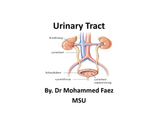
Urinary Tract By. Dr Mohammed Faez MSU
Urinary System ,[object Object]
Perform all functions except actual excretion.
Two Ureters
Convey urine from Kidneys to Urinary Bladder
Urinary Bladder
Holds Urine until excretion
Urethra
Conveys urine from bladder to outside of body.,[object Object]
The kidneys The right kidney lies approximately 1 cm lower than the left because of the large size of the right lobe of the liver. The lateral surface is convex and the medial surface is concave, with a vertical cleft called the renal hilusleading to the renal sinus Ureters, renal blood vessels, lymphatics, and nerves enter and exit at thehilus
The kidneys The hilum of the kidney is situated medially and transmits from front to back the: renal vein, renal artery, ureteric pelvis as well as lymphatics and sympathetic vasomotor nerves.  Each kidney consists of an outer renal cortex and an inner renal medulla. The renal cortex is a continuous band of pale tissue that completely surrounds the renal medulla. Extensions of the renal cortex (the renal columns) project into the inner aspect of the kidney, dividing the renal medulla into discontinuous aggregations of triangular-shaped tissue (the renal pyramids).
The kidneys The renal pelvis divides into two or three major calices and these, in turn, divide into minor calices which receive urine from the renal pyramids by way of the papillae.
Renal Artery and Vein      *usually arise at L2 Capsule Pelvis Major Calyx Cortex Ureter Medulla Minor Calyx The kidneys
The kidneys The kidneys have the following coverings : Fibrous capsule: This surrounds the kidney and is closely applied to its outer surface that prevents kidney infection. Perirenal fat: This covers the fibrous capsule that cushions the kidney. Renal fascia: This is a condensation of connective tissue that lies outside the perirenal fat and encloses the kidneys and suprarenal glands; it is continuous laterally with the fascia transversalis. Pararenal fat: This lies external to the renal fascia and is often in large quantity. It forms part of the retroperitoneal fat
Right Kidney Anatomical Relations Anteriorly:  The suprarenal gland, the liver, the second part of the duodenum, and the right colic flexure Posteriorly:  The diaphragm; the costodiaphragmatic recess of the pleura; the 12th rib; and the psoas, quadratuslumborum, and transversusabdominis muscles.       The subcostal (T12), iliohypogastric, and ilioinguinal nerves (L1) run downward and laterally.
Left Kidney Anatomical Relations Anteriorly:  The suprarenal gland, the spleen, the stomach, the pancreas, the left colic flexure, and coils of jejunum. Posteriorly:  The diaphragm; the costodiaphragmatic recess of the pleura; the 11th (the left kidney is higher) and 12th ribs; and the psoas, quadratuslumborum, and transversusabdominis muscles. The subcostal (T12), iliohypogastric, and ilioinguinal nerves (L1) run downward and laterally.
Posterior Anatomical Relations
Arterial Blood Supply of kidney Renal artery A single large renal artery, a lateral branch of the abdominal aorta, supplies each kidney.  These vessels usually arise just inferior to the origin of the superior mesenteric artery between vertebrae L1and L2. The left renal artery usually arises a little higher than the right, and the right renal artery is longer and passes posterior to the inferior vena cava.
Arterial Blood Supply of kidney They are distributed to different segments or areas of the kidney. Lobar arteries arise from each segmental artery, one for each renal pyramid. Before entering the renal substance, each lobar artery gives off two or three interlobar arteries. The interlobar arteries run toward the cortex on each side of the renal pyramid. At the junction of the cortex and the medulla, the interlobar arteries give off the arcuate arteries.
Venous drainge of kidney Renal Vein  The renal vein emerges from the hilum in front of the renal artery and drains into the inferior vena cava. The left renal vein is long as it courses in front of the aorta to drain into the IVC. The right renal artery passes behind the IVC.
Lymphatic Drainage of kidney Lymph drains to the para-aortic lymph nodes.
Nerve Supply of kidney The nerve supply is the renal sympathetic plexus. The afferent fibers that travel through the renal plexus enter the spinal cord in the 10th, 11th, and 12th thoracic nerves.
The ureter The two ureters are muscular tubes that extend from the kidneys to the posterior surface of the urinary bladder . Each ureter measures about 10 in. (25 cm) long and resembles the esophagus (also 10 in. long)
The ureter The ureter has three constrictions along its course:  where the renal pelvis joins the ureter,  where it is kinked as it crosses the pelvic brim, where it pierces the bladder wall.
Course of the Ureter The ureter emerges from the renal pelvis at the hilum, It passes along the medial part of psoas major behind, but adherent to, the peritoneum. It then crosses the common iliac bifurcation anterior to the sacro-iliac joint and courses over the lateral wall of the pelvis to the ischial spine.
Course of the Ureter At the ischial spine the ureter passes forwards and medially to enter the bladder obliquely. The intravesical portion of the ureter is approximately 2 cm long and its passage through the bladder wall produces a sphincter-like effect.  In the male the ureter is crossed superficially near its termination by the vas deferens.  In the female the ureter passes above the lateral fornix of the vagina but below the broad ligament and uterine vessels.
Relations of Right Ureter Anteriorly: The duodenum, the terminal part of the ileum, the right colic and ileocolic vessels, the right testicular or ovarian vessels, and the root of the mesentery of the small intestine. Posteriorly: The right psoas muscle, which separates it from the lumbar transverse processes, and the bifurcation of the right common iliac artery
Relations of Left Ureter Anteriorly:  The sigmoid colon and sigmoid mesocolon, the left colic vessels, and the left testicular or ovarian vessels. The inferior mesenteric vein lies along the medial side of the left ureter Posteriorly:  The left psoas muscle, which separates it from the lumbar transverse processes, and the bifurcation of the left common iliac artery.
Blood supply of Ureter Upper end receives blood supply from the renal arteries, The middle part may receive branches from the abdominal aorta, the testicular or ovarian arteries, and the common iliac arteries, In the pelvic cavity, the ureters are supplied by one or more arteries from branches of the internal iliac arteries and inferior vesical arteries.
Lymphatic Drainage of The Ureters Lymphatic drainage of the ureters follows a pattern similar to that of the arterial supply: The upper part of each ureter drains to the lumbar nodes;  The middle part of each ureter drains to lymph nodes associated with the common iliac vessels;  The inferior part of each ureter drains to lymph nodes associated with the external and internal iliac vessels.
Uretericinnervation Uretericinnervation is from the renal, aortic, superior hypogastric, and inferior hypogastric plexuses through nerves that follow the blood vessels. Visceral efferent fibers come from both sympathetic and parasympathetic sources, while visceral afferent fibers return to T11 to L2 spinal cord levels.
Urinary Bladder  Urinary bladder is a musculomembranous sac which acts as a reservoir for the urine. The bladder is the most anterior element of the pelvic viscera.  It is entirely situated in the pelvic cavity when empty, it expands superiorly into the abdomen when full.
Urinary Bladder  The empty bladder is shaped like a three-sided pyramid. It has an apex, a base, a superior surface, and two inferolateral surfaces.
Urinary Bladder  The apex of the bladder is directed toward the top of the pubic symphysis; a structure known as the median umbilical ligament (a remnant of the embryologic urachus that contributes to the formation of the bladder) continues from it superiorly up the anterior abdominal wall to the umbilicus.
Urinary Bladder  The base of the bladder is shaped like an inverted triangle and faces posteroinferiorly. The two ureters enter the bladder at each of the upper corners of the base, and the urethra drains inferiorly from the lower corner of the base.  The smooth triangular area between the openings of the ureters and urethra on the inside of the bladder is known as the trigone
Urinary Bladder  The inferolateral surfacesof the bladder are cradled between the levatorani muscles of the pelvic diaphragm and the adjacent obturatorinternus muscles above the attachment of the pelvic diaphragm.  The superior surface is slightly domed when the bladder is empty.  It balloons upward as the bladder fills.
Neck of bladder  The neck of the bladder surrounds the origin of the urethra at the point where the two inferolateral surfaces and the base intersect. The neck is the most inferior part of the bladder and also the most 'fixed' part.
Urinary Bladder: Location Posterior to pubic symphysis In females is anterior to vagina & inferior to uterus
Urinary Bladder: Location Posterior to pubic symphysis In males lies anterior to rectum
Blood Supply of Urinary Bladder  Its blood supply from the superior and inferior vesicular arteries. These arteries are tributaries of the internal iliac arteries.
Innervations of Urinary Bladder The pelvic plexus is supplying the urinary bladder with autonomic nerves.  The sympathetic innervation is directed to the blood vessels, urethral openings, and the trigone. The last thoracic and L1,2 nerves create the necessary innervation to the bladder.  Parasympathetic innervation is derived from S2,3 and 4 nerves. These are aimed at serving the detrusor muscle.  The pelvic spinal nerves are responsible for responding to the sensory response of a full bladder, which responds to the impulses sent via the central nervous system.
Urethra The urethra begins at the base of the bladder and ends with an external opening in the perineum. The urethra differs significantly in women and men.
Urethra In women The urethra is short, being about 4 cm long.  It travels a slightly curved course as it passes inferiorly through the pelvic floor into the perineum, where it passes through the deep perineal pouch and perineal membrane before opening in the vestibule that lies between the labia minora.

Contenu connexe

Tendances (20)

Anatomy of Pancreas
Anatomy of PancreasAnatomy of Pancreas
Anatomy of Pancreas
 
urinary system(urinary tract)
urinary system(urinary tract)urinary system(urinary tract)
urinary system(urinary tract)
 
Urinary System
Urinary SystemUrinary System
Urinary System
 
Extrahepatic Biliary Apparatus
Extrahepatic Biliary ApparatusExtrahepatic Biliary Apparatus
Extrahepatic Biliary Apparatus
 
Urinary+system+anatomy+and+physiology
Urinary+system+anatomy+and+physiologyUrinary+system+anatomy+and+physiology
Urinary+system+anatomy+and+physiology
 
urinary Bladder.ppt
urinary Bladder.ppturinary Bladder.ppt
urinary Bladder.ppt
 
Urinary bladder and urethra
Urinary bladder  and urethraUrinary bladder  and urethra
Urinary bladder and urethra
 
Urethra
UrethraUrethra
Urethra
 
Liver anatomy
Liver anatomyLiver anatomy
Liver anatomy
 
Kidney structure (anatomy)
Kidney structure (anatomy)Kidney structure (anatomy)
Kidney structure (anatomy)
 
Ureters
UretersUreters
Ureters
 
Anatomy Of Urinary Bladder
Anatomy Of Urinary BladderAnatomy Of Urinary Bladder
Anatomy Of Urinary Bladder
 
Urinary system
Urinary systemUrinary system
Urinary system
 
anatomy of spleen
anatomy of spleenanatomy of spleen
anatomy of spleen
 
Anatomy of liver
Anatomy of liver Anatomy of liver
Anatomy of liver
 
Physiology of Urine Formation
Physiology of Urine Formation Physiology of Urine Formation
Physiology of Urine Formation
 
Female reproductive system
Female reproductive system Female reproductive system
Female reproductive system
 
Urinary System
Urinary SystemUrinary System
Urinary System
 
STRUCTURE OF HUMAN KIDNEY
STRUCTURE OF HUMAN KIDNEYSTRUCTURE OF HUMAN KIDNEY
STRUCTURE OF HUMAN KIDNEY
 
Male reproductive system
Male reproductive systemMale reproductive system
Male reproductive system
 

En vedette

Basic concepts in urogynaecology
Basic concepts in urogynaecologyBasic concepts in urogynaecology
Basic concepts in urogynaecologyNeha Jain
 
Bladder outlet obstruction in children
Bladder outlet obstruction in childrenBladder outlet obstruction in children
Bladder outlet obstruction in childrenVernon Pashi
 
Engineering a bioartificial kidney utilizing a decellularized matrix
Engineering a bioartificial kidney utilizing a decellularized matrixEngineering a bioartificial kidney utilizing a decellularized matrix
Engineering a bioartificial kidney utilizing a decellularized matrixRegine Labog
 
Structure and development of Urinary bladder by Dr. Tatheer
Structure and development of Urinary bladder by Dr. TatheerStructure and development of Urinary bladder by Dr. Tatheer
Structure and development of Urinary bladder by Dr. TatheerSMS_2015
 
Gall Bladder & Pancreas.
Gall Bladder & Pancreas.Gall Bladder & Pancreas.
Gall Bladder & Pancreas.Saadiyah Naeemi
 
Constrictions of ureter
Constrictions of ureterConstrictions of ureter
Constrictions of ureterMBBS IMS MSU
 
Digestive system of poultry (avian physiology)
Digestive system of poultry (avian physiology)Digestive system of poultry (avian physiology)
Digestive system of poultry (avian physiology)mithu mehr
 
Phimosis, paraphimosis & circumcision
Phimosis, paraphimosis & circumcisionPhimosis, paraphimosis & circumcision
Phimosis, paraphimosis & circumcisionK P
 
Blood supply, lymphatic drainage and nerves of the gastrointestinal system
Blood supply, lymphatic drainage and nerves of the gastrointestinal systemBlood supply, lymphatic drainage and nerves of the gastrointestinal system
Blood supply, lymphatic drainage and nerves of the gastrointestinal systemkhaledshora
 
Basic Vascular Access Ice Ppt Presentation.Ppt2
Basic Vascular Access Ice Ppt Presentation.Ppt2Basic Vascular Access Ice Ppt Presentation.Ppt2
Basic Vascular Access Ice Ppt Presentation.Ppt2dkingswmn
 
Pelvic organ prolapse gynaecology ppt
Pelvic organ prolapse gynaecology ppt Pelvic organ prolapse gynaecology ppt
Pelvic organ prolapse gynaecology ppt TONY SCARIA
 
Renal Histo-Pathology (I) - Normal Kidney Light Microscopy - Dr. Gawad
Renal Histo-Pathology (I) - Normal Kidney Light Microscopy - Dr. GawadRenal Histo-Pathology (I) - Normal Kidney Light Microscopy - Dr. Gawad
Renal Histo-Pathology (I) - Normal Kidney Light Microscopy - Dr. GawadNephroTube - Dr.Gawad
 

En vedette (19)

Basic concepts in urogynaecology
Basic concepts in urogynaecologyBasic concepts in urogynaecology
Basic concepts in urogynaecology
 
Bladder outlet obstruction in children
Bladder outlet obstruction in childrenBladder outlet obstruction in children
Bladder outlet obstruction in children
 
Engineering a bioartificial kidney utilizing a decellularized matrix
Engineering a bioartificial kidney utilizing a decellularized matrixEngineering a bioartificial kidney utilizing a decellularized matrix
Engineering a bioartificial kidney utilizing a decellularized matrix
 
Trus biopsy prostate
Trus biopsy prostateTrus biopsy prostate
Trus biopsy prostate
 
Renal haemodynamics
Renal haemodynamicsRenal haemodynamics
Renal haemodynamics
 
Structure and development of Urinary bladder by Dr. Tatheer
Structure and development of Urinary bladder by Dr. TatheerStructure and development of Urinary bladder by Dr. Tatheer
Structure and development of Urinary bladder by Dr. Tatheer
 
Bladder stones
Bladder stones Bladder stones
Bladder stones
 
Gall Bladder & Pancreas.
Gall Bladder & Pancreas.Gall Bladder & Pancreas.
Gall Bladder & Pancreas.
 
Constrictions of ureter
Constrictions of ureterConstrictions of ureter
Constrictions of ureter
 
Digestive system of poultry (avian physiology)
Digestive system of poultry (avian physiology)Digestive system of poultry (avian physiology)
Digestive system of poultry (avian physiology)
 
Bladder calculi
Bladder calculiBladder calculi
Bladder calculi
 
Development of Kidney
Development of KidneyDevelopment of Kidney
Development of Kidney
 
Phimosis, paraphimosis & circumcision
Phimosis, paraphimosis & circumcisionPhimosis, paraphimosis & circumcision
Phimosis, paraphimosis & circumcision
 
Cystocele
CystoceleCystocele
Cystocele
 
Blood supply, lymphatic drainage and nerves of the gastrointestinal system
Blood supply, lymphatic drainage and nerves of the gastrointestinal systemBlood supply, lymphatic drainage and nerves of the gastrointestinal system
Blood supply, lymphatic drainage and nerves of the gastrointestinal system
 
Basic Vascular Access Ice Ppt Presentation.Ppt2
Basic Vascular Access Ice Ppt Presentation.Ppt2Basic Vascular Access Ice Ppt Presentation.Ppt2
Basic Vascular Access Ice Ppt Presentation.Ppt2
 
Pelvic organ prolapse gynaecology ppt
Pelvic organ prolapse gynaecology ppt Pelvic organ prolapse gynaecology ppt
Pelvic organ prolapse gynaecology ppt
 
Genito Urinary System
Genito Urinary SystemGenito Urinary System
Genito Urinary System
 
Renal Histo-Pathology (I) - Normal Kidney Light Microscopy - Dr. Gawad
Renal Histo-Pathology (I) - Normal Kidney Light Microscopy - Dr. GawadRenal Histo-Pathology (I) - Normal Kidney Light Microscopy - Dr. Gawad
Renal Histo-Pathology (I) - Normal Kidney Light Microscopy - Dr. Gawad
 

Similaire à Urinary tract

Anatomy-Urinary-System-part-I.pptx
Anatomy-Urinary-System-part-I.pptxAnatomy-Urinary-System-part-I.pptx
Anatomy-Urinary-System-part-I.pptxRexSel1
 
Anatomi Sistem Perkemihan atau Urinaria.pdf
Anatomi Sistem Perkemihan atau Urinaria.pdfAnatomi Sistem Perkemihan atau Urinaria.pdf
Anatomi Sistem Perkemihan atau Urinaria.pdfLuthfiHamda
 
6. ANATOMY OF THE KIDNEY, URETER & POSTERIOR.pdf
6. ANATOMY OF THE KIDNEY, URETER & POSTERIOR.pdf6. ANATOMY OF THE KIDNEY, URETER & POSTERIOR.pdf
6. ANATOMY OF THE KIDNEY, URETER & POSTERIOR.pdfmarkmuiruri581
 
Anatomy of Urinary system
Anatomy of Urinary systemAnatomy of Urinary system
Anatomy of Urinary systemdrsukriti1
 
Urinary System.pptx
Urinary System.pptxUrinary System.pptx
Urinary System.pptxAklilu26
 
Male reproductive system
Male reproductive systemMale reproductive system
Male reproductive systemMBBS IMS MSU
 
anatomy of Kidney and suprarenal gland
anatomy of Kidney and suprarenal glandanatomy of Kidney and suprarenal gland
anatomy of Kidney and suprarenal glandMohamed El Fiky
 
Anatomy of small and large intestine
Anatomy of  small and large intestineAnatomy of  small and large intestine
Anatomy of small and large intestineDr. Mohammad Mahmoud
 
6 large intestine
6 large intestine6 large intestine
6 large intestineomthakur30
 

Similaire à Urinary tract (20)

Anatomy-Urinary-System-part-I.pptx
Anatomy-Urinary-System-part-I.pptxAnatomy-Urinary-System-part-I.pptx
Anatomy-Urinary-System-part-I.pptx
 
Anatomi Sistem Perkemihan atau Urinaria.pdf
Anatomi Sistem Perkemihan atau Urinaria.pdfAnatomi Sistem Perkemihan atau Urinaria.pdf
Anatomi Sistem Perkemihan atau Urinaria.pdf
 
6. ANATOMY OF THE KIDNEY, URETER & POSTERIOR.pdf
6. ANATOMY OF THE KIDNEY, URETER & POSTERIOR.pdf6. ANATOMY OF THE KIDNEY, URETER & POSTERIOR.pdf
6. ANATOMY OF THE KIDNEY, URETER & POSTERIOR.pdf
 
Male Pelvic Viscera
Male Pelvic VisceraMale Pelvic Viscera
Male Pelvic Viscera
 
ANATOMY OF URETERS.pptx
ANATOMY OF URETERS.pptxANATOMY OF URETERS.pptx
ANATOMY OF URETERS.pptx
 
Anatomy of Urinary system
Anatomy of Urinary systemAnatomy of Urinary system
Anatomy of Urinary system
 
Urinary System.pptx
Urinary System.pptxUrinary System.pptx
Urinary System.pptx
 
Kidney
KidneyKidney
Kidney
 
Urinary system
Urinary systemUrinary system
Urinary system
 
Male reproductive system
Male reproductive systemMale reproductive system
Male reproductive system
 
Kidney
KidneyKidney
Kidney
 
Ureter
UreterUreter
Ureter
 
Urinary System
Urinary SystemUrinary System
Urinary System
 
Renal anatomy
Renal anatomyRenal anatomy
Renal anatomy
 
Renal anatomy
Renal anatomyRenal anatomy
Renal anatomy
 
Anatomy of Ureter
Anatomy of UreterAnatomy of Ureter
Anatomy of Ureter
 
pancreas-180218085812.pdf
pancreas-180218085812.pdfpancreas-180218085812.pdf
pancreas-180218085812.pdf
 
anatomy of Kidney and suprarenal gland
anatomy of Kidney and suprarenal glandanatomy of Kidney and suprarenal gland
anatomy of Kidney and suprarenal gland
 
Anatomy of small and large intestine
Anatomy of  small and large intestineAnatomy of  small and large intestine
Anatomy of small and large intestine
 
6 large intestine
6 large intestine6 large intestine
6 large intestine
 

Plus de MBBS IMS MSU

Hema practical 05 hema staining
Hema practical 05 hema stainingHema practical 05 hema staining
Hema practical 05 hema stainingMBBS IMS MSU
 
Hema practical 03 coagulation
Hema practical 03 coagulationHema practical 03 coagulation
Hema practical 03 coagulationMBBS IMS MSU
 
Hema practical 02 hematology
Hema practical 02 hematologyHema practical 02 hematology
Hema practical 02 hematologyMBBS IMS MSU
 
Pharmacology anticoagulation
Pharmacology   anticoagulationPharmacology   anticoagulation
Pharmacology anticoagulationMBBS IMS MSU
 
Microbiology hiv-yf
Microbiology   hiv-yfMicrobiology   hiv-yf
Microbiology hiv-yfMBBS IMS MSU
 
Forensic medicine firearms and firearm injuries
Forensic medicine   firearms and firearm injuriesForensic medicine   firearms and firearm injuries
Forensic medicine firearms and firearm injuriesMBBS IMS MSU
 
Forensic medicine firearms and firearm injuries
Forensic medicine   firearms and firearm injuriesForensic medicine   firearms and firearm injuries
Forensic medicine firearms and firearm injuriesMBBS IMS MSU
 
Forensic medicine firearms and firearm injuries
Forensic medicine   firearms and firearm injuriesForensic medicine   firearms and firearm injuries
Forensic medicine firearms and firearm injuriesMBBS IMS MSU
 
Forensic medicine changes after death
Forensic medicine   changes after deathForensic medicine   changes after death
Forensic medicine changes after deathMBBS IMS MSU
 
Pharmacology cvs medicine
Pharmacology   cvs medicinePharmacology   cvs medicine
Pharmacology cvs medicineMBBS IMS MSU
 
Pharmacology antiarrhythmias
Pharmacology   antiarrhythmiasPharmacology   antiarrhythmias
Pharmacology antiarrhythmiasMBBS IMS MSU
 
Pharmacology angina
Pharmacology   anginaPharmacology   angina
Pharmacology anginaMBBS IMS MSU
 
Pathology hematology 3
Pathology   hematology 3Pathology   hematology 3
Pathology hematology 3MBBS IMS MSU
 
Forensic medicine the medico-legal autopsy
Forensic medicine   the medico-legal autopsyForensic medicine   the medico-legal autopsy
Forensic medicine the medico-legal autopsyMBBS IMS MSU
 
Forensic medicine post mortem artefact
Forensic medicine   post mortem artefactForensic medicine   post mortem artefact
Forensic medicine post mortem artefactMBBS IMS MSU
 
Pharmacology anemia and its treatment
Pharmacology   anemia and its treatmentPharmacology   anemia and its treatment
Pharmacology anemia and its treatmentMBBS IMS MSU
 
Pharmacology neuromuscular blockers & anemia
Pharmacology   neuromuscular blockers & anemiaPharmacology   neuromuscular blockers & anemia
Pharmacology neuromuscular blockers & anemiaMBBS IMS MSU
 
Pharmacology - Parkinsonism
Pharmacology - ParkinsonismPharmacology - Parkinsonism
Pharmacology - ParkinsonismMBBS IMS MSU
 
Forensic medicine medical negligence
Forensic medicine   medical negligenceForensic medicine   medical negligence
Forensic medicine medical negligenceMBBS IMS MSU
 
Forensic medicine medical negligence 2-bolam principle
Forensic medicine   medical negligence 2-bolam principleForensic medicine   medical negligence 2-bolam principle
Forensic medicine medical negligence 2-bolam principleMBBS IMS MSU
 

Plus de MBBS IMS MSU (20)

Hema practical 05 hema staining
Hema practical 05 hema stainingHema practical 05 hema staining
Hema practical 05 hema staining
 
Hema practical 03 coagulation
Hema practical 03 coagulationHema practical 03 coagulation
Hema practical 03 coagulation
 
Hema practical 02 hematology
Hema practical 02 hematologyHema practical 02 hematology
Hema practical 02 hematology
 
Pharmacology anticoagulation
Pharmacology   anticoagulationPharmacology   anticoagulation
Pharmacology anticoagulation
 
Microbiology hiv-yf
Microbiology   hiv-yfMicrobiology   hiv-yf
Microbiology hiv-yf
 
Forensic medicine firearms and firearm injuries
Forensic medicine   firearms and firearm injuriesForensic medicine   firearms and firearm injuries
Forensic medicine firearms and firearm injuries
 
Forensic medicine firearms and firearm injuries
Forensic medicine   firearms and firearm injuriesForensic medicine   firearms and firearm injuries
Forensic medicine firearms and firearm injuries
 
Forensic medicine firearms and firearm injuries
Forensic medicine   firearms and firearm injuriesForensic medicine   firearms and firearm injuries
Forensic medicine firearms and firearm injuries
 
Forensic medicine changes after death
Forensic medicine   changes after deathForensic medicine   changes after death
Forensic medicine changes after death
 
Pharmacology cvs medicine
Pharmacology   cvs medicinePharmacology   cvs medicine
Pharmacology cvs medicine
 
Pharmacology antiarrhythmias
Pharmacology   antiarrhythmiasPharmacology   antiarrhythmias
Pharmacology antiarrhythmias
 
Pharmacology angina
Pharmacology   anginaPharmacology   angina
Pharmacology angina
 
Pathology hematology 3
Pathology   hematology 3Pathology   hematology 3
Pathology hematology 3
 
Forensic medicine the medico-legal autopsy
Forensic medicine   the medico-legal autopsyForensic medicine   the medico-legal autopsy
Forensic medicine the medico-legal autopsy
 
Forensic medicine post mortem artefact
Forensic medicine   post mortem artefactForensic medicine   post mortem artefact
Forensic medicine post mortem artefact
 
Pharmacology anemia and its treatment
Pharmacology   anemia and its treatmentPharmacology   anemia and its treatment
Pharmacology anemia and its treatment
 
Pharmacology neuromuscular blockers & anemia
Pharmacology   neuromuscular blockers & anemiaPharmacology   neuromuscular blockers & anemia
Pharmacology neuromuscular blockers & anemia
 
Pharmacology - Parkinsonism
Pharmacology - ParkinsonismPharmacology - Parkinsonism
Pharmacology - Parkinsonism
 
Forensic medicine medical negligence
Forensic medicine   medical negligenceForensic medicine   medical negligence
Forensic medicine medical negligence
 
Forensic medicine medical negligence 2-bolam principle
Forensic medicine   medical negligence 2-bolam principleForensic medicine   medical negligence 2-bolam principle
Forensic medicine medical negligence 2-bolam principle
 

Urinary tract

  • 1. Urinary Tract By. Dr Mohammed Faez MSU
  • 2.
  • 3. Perform all functions except actual excretion.
  • 5. Convey urine from Kidneys to Urinary Bladder
  • 7. Holds Urine until excretion
  • 9.
  • 10. The kidneys The right kidney lies approximately 1 cm lower than the left because of the large size of the right lobe of the liver. The lateral surface is convex and the medial surface is concave, with a vertical cleft called the renal hilusleading to the renal sinus Ureters, renal blood vessels, lymphatics, and nerves enter and exit at thehilus
  • 11. The kidneys The hilum of the kidney is situated medially and transmits from front to back the: renal vein, renal artery, ureteric pelvis as well as lymphatics and sympathetic vasomotor nerves. Each kidney consists of an outer renal cortex and an inner renal medulla. The renal cortex is a continuous band of pale tissue that completely surrounds the renal medulla. Extensions of the renal cortex (the renal columns) project into the inner aspect of the kidney, dividing the renal medulla into discontinuous aggregations of triangular-shaped tissue (the renal pyramids).
  • 12. The kidneys The renal pelvis divides into two or three major calices and these, in turn, divide into minor calices which receive urine from the renal pyramids by way of the papillae.
  • 13. Renal Artery and Vein *usually arise at L2 Capsule Pelvis Major Calyx Cortex Ureter Medulla Minor Calyx The kidneys
  • 14. The kidneys The kidneys have the following coverings : Fibrous capsule: This surrounds the kidney and is closely applied to its outer surface that prevents kidney infection. Perirenal fat: This covers the fibrous capsule that cushions the kidney. Renal fascia: This is a condensation of connective tissue that lies outside the perirenal fat and encloses the kidneys and suprarenal glands; it is continuous laterally with the fascia transversalis. Pararenal fat: This lies external to the renal fascia and is often in large quantity. It forms part of the retroperitoneal fat
  • 15.
  • 16. Right Kidney Anatomical Relations Anteriorly: The suprarenal gland, the liver, the second part of the duodenum, and the right colic flexure Posteriorly: The diaphragm; the costodiaphragmatic recess of the pleura; the 12th rib; and the psoas, quadratuslumborum, and transversusabdominis muscles. The subcostal (T12), iliohypogastric, and ilioinguinal nerves (L1) run downward and laterally.
  • 17. Left Kidney Anatomical Relations Anteriorly: The suprarenal gland, the spleen, the stomach, the pancreas, the left colic flexure, and coils of jejunum. Posteriorly: The diaphragm; the costodiaphragmatic recess of the pleura; the 11th (the left kidney is higher) and 12th ribs; and the psoas, quadratuslumborum, and transversusabdominis muscles. The subcostal (T12), iliohypogastric, and ilioinguinal nerves (L1) run downward and laterally.
  • 19.
  • 20. Arterial Blood Supply of kidney Renal artery A single large renal artery, a lateral branch of the abdominal aorta, supplies each kidney. These vessels usually arise just inferior to the origin of the superior mesenteric artery between vertebrae L1and L2. The left renal artery usually arises a little higher than the right, and the right renal artery is longer and passes posterior to the inferior vena cava.
  • 21. Arterial Blood Supply of kidney They are distributed to different segments or areas of the kidney. Lobar arteries arise from each segmental artery, one for each renal pyramid. Before entering the renal substance, each lobar artery gives off two or three interlobar arteries. The interlobar arteries run toward the cortex on each side of the renal pyramid. At the junction of the cortex and the medulla, the interlobar arteries give off the arcuate arteries.
  • 22.
  • 23. Venous drainge of kidney Renal Vein The renal vein emerges from the hilum in front of the renal artery and drains into the inferior vena cava. The left renal vein is long as it courses in front of the aorta to drain into the IVC. The right renal artery passes behind the IVC.
  • 24.
  • 25. Lymphatic Drainage of kidney Lymph drains to the para-aortic lymph nodes.
  • 26. Nerve Supply of kidney The nerve supply is the renal sympathetic plexus. The afferent fibers that travel through the renal plexus enter the spinal cord in the 10th, 11th, and 12th thoracic nerves.
  • 27. The ureter The two ureters are muscular tubes that extend from the kidneys to the posterior surface of the urinary bladder . Each ureter measures about 10 in. (25 cm) long and resembles the esophagus (also 10 in. long)
  • 28. The ureter The ureter has three constrictions along its course: where the renal pelvis joins the ureter, where it is kinked as it crosses the pelvic brim, where it pierces the bladder wall.
  • 29. Course of the Ureter The ureter emerges from the renal pelvis at the hilum, It passes along the medial part of psoas major behind, but adherent to, the peritoneum. It then crosses the common iliac bifurcation anterior to the sacro-iliac joint and courses over the lateral wall of the pelvis to the ischial spine.
  • 30. Course of the Ureter At the ischial spine the ureter passes forwards and medially to enter the bladder obliquely. The intravesical portion of the ureter is approximately 2 cm long and its passage through the bladder wall produces a sphincter-like effect. In the male the ureter is crossed superficially near its termination by the vas deferens. In the female the ureter passes above the lateral fornix of the vagina but below the broad ligament and uterine vessels.
  • 31. Relations of Right Ureter Anteriorly: The duodenum, the terminal part of the ileum, the right colic and ileocolic vessels, the right testicular or ovarian vessels, and the root of the mesentery of the small intestine. Posteriorly: The right psoas muscle, which separates it from the lumbar transverse processes, and the bifurcation of the right common iliac artery
  • 32. Relations of Left Ureter Anteriorly: The sigmoid colon and sigmoid mesocolon, the left colic vessels, and the left testicular or ovarian vessels. The inferior mesenteric vein lies along the medial side of the left ureter Posteriorly: The left psoas muscle, which separates it from the lumbar transverse processes, and the bifurcation of the left common iliac artery.
  • 33.
  • 34. Blood supply of Ureter Upper end receives blood supply from the renal arteries, The middle part may receive branches from the abdominal aorta, the testicular or ovarian arteries, and the common iliac arteries, In the pelvic cavity, the ureters are supplied by one or more arteries from branches of the internal iliac arteries and inferior vesical arteries.
  • 35. Lymphatic Drainage of The Ureters Lymphatic drainage of the ureters follows a pattern similar to that of the arterial supply: The upper part of each ureter drains to the lumbar nodes; The middle part of each ureter drains to lymph nodes associated with the common iliac vessels; The inferior part of each ureter drains to lymph nodes associated with the external and internal iliac vessels.
  • 36. Uretericinnervation Uretericinnervation is from the renal, aortic, superior hypogastric, and inferior hypogastric plexuses through nerves that follow the blood vessels. Visceral efferent fibers come from both sympathetic and parasympathetic sources, while visceral afferent fibers return to T11 to L2 spinal cord levels.
  • 37. Urinary Bladder Urinary bladder is a musculomembranous sac which acts as a reservoir for the urine. The bladder is the most anterior element of the pelvic viscera. It is entirely situated in the pelvic cavity when empty, it expands superiorly into the abdomen when full.
  • 38. Urinary Bladder The empty bladder is shaped like a three-sided pyramid. It has an apex, a base, a superior surface, and two inferolateral surfaces.
  • 39. Urinary Bladder The apex of the bladder is directed toward the top of the pubic symphysis; a structure known as the median umbilical ligament (a remnant of the embryologic urachus that contributes to the formation of the bladder) continues from it superiorly up the anterior abdominal wall to the umbilicus.
  • 40. Urinary Bladder The base of the bladder is shaped like an inverted triangle and faces posteroinferiorly. The two ureters enter the bladder at each of the upper corners of the base, and the urethra drains inferiorly from the lower corner of the base. The smooth triangular area between the openings of the ureters and urethra on the inside of the bladder is known as the trigone
  • 41. Urinary Bladder The inferolateral surfacesof the bladder are cradled between the levatorani muscles of the pelvic diaphragm and the adjacent obturatorinternus muscles above the attachment of the pelvic diaphragm. The superior surface is slightly domed when the bladder is empty. It balloons upward as the bladder fills.
  • 42. Neck of bladder The neck of the bladder surrounds the origin of the urethra at the point where the two inferolateral surfaces and the base intersect. The neck is the most inferior part of the bladder and also the most 'fixed' part.
  • 43. Urinary Bladder: Location Posterior to pubic symphysis In females is anterior to vagina & inferior to uterus
  • 44. Urinary Bladder: Location Posterior to pubic symphysis In males lies anterior to rectum
  • 45. Blood Supply of Urinary Bladder Its blood supply from the superior and inferior vesicular arteries. These arteries are tributaries of the internal iliac arteries.
  • 46. Innervations of Urinary Bladder The pelvic plexus is supplying the urinary bladder with autonomic nerves. The sympathetic innervation is directed to the blood vessels, urethral openings, and the trigone. The last thoracic and L1,2 nerves create the necessary innervation to the bladder. Parasympathetic innervation is derived from S2,3 and 4 nerves. These are aimed at serving the detrusor muscle. The pelvic spinal nerves are responsible for responding to the sensory response of a full bladder, which responds to the impulses sent via the central nervous system.
  • 47.
  • 48. Urethra The urethra begins at the base of the bladder and ends with an external opening in the perineum. The urethra differs significantly in women and men.
  • 49. Urethra In women The urethra is short, being about 4 cm long. It travels a slightly curved course as it passes inferiorly through the pelvic floor into the perineum, where it passes through the deep perineal pouch and perineal membrane before opening in the vestibule that lies between the labia minora.
  • 50. Urethra In men The urethra is long, about 20 cm, and bends twice along its course. Beginning at the base of the bladder and passing inferiorly through the prostate, it passes through the deep perineal pouch and perineal membrane and immediately enters the root of the penis. The urethra exits the deep perineal pouch, it bends forward to course anteriorly in the root of the penis. When the penis is flaccid, the urethra makes another bend, this time inferiorly, when passing from the root to the body of the penis. During erection, the bend between the root and body of the penis disappears.
  • 51. Urethra The urethra in men is divided into: preprostatic, prostatic, membranous, and spongy (penile) parts.
  • 52. Parts of the Urethra Preprostatic part The preprostatic part of the urethra is about 1 cm long. It extends from the base of the bladder to the prostate, and is associated with a circular cuff of smooth muscle fibers (the internal urethral sphincter). Contraction of this sphincter prevents retrograde movement of semen into the bladder during ejaculation. Prostatic part The prostatic part of the urethra is 3-4 cm long and is surrounded by the prostate. In this region, the lumen of the urethra is marked by a longitudinal midline fold of mucosa (the urethral crest). (Read more)
  • 53. Parts of the Urethra Membranous part The membranous part of the urethra is narrow and passes through the deep perineal pouch. During its transit through this pouch, the urethra, in both men and women, is surrounded by skeletal muscle of the external urethral sphincter. Spongy (Penile) urethra The spongy urethra is surrounded by erectile tissue (the corpus spongiosum) of the penis. It is enlarged to form a bulb at the base of the penis and again at the end of the penis to form the navicularfossa). The external urethral orifice is the sagittal slit at the end of the penis.