call girls in Kamla Market (DELHI) 🔝 >༒9953330565🔝 genuine Escort Service 🔝✔️✔️
The fetal venous system, Part II
1. Ultrasound Obstet Gynecol 2010; 36: 93–111
Published online in Wiley InterScience (www.interscience.wiley.com). DOI: 10.1002/uog.7622
The fetal venous system, Part II: ultrasound evaluation
of the fetus with congenital venous system malformation
or developing circulatory compromise
S. YAGEL*#, Z. KIVILEVITCH†#, S. M. COHEN*, D. V. VALSKY*, B. MESSING*, O. SHEN*
and R. ACHIRON‡
*Obstetrics and Gynecology Ultrasound Center, Hadassah-Hebrew University Medical Centers, Mt Scopus, Jerusalem, †Ultrasound Unit,
Negev Medical Center, Macabbi Health Services, Beer Sheba and ‡Ultrasound Unit, Department of Obstetrics and Gynecology, Chaim
Sheba Medical Center, Tel Hashomer and Sackler School of Medicine, Tel Aviv, Israel
K E Y W O R D S: 3D/4DUS; fetal physiology; fetal venous system; IUGR; prenatal diagnosis; venous Doppler; venous
malformations
ABSTRACT a classification of fetal venous system anomalies that
expands on the four major embryonic groups mentioned
The human fetal venous system is well-recognized as a
above.
target for investigation in cases of circulatory compromise,
and a broad spectrum of malformations affecting this
system has been described. In Part I of this review,
we described the normal embryology, anatomy and Classification system of fetal venous system anomalies
physiology of this system, essential to the understanding
of structural anomalies and the sequential changes A. Cardinal veins
encountered in intrauterine growth restriction and other a. Complex malformations: heterotaxy syndromes.
developmental disorders. In Part II we review the etiology b. Isolated malformations: e.g. persistent left superior
and sonographic appearance of malformations of the vena cava (PLSVC) or double superior vena cava
human fetal venous system, discuss the pathophysiology (SVC), interrupted inferior vena cava, persistent
of the system and describe venous Doppler investigation left inferior vena cava, double inferior vena cava.
in the fetus with circulatory compromise. Copyright B. Umbilical veins
2010 ISUOG. Published by John Wiley & Sons, Ltd. a. Primary failure to create critical anastomoses:
abnormal connection of umbilical vein (UV) with
agenesis of ductus venosus (DV) (with intra- or
CONGENITAL ANOMALIES extrahepatic systemic shunt of the UV).
O F T H E F E T A L V E N O U S S Y S T E M: b. Persistent right UV with or without left UV
ETIOLOGY AND SONOGRAPHIC and/or DV.
APPEARANCE c. UV varix.
C. Vitelline veins
Abnormal development of the fetal venous system can a. Primary failure to create critical anastomoses
stem from any of its four embryonic systems: the i. Complete agenesis of portal system (portosys-
umbilical, vitelline, cardinal and pulmonary systems. We temic shunt)
speculate that the normal developmental course of the ii. Partial agenesis of right or left or both portal
fetal venous system may be disturbed in two ways: (a) by branches (portohepatosystemic shunt)
primary failure of a system or part of a system to form or to D. Anomalous pulmonary venous connection
create critical anastomoses, or (b) by secondary occlusion a. Total anomalous pulmonary venous connection
of an already transformed system. We1,2 have proposed b. Partial anomalous pulmonary venous connection
Correspondence to: Prof. S. Yagel, Hadassah University Hospital, Mt Scopus - Obstetrics and Gynecology, PO Box 24035, Jerusalem,
Mt. Scopus 91240, Israel (e-mail: simcha.yagel@gmail.com)
#S.Y. and Z.K. contributed equally to this article.
Accepted: 5 February 2010
Copyright 2010 ISUOG. Published by John Wiley & Sons, Ltd. REVIEW
2. 94 Yagel et al.
Cardinal veins
Heterotaxy syndromes
Heterotaxy syndromes, or incomplete errors of lateral-
ization, involve abnormal placement of some organs due
to a failure to establish normal left–right patterning. It
is important to differentiate these anomalies from com-
plete situs inversus, in which all organs (visceral and
thoracic) are rotated to the opposite side, and which is
usually asymptomatic. Therefore, the first step in evaluat-
ing venous system malformations is to determine the fetal
visceral and thoracic situs.
The incidence of heterotaxy syndromes in newborns is
approximately 1 : 10003,4 , and they constitute 2–4% of
all congenital heart diseases (CHD)5 . These syndromes
present in two main forms: asplenia and polysplenia
syndromes. Asplenia is characterized by predominant
right-sidedness and right atrial isomerism, resulting in
a fetus whose left side is a mirror image of its right side.
Congenital heart malformations are frequent (occurring
in 50–100% of cases) and severe and represent the most
important prognostic factors. The most frequent venous
system malformations associated with asplenia include:
PLSVC, anomalous pulmonary venous return and left
sided inferior vena cava (IVC), resulting in a characteristic
juxtaposition of the abdominal aorta and IVC.
Polysplenia is characterized by predominant left-
sidedness and left atrial isomerism, resulting in a fetus
whose right side is a mirror image of its left side.
Associated cardiac malformations are rare and less severe
than in asplenia, the most common being atrioventricular
septal defect with complete heart block. The characteristic
venous malformation is an interrupted IVC with azygos
continuation to the SVC. This anomaly arises from a
failure to form the right subcardinal–hepatic anastomosis,
resulting in absence of the hepatic segment of the IVC.
The sonographic landmark is a dilated azygos vein
alongside the aorta on the abdominal circumference
plane and four-chamber view plane, and atrioventricular
block bradycardia. In the sagittal abdominal plane the
descending aorta and azygos vein run side-by-side with
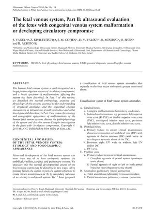
opposite directions of flow (Figure 1). Figure 1 Interrupted inferior vena cava with azygos continuation
Interrupted IVC has also been reported as an isolated imaged in high-definition power flow Doppler (HDPD) (a) and in
entity6 – 10 . In such cases it is usually clinically silent. B-flow (b). Note the dilated azygos vein (Az) draining into the
superior vena cava (SVC), with flow in the opposite direction to the
flow in the aorta (Ao). Compare with insets showing HDPD and
Persistent left superior vena cava (PLSVC) B-flow images of the normal heart and great vessels. DV, ductus
venosus; LHV, left hepatic vein. (Insets reproduced with permission
PLSVC is a known variant of venous return observed from Yagel et al.75 ).
in 0.3% of adults without cardiac malformation and in
approximately 4% of adults with CHD11 . Galindo et al.12
found a prevalence of 0.2% in fetuses with normal hearts, innominate vein) (Figure 2). The vessel most often drains
and 9% of fetuses with cardiac anomalies, in a population into the coronary sinus13,14 . It is diagnosed by fetal
referred for echocardiographic examination in a tertiary echocardiography on observation of a dilated coronary
center, calculating an odds ratio for CHD of 49.9 for a sinus (though this may not always be present) and an
fetus with PLSVC. extraneous vessel identified to the left of the ductal arch in
PLSVC is a remnant of the proximal segment of the the three-vessels and trachea (3VT) view of the fetal heart.
left anterior cardinal vein (LACV), resulting from failure In very rare instances, PLSVC may be seen with absent
of the LACV to atrophy following formation of the right SVC15 ; in this case the 3VT view shows three vessels:
oblique anastomosis with the right cardinal vein (the the LSVC, ductal arch and aortic arch16 . Multiplanar
Copyright 2010 ISUOG. Published by John Wiley & Sons, Ltd. Ultrasound Obstet Gynecol 2010; 36: 93–111.
3. The fetal venous system, Part II 95
Figure 2 Persistent left superior vena cava imaged in gray-scale (a) and with color Doppler (b). In (c) the dilated coronary sinus is visible (CS
and arrows), where the persistent left superior vena cava (LSVC) drains into it. Ao, aorta; MPA, main pulmonary artery; RSVC, right
superior vena cava; Tr, trachea.
reconstruction mode in 4D ultrasound has been shown to anomalies, and syndromes such as Noonan syndrome. It
be useful in diagnosis of the PLSVC anomaly17 . is associated with agenesis of the portal vein in as many as
When isolated, PLSVC usually has no clinical sig- 50% of cases, and may be associated in other cases with
nificance; however, it has been reported to occur in partial or total agenesis of the portal system. Hydrops
isolation in only 9% of cases. It is more often seen in and generalized edema occur in 33–52% of cases30,31 ,
association with other anomalies: both cardiac (23% of and may require postnatal device occlusion29 . DV agen-
PLSVC cases), including atrioventricular septal defect, esis presents as an isolated finding in only 35–59% of
double-outlet right ventricle, left outflow tract obstructive cases. In such cases, however, 80–100% have normal
anomalies, conotruncal anomalies and ventricular septal outcome30 – 32 . While only sporadic cases were reported in
defect, and other extracardiac malformations, includ- neonates before the advent of ultrasound, prenatal sono-
ing heterotaxy syndrome (41–45%), esophageal atresia, graphic diagnosis of DV agenesis is feasible and many case
diaphragmatic hernia, IVC malformations, complex mal- series have appeared in the literature2 . Among fetuses with
formation syndromes and chromosomal anomalies. There no or minor associated anomalies, location of the umbili-
may be significant morbidity and mortality, arising from cal drainage site seems to impact on prognosis, with a bet-
the associated anomalies rather than the lesion itself12,18 . ter outcome expected in cases without liver bypass22,31 – 34 .
Other very rare anomalies of the vena cava have Jaeggi et al.22 reported 12 cases of agenesis of the DV
been reported in the pediatric literature and extensively with extrahepatic shunt and reviewed a further 17 from
reviewed19 . the literature. They reported a mortality rate of 17% from
congestive heart failure in cases with extrahepatic shunt.
Associated malformations were reported in 87% of the
Umbilical veins cases, single umbilical artery being the most common.
Berg et al.31 reported 23 cases of agenesis of the DV,
Anomalies of the umbilical and portal veins constitute
four with extrahepatic drainage and 19 with drainage
the largest group of congenital venous anomalies detected
of the umbilical vein into the portal sinus. Fifteen of
in-utero. They include three main entities: agenesis of
the 23 fetuses had associated chromosomal or structural
the DV with extrahepatic umbilicosystemic shunt or with
anomalies that impacted outcome. Among the subgroup
intrahepatic umbilicohepatic shunt; persistent right UV
of fetuses (8/23) with no or minor associated malfor-
(PRUV) with or without intact DV; UV varix.
mations, outcome was significantly better among those
without liver bypass. They reported a total of 19 cases
Agenesis of the ductus venosus with intrahepatic drainage and no or minor associated
anomalies, all of which survived, while only 20/29 with
Agenesis of the DV results from failure to form the ‘criti- extrahepatic drainage and no or minor associated anoma-
cal anastomosis’: no connection is established between the lies survived. Fetuses with extrahepatic drainage have
UV and DV, and this in turn leads to shunting of umbilical a higher risk of congestive heart failure, even in the
blood through an aberrant vessel that may flow either into absence of cardiovascular anomaly. In reviewing their and
extrahepatic veins such as the iliac vein, IVC, SVC, right published cases the authors determined that findings of
atrium20 – 27 or coronary sinus28 , or via an intrahepatic cardiac malformations, complex non-chromosomal mal-
venous network (umbilicohepatic shunt), through the por- formation syndromes and hydrops were predictive of
tal sinus to the hepatic sinusoid (umbilicoportal–hepatic in-utero or neonatal demise, whether drainage was intra-
shunt) or even directly into the right atrium (Figure 3). The or extrahepatic31 .
prevalence of the anomaly has been estimated at 6 : 1000 From our observations we suggest that the parameter
fetal examinations29 . It is often (in 24–65% of cases) that most influences outcome in cases of agenesis of the
associated with cardiac, extracardiac and chromosomal DV is the development of the portal system. We recently
Copyright 2010 ISUOG. Published by John Wiley & Sons, Ltd. Ultrasound Obstet Gynecol 2010; 36: 93–111.
4. 96 Yagel et al.
presented data that support the notion that if the shunt from 1 : 250 to 1 : 57037,39 – 42 . It may replace the left
is a narrower, ductus-like connection, some or all of the UV, or be found as an intrahepatic supernumerary
portal system will have developed. This in turn impacts vein, connecting to the right portal vein. It may also
on prognosis35 . bypass the liver, causing aberrant drainage of blood into
the IVC or right atrium40,43,44 . It has been suggested
Persistent right umbilical vein (PRUV) that primary or secondary occlusion by thromboembolic
events arising from the placenta may lead to early
In the course of normal embryonic development, the
right UV degenerates and the left UV is left to carry streaming of blood through the right UV to cause this
blood from the placenta to the fetus. Failure of right UV anomaly45 . Teratogenic agents such as retinoic acid or
involution results in the PRUV anomaly1,36 – 39 (Figure 4). deficient folate induced the PRUV anomaly in rats46 .
PRUV is the most frequently detected fetal venous system Secondary formation of UV anomalies may result from
anomaly. Its prevalence has been estimated to range thromboembolic events occluding the DV or other veins.
Figure 3 (Continued over).
Copyright 2010 ISUOG. Published by John Wiley & Sons, Ltd. Ultrasound Obstet Gynecol 2010; 36: 93–111.
5. The fetal venous system, Part II 97
Figure 3 (Continued) Variations of agenesis of the ductus venosus (DV).
(a–c) Case of agenesis of the DV with narrow extrahepatic shunt from the
umbilical vein (UV) to the inferior vena cava (IVC), imaged with high-
definition power flow Doppler (HDPD) (a,c) and B-flow (b). In (c) the
appropriately developed portal system is shown. (Az, azygos vein; LHV,
left hepatic vein; LPV, left portal vein; MHV, main hepatic vein; RPVa and
p, anterior and posterior branches of right portal vein; St, stomach.) (d–g)
Complex case of agenesis of the DV with extrahepatic shunt, complicated
with interrupted IVC. (d) HDPD image showing the UV draining into the
shunt, between the IVC and Az. Note the Az is behind the aorta (red)
Int-I, internal iliac vein. (e) B-flow image which helps to demonstrate the
interrupted IVC with Az continuation, and the point of communication
of the UV to the IVC at this connection. Note that there are almost no
blood vessels in the area of the liver, which further suggests agenesis of
the portal system. (f,g) Tomographic ultrasound imaging showing
sequential sagittal planes, confirming the agenesis of the DV and inter-
rupted IVC. The image in (f) is slightly lateral to the left of that in (g).
AoA, aortic arch; dAo, descending aorta; Az, azygos; CT, celiac trunk;
DA, ductus arteriosus; MPA, main pulmonary artery. (h,i) Another case of agenesis of the DV with intrahepatic shunt of the UV to the right
hepatic vein (RHV), imaged in HDPD and B-flow. (j) 4D ultrasound B-flow image of absent DV, with UV drainage to the right atrium (RA).
Ao, aorta; HV, hepatic veins; UC, umbilical cord. (Reproduced with permission from Yagel et al.134.) (k) Agenesis of the DV with partial
agenesis of the portal system. Inversion mode in three-dimensional rendering demonstrating the umbilicoiliac shunt (carets). UA, umbilical
artery. (Reproduced with permission from Achiron et al.55).
Figure 4 Typical appearance of persistent right umbilical vein (UV). (a) The UV is seen passing laterally right of the gall bladder (GB).
(b) The portal vein presents a mirror image of its normal anatomical configuration. The left portal vein (LPV) assumes the right portal vein
(RPV) bifurcation, and its lateral branches change sides, the right side having the inferior (LPVi) and superior (LPVs) branches, and the left
side having only one lateral branch. The ductus venosus (DV) has its origin to the left of the UV axis. (c) The main portal vein (MPV) is
connected to the RPV in a more inferior plane relative to its normal position. LPVm, left portal vein medial branch; ST, stomach; SP, spine;
Ao, aorta.
Echogenic foci situated within the fetal liver suggest this with multiple associated anomalies47 . While early reports
etiology. seemed to indicate that PRUV was a worrisome finding
Two of the 74 cases of PRUV reviewed by De Catte in prenatal ultrasound43,45 , accumulated experience has
et al. had Noonan syndrome, and one had trisomy 18, all shown that in the absence of other anomalies intrahepatic
Copyright 2010 ISUOG. Published by John Wiley & Sons, Ltd. Ultrasound Obstet Gynecol 2010; 36: 93–111.
6. 98 Yagel et al.
PRUV with normal DV connection is usually a normal
anatomical variant with no clinical significance37,39,40,42 .
Ultrasound identification of PRUV is made by visualizing
the aberrant vein passing laterally to the right of the gall
bladder in the plane of measurement of the abdominal
circumference (Figure 4a).
Fetal intra-abdominal umbilical vein varix
Umbilical vein varix is a focal dilatation of vein. It is an
uncommon finding, with an estimated intrauterine inci-
dence of about 1 : 100048 . Most reported UV varices are of
the intra-abdominal UV, but extra-abdominal UV varices
have been reported49 . Fetal intra-abdominal UV (FIUV)
varix is diagnosed when a sonographically anechoic cystic
mass is seen between the abdominal wall and lower liver
edge. Color Doppler ultrasonography helps to distinguish
this vascular anomaly from other cystic lesions in this area
(Figure 5). The diameter of most varices ranges between 8
and 14 mm50 and FIUV varix has been variously defined
as an intra-abdominal UV diameter at least 1.5 times
greater than the diameter of the intrahepatic UV51 , or as
an intra-abdominal UV diameter exceeding 9 mm52 .
In a review of 91 cases, Fung et al.53 reported that
68% presented as an isolated finding, and of these 74%
had a normal outcome. However, 8.1% (5/62) resulted
in intrauterine death in which no specific cause could
be identified and one (1.6%) case had trisomy 21. In
the group with associated sonographic findings, only
27.6% had normal outcome. Congenital abnormalities Figure 5 Fetal intra-abdominal umbilical vein (UV) varix: an
and syndromes were confirmed after birth in 20.7% anechoic cystic mass (calipers) is seen between the abdominal wall
(6/29), while 27.6% had chromosomal abnormalities. and the lower liver edge in a longitudinal section (a) and imaged
The overall incidence of aneuploidy was 9.9%, the with high-definition power flow Doppler (b). The varix is shown to
majority being trisomy 21 or 18. The incidence of sudden be 1.5 times the diameter of the UV. Bl, bladder.
intrauterine demise was 5.5%, occurring between 29 and
38 weeks of gestation. into two types: Type I, in which there is complete diver-
The diameter of the UV at first diagnosis and the max- sion of the portal blood into the vena cava (portosystemic
imum diameter of the UV over the course of pregnancy shunt); and Type II, in which the portal veins are pre-
were not found to be related to the occurrence of obstet- served but a portion of portal blood is diverted into the
ric complications53 . Diagnosis of FIUV varix warrants a systemic venous circulation through the liver (portohep-
detailed anatomical search for additional anomalies, kary- atic shunt). Both types were further classified into two
otyping, echocardiography, and close monitoring of the subtypes: Subtype a, in which the splenic vein (SpV) and
fetus for sonographic signs of hemodynamic disturbance. superior mesenteric vein (SMV) do not join to form a
Fetal heart monitoring is advised from 28 weeks of gesta- confluence, and thus there is no anatomical portal vein;
tion. The optimal timing for delivery remains the subject and Subtype b, in which the SpV and SMV do join to
of debate. In light of the apparent high risk of unfavor- form a confluence that may shunt to the IVC, renal vein,
able outcome, including sudden intrauterine demise even iliac vein, azygos vein or right atrium60 .
in isolated FIUV varix, the option of induction of labor Complete absence of the portal venous system (CAPVS)
when lung maturity is ascertained may be considered54 . is an extreme example of total failure of the vitelline
veins to transform into the portal system, i.e. there is
primary failure to form the critical anastomosis with the
Vitelline veins hepatic sinusoids or UVs (Figures 6 and 7). As a result,
the enterohepatic circulation is disturbed and the portal
Complete or incomplete agenesis of the portal system venous blood is shunted systemically. Liver development
is supplied by the hepatic arteries. Mesenteric and splenic
Anomalies of the vitelline veins are extremely rare, and venous blood may drain directly into the IVC, renal veins
few cases during fetal life have been reported55 – 59 . Mor- or hepatic veins, or via the caput medusa to the heart61 .
gan and Superina60 proposed classifying portal agenesis Associated anomalies are frequent. Northrup et al.62
Copyright 2010 ISUOG. Published by John Wiley & Sons, Ltd. Ultrasound Obstet Gynecol 2010; 36: 93–111.
7. The fetal venous system, Part II 99
reported heterotaxy–polysplenia in 25% of their cases,
CHD in 30% (most frequently atrial septal defect and/or
ventricular septal defect) and Goldenhar syndrome in
10%. Achiron et al.55 reported associated malformations,
including trisomy 21, in four of their five cases.
Incomplete absence of the portal venous system
(IAPVS) or partial failure to form critical anastomoses
may represent a more benign form of vitelline vein
abnormality, and may result in agenesis of the right
portal system, with a persistent left vitelline vein connected
directly to the hepatic vein (portohepatic shunt) and an
absent or preserved DV (Figure 8). Neonatal spontaneous
resolution may occur. Achiron et al.55 reported four
cases, none with associated malformations, in three of
which the shunts resolved spontaneously postnatally55 .
Prognosis of IAPVS depends on the presence of associated
malformations and the development of hemodynamic
imbalance with signs of heart failure, cardiomegaly
and hydrops. As opposed to CAPVS, IAPVS is rarely
associated with other malformations, and hemodynamic
compromise depends on the intrahepatic shunt ratio,
leading to IUGR63 .
Hepatic bypass of the portal circulation is known
to cause the so-called hepatic artery buffer effect,
which compensates for the decreased portal supply
by increasing blood flow in the hepatic artery. This
results in a prominent Doppler signal55 , increased blood
flow velocity, and decreased pulsatility index (PI) in
the hepatic artery64 . Long term metabolic sequelae
of portosystemic shunting have been reported in the
postnatal literature and include: hypergalactosemia65 ,
hyperbilirubinemia, hyperammonemia66 , liver masses
including focal nodular hyperplasia, adenoma, hepa-
toblastoma and hepatocellular carcinoma, and rarely
encephalopathy67,68 .
Anomalous pulmonary venous connection
Anomalous pulmonary venous connection is a group
of rare anomalies that may present in total or
partial form. The combined incidence of anoma-
lous pulmonary venous connection (APVC) has been
reported as 6.8 : 100 000 livebirths69 or 4.9% of all
CHD70 .
Partial anomalous pulmonary venous connection
Partial anomalous pulmonary venous connection
(PAPVC) is a group of anomalies involving one or more,
Figure 6 (a) Tomographic ultrasound imaging (TUI) of the normal but not all, of the pulmonary veins connecting to the
portal system. LPV, left portal vein; MPV, main portal vein; RAPV,
right anterior portal vein; RPPV, right posterior portal vein.
right atrium or a tributary (Figure 9)71 . An atrial septal
(b) TUI in a case of complete agenesis of the portal venous system. defect is often present. There are several variations of this
The arrow indicates the only remnant of the portal system. St, anomaly70,72 – 74 .
stomach. (c) High-definition power flow Doppler image of the
sagittal plane showing that the ductus venosus (DV) is present; (1) Right pulmonary veins to SVC (Figure 9a): usually
thus, the anomaly is not secondary to absent DV in this instance.
The circle indicates the umbilical cord. Ao, aorta; CT, celiac trunk; the right upper and middle lung lobes drain into the
IVC, inferior vena cava; LHV, left hepatic vein, UA, umbilical SVC and atrial septal defect is present; occasionally
artery; UV, umbilical vein. the atrial septum is intact.
Copyright 2010 ISUOG. Published by John Wiley & Sons, Ltd. Ultrasound Obstet Gynecol 2010; 36: 93–111.
8. 100 Yagel et al.
Figure 7 A case of complete agenesis of the portal venous system. (a) Transverse view of the fetal abdomen showing, beyond the stomach
(St), the spleen (Sp), with the splenic vein coursing towards the right side of the body (blue) to form a confluence with the main portal vein.
Note absence of the afferent intrahepatic venous system and enlarged IVC when compared with the aorta (red, arrow). (b) Pulsed Doppler
insonation of the splenic vein–portal system junction depicting triphasic flow compatible with a systemic venous shunt. (c,d) Venography
performed post-termination. In (c), note the umbilico-IVC shunt (arrow); (d) was taken 1 min later: the splenic vein (long black arrow) is
shown to be confluent with the renal vein (short arrow) and with the superior mesenteric vein (double headed arrow), forming a conduit
(dotted arrow, c) which became enlarged (white arrow) before shunting into the IVC. (Reproduced with permission from Achiron et al.55 ).
(2) Right pulmonary veins to right atrium: veins from all veins form a characteristic ‘fir-tree’ pattern; the atrial
right lung lobes drain directly into the right atrium, septum is intact. This variation is often seen with right
which can be dilated to as much as twice its normal lung hypoplasia, bronchial system anomalies, cardiac
size; there is usually an atrial septal defect. dextroposition, right pulmonary artery hypoplasia,
(3) Right pulmonary veins to IVC (Figure 9b): all or some anomalous arterial connection between the aorta and
right lung lobes are drained into the IVC; right lung right lung and other cardiac anomalies.
Copyright 2010 ISUOG. Published by John Wiley & Sons, Ltd. Ultrasound Obstet Gynecol 2010; 36: 93–111.
9. The fetal venous system, Part II 101
Echocardiographic diagnosis of PAPVC is made by
visualizing pulmonary veins connecting to the structure
involved; Doppler studies are invaluable in identification
and classification of this group of anomalies, and three-
dimensional (3D)/4D ultrasound modalities may have
added value in their diagnosis74 – 76 .
Total anomalous pulmonary venous connection
Total anomalous pulmonary venous connection (TAPVC)
involves complete disconnection between the pulmonary
veins and the left atrium (Figure 10)71 . All the pulmonary
veins drain into the right atrium or systemic veins. One-
third of cases present with an associated anomaly, while
two thirds are isolated. TAPVC can be classified into four
types71 :
Type I: anomalous connection at the supracardiac level;
Type II: anomalous connection at the cardiac level;
Type III: anomalous connection at the infracardiac level;
Type IV: connections at two or more of supracardiac,
cardiac and infracardiac levels.
Alternatively, this anomaly can be classed into two
groups:
(1) supradiaphragmatic type: pulmonary veins are con-
nected to: the left innominate vein by the characteristic
vertical vein, or the coronary sinus, the right atrium,
or the SVC, without pulmonary venous obstruction;
(2) infradiaphragmatic type: pulmonary veins drain into
the portal vein or hepatic veins, with pulmonary
venous obstruction.
There is usually an atrial septal defect. An obstruction
in the pulmonary venous channel will impact the
prognosis of TAPVC. One third of patients have
Figure 8 Incomplete absence of the portal venous system (IAPVS). an associated major anomaly, including cor bilocular,
Two different forms are shown: (a) absent right portal vein and single ventricle, truncus arteriosus, transposition of the
portal sinus, showing X-shaped configuration formed by the great arteries, pulmonary atresia, aortic coarctation,
umbilical vein (UV), left portal vein (LPV) branches and the ductus
hypoplastic left ventricle, or anomalies of the systemic
venosus (DV), in the standard transverse plane for abdominal
circumference measurement; (b) dilated aberrant ‘horseshoe veins.
shaped’ arrangement of vessels encircling the right hepatic lobe. Ao, TAPVC should be suspected when pulmonary veins
aorta; IVC, inferior vena cava; LPVi, left portal vein inferior cannot be visualized entering the left atrium in the
branch; LPVm, left portal vein medial branch; SP, spine; four-chamber apical view plane, with a wide gap
St, stomach.
between the posterior wall of the left atrium and the
descending aorta. In the supracardiac type, the right
(4) Left pulmonary veins to left innominate vein atrium volume overload causes enlargement of the
(Figure 9c): veins from only the upper left lobe, or atrium and a bowing leftwards of the interatrial septum.
all of the left lung, drain into the left innominate The common pulmonary channel may be visualized
vein via an anomalous vertical vein; atrial septal posterior to the left atrium with color Doppler, and
defect is usually present. Left pulmonary drainage this can be followed to the site of its connection.
may alternately be to the coronary sinus, IVC, right The ascending vertical vein to the left innominate vein
SVC, right atrium or left subclavian vein, and may be can be seen in the 3VT view as an additional vessel
associated with cardiac defects or polysplenia/asplenia to the left of the pulmonary artery. Doppler studies
or Turner or Noonan syndromes. show the direction of blood flow in this vessel is
(5) Left pulmonary veins to coronary sinus (Figure 9d), cephalad, opposite to that in the SVC. In the infracardiac
IVC, right SVC, right atrium or left subclavian vein; type, the descending vertical vein can be diagnosed
very rarely, the right pulmonary veins may connect to in a transverse plane of the abdomen as an extra
the azygos vein or coronary sinus. vessel in front of the descending aorta. In the sagittal
Copyright 2010 ISUOG. Published by John Wiley & Sons, Ltd. Ultrasound Obstet Gynecol 2010; 36: 93–111.
10. 102 Yagel et al.
(a) (b) S.V.C.
L. Inn. V.
S.V.C.
R.P.V.
L.P.V. L.P.V.
R.A.
R.A. L.A. L.A.
C.S.
C.S.
I.V.C.
L.V.
R.P.V. R.V. L.V.
R.V.
I.V.C.
(c) (d)
S.V.C.
V.
S.V.C. L. Inn.
R.P.V.
v.v. L.P.V.
R.A.
R.P.V. L.P.V. C.S.
L.A.
R.A.
C.S. L.A.
I.V.C.
L.V.
R.V.
I.V.C.
L.V.
R.V.
Figure 9 Common forms of partial anomalous pulmonary venous connection. (a) Anomalous connection of the right pulmonary veins
(R.P.V.) to the superior vena cava (S.V.C.). There is usually a high or sinus venous defect. (b) Anomalous connections of the right
pulmonary veins to the inferior vena cava (I.V.C.). The right lung commonly drains by one pulmonary vein without its usual anatomical
divisions. Parenchymal abnormalities of the right lung are common and the atrial septum is usually intact. (c) Anomalous connection of the
left pulmonary veins (L.P.V.) to the left innominate vein (L. Inn. V.) by way of a vertical vein (v.v.). There may be an additional left-to-right
shunt through the atrial septal defect. (d) Anomalous connection of the L.P.V. to the coronary sinus (C.S.). L.A., left atrium; L.V., left
ventricle; R.A., right atrium; R.V., right ventricle. (Reproduced with permission from Krabill and Lucas71 (Krabill KA, Lucas RV. In Heart
Disease in Infants, Children, and Adolescents (5th edn), Moss AJ, Adams FH, Emmanouilides GC (eds). Williams and Wilkins: Baltimore,
1995; 841–849).)
plane, the vertical vein courses cephalad between the and the development of the pulmonary vascular bed.
IVC and the descending aorta, crossing the diaphragm Isolated TAPVC, if detected and corrected surgically
(Figure 11). early in life, has an excellent outcome. Prognosis is
Doppler evaluation is key to the characteriza- poor when it is associated with other cardiac lesions
tion of TAPVC to identify the anomalous connec- and portal vein obstruction. In cases of TAPVC with
tion. Turbulent flow will reveal the location of any obstruction in the anomalous venous channel, the
obstruction and the degree of obstruction can be neonate usually dies in the first weeks of postnatal
quantified70 – 72 . life71,77 .
Prognosis THE FETAL VENOUS SYSTEM
IN PATHOLOGICAL CONDITIONS
Prognosis in cases of PAPVC and TAPVC is affected by the
presence of any cardiac or extracardiac malformations, In the physiology section of Part I of this review we
the presence and size of the interatrial connection and any emphasized the unique role of the venous system in
obstructive lesion in the anomalous connecting vessels, maintaining a positive umbilicocaval pressure gradient.
Copyright 2010 ISUOG. Published by John Wiley & Sons, Ltd. Ultrasound Obstet Gynecol 2010; 36: 93–111.
11. The fetal venous system, Part II 103
(a) S.V.C. L. Inn. V. (b) S.V.C.
v.v. C.P.V.
R.P.V. C.P.V. L.P.V.
R.A.
L.A.
R.A. C.S.
C.S. L.A.
I.V.C L.V.
I.V.C.
R.V.
R.V. L.V.
(c) (d)
S.V.C.
S.V.C.
C.P.V.
R.A. L.P.V. R.A.
R.P.V. C.S. L.A.
C.S.
I.V.C.
I.V.C.
L.V. L.V.
R.V.
R.V.
I.V.C.
R.H. L.H.
D.V.
L.P.
R.P. P.V. S.V.
S.M.V.
Figure 10 Common forms of total anomalous pulmonary venous connection. (a) Anomalous connection of the pulmonary veins to the left
innominate vein (L. Inn. V.) by way of a vertical vein (V.V.). (b) Anomalous connection to the coronary sinus (C.S.): the pulmonary veins
join to form a confluence designated as the common pulmonary vein (C.P.V.), which connects to the C.S. (c) Anomalous connection to the
right atrium (R.A.). The left and right pulmonary veins (L.P.V. and R.P.V.) usually enter the R.A. separately. (d) Anomalous connection to
the portal vein (P.V.). The pulmonary veins form a confluence, from which an anomalous channel arises. This connects to the P.V., which
communicates with the inferior vena cava (I.V.C.) by way of the ductus venosus (D.V.) or the hepatic sinusoids. L.A., left atrium; L.H., left
hepatic vein; L.P., left portal vein; L.V., left ventricle; R.H., right hepatic vein; R.P., right left portal vein; R.V., right ventricle; S.M.V,
superior mesenteric vein; S.V., splenic vein; S.V.C., superior vena cava. (Reproduced with permission from Krabill and Lucas71 (Krabill KA,
Lucas RV. In Heart Disease in Infants, Children, and Adolescents (5th edn), Moss AJ, Adams FH, Emmanouilides GC (eds). Williams and
Wilkins: Baltimore, 1995; 841–849).)
This assures a low preload index, which is essential for gradual and sequential, in correlation with the severity of
optimal cardiac drainage. Cardiac output progressively the hypoxic insult, hence creating an efficient monitoring
increases and resistance of the placental vascular bed tool of evolving fetal distress.
decreases while the umbilicocaval pressure gradient The most frequent pathological condition affecting the
is maintained steady during the third trimester of fetoplacental hemodynamic balance is placental dysfunc-
pregnancy78 . Any situation hampering this hemodynamic tion. Other etiologies are cardiac (malformations, arrhyth-
balance initiates compensatory mechanisms in the arterial mias, or increased preload blood volume) and conditions
and venous systems. These compensatory mechanisms are resulting in elevation of ventricular end-diastolic pressure,
Copyright 2010 ISUOG. Published by John Wiley & Sons, Ltd. Ultrasound Obstet Gynecol 2010; 36: 93–111.
12. 104 Yagel et al.
Figure 11 Total anomalous pulmonary venous connection (infracardiac type) imaged in the sagittal plane. Tomographic ultrasound imaging
was applied in post-processing and clearly shows the vertical vein draining into one of the hepatic veins. Ao, aorta; DV, ductus venosus;
LHV, left hepatic vein; VV, vertical vein.
atrial pressure and, consequently, precordial venous pres- resistance indices (RI) of these two compartments has been
sure. We focus here on uteroplacental insufficiency and used clinically as a monitoring tool of fetal compensatory
the gradual hemodynamic changes that accompany its mechanisms and the magnitude of hypoxic insult85 – 87 .
compromised vascular function. The implications for the venous system of these changes
Reduced placental supply to the fetus initiates multiple in resistance in the different compartments are four-fold:
compensatory vasodilatory mechanisms involving various reduced blood supply from the placenta to the liver, an
arterial and venous systems. In the arterial system, the increased proportion (also per unit of weight) of cardiac
organ most widely studied and clinically evaluated is output diverted to the fetal body and consequently to
the brain (brain-sparing effect)79,80 . Evaluation of various the IVC, increased blood flow through the SVC resulting
parts of the arterial circulation in the fetus with circulatory from the brain-sparing effect, and an increased afterload
compromise has been widely performed and reviewed on the right ventricle. These hemodynamic changes lead
elsewhere81 – 84 . to increased preload indices that hamper the supply of
The cascade of evolving circulatory compromise high-quality blood flow from the placenta.
in uteroplacental insufficiency is characterized by hemo- Venous compensatory mechanisms aim to improve the
dynamic changes in the placenta, heart and brain. The placental supply to the heart by increasing the proportion
fetal circulation may be viewed as comprising two com- of DV shunting from the portal sinus. Human Doppler
partments, with the division at the aortic isthmus: the right studies have reported variable results. Bellotti et al.88
ventricle/placenta, having high resistance, and the left ven- found that the percentage of UV blood flow shunted
tricle/brain, having low resistance. The ratio between the through the DV increased from 26.5% in normally
Copyright 2010 ISUOG. Published by John Wiley & Sons, Ltd. Ultrasound Obstet Gynecol 2010; 36: 93–111.
13. The fetal venous system, Part II 105
grown fetuses to 90.3% in growth-restricted fetuses, with of adverse outcome among growth-restricted fetuses
a consequent increased normalized blood flow volume are cardiomegaly, monophasic atrioventricular filling or
from 30.8 mL/min/kg to 41.3 mL/min/kg. Others showed holosystolic tricuspid regurgitation, and pulsations in
more moderate changes. Tchirikov et al.89 reported an the UV96 , while among hydropic fetuses they were
increase from 43% to 62%, and Kiserud et al.90 reported UV and DV Doppler94 . This concurs with previ-
an increase from 25% in normally grown to a mean of ous studies showing that in the temporal sequence
39% in IUGR fetuses. They found that the degree of of events prior to the appearance of abnormal fetal
shunting in IUGR fetuses is positively correlated with the heart rate pattern, atonia or fetal demise, the arte-
severity of placental insufficiency as reflected by the UA rial Doppler changes precede those of the venous
diastolic flow. A significant increase in shunting to the DV system97 but are too early a sign to be used to
was found only in IUGR fetuses with UA-PI above the time the decision to deliver. This was particularly evi-
97.5th percentile (35% blood shunted) and in those with dent in cases of early-onset IUGR, before 32 weeks of
absent/reversed diastolic flow (57% shunted)90 . gestation84,98 .
Preferential flow through the DV results from reciprocal Adverse perinatal outcome is related to gestational
hemodynamic changes inside the liver. The presence of a age at delivery and the degree of placental insufficiency.
sphincter in the isthmic portion of the DV has never been Decision-making concerning the time of delivery aims to
confirmed91 . strike a balance between fetal injury related to prematu-
Tchirikov et al.92 showed in vitro that rings derived rity, and that related to placental insufficiency99 . Results
from portal venous branches developed more force of the GRIT study100 indicated no significant difference
in reaction to catecholamines than did vascular rings in terms of stillbirth rate when delivery was immediate.
obtained from the isthmic portion of the DV. In hypoxia, However, this was refuted by findings at the 2-year follow
increased plasma catecholamine levels may evoke an up: the rate of cerebral palsy in the immediate delivery
increase in the DV shunting rate through differential group was as expected (10%), while in the delayed deliv-
contraction of the portal venous branches as compared to ery group it was 0%, suggesting that deferred delivery
the DV. This effect may come into play, in addition to the could be protective management101 . In a prospective mul-
active nitric oxide-mediated DV distension, which was ticenter study, Baschat et al.83 found that gestational age
shown experimentally92 . This intrahepatic shift of blood at delivery is the best predictor for intact survival until
from the portal system to the DV during hypoxia was 29 weeks or 800 g estimated fetal weight, with sensitivities
supported by an in-vivo Doppler study. Kessler et al.93 of 68% and 72%, respectively. Above these thresholds,
demonstrated that in IUGR fetuses with UA-PI above DV Doppler parameters were shown to be the primary car-
the 97.5th percentile, total liver perfusion is decreased diovascular factor in predicting intact survival, and as such
equally to the left and right lobes. However, as placental are the only parameters to use as a trigger for delivery.
insufficiency increased (as measured by the oxygen partial
pressure in the UA), the proportion of left portal vein
(LPV) contribution to the right lobe decreased, being
Doppler parameters Biophysical parameters
replaced by increased flow from the main portal vein.
In summary, placental vasculopathy leads to reduced Decreased UV
Early
umbilical flow to the heart. This initiates an intrahepatic Decreased
response flow volume Hypoxia
fetal growth
compensatory mechanism, which consists of shifting
blood from the liver to the DV. The liver plays a central Increased
role in regulating fetal growth. Reduced liver perfusion UA resistance Decreased
AFI
may provide the first clinical evidence of placental
insufficiency, restricted fetal growth and decreased fetal Decreased
MPV resistance
weight. Decreased
Decision point fetal movement
Increased
venous pulsation
DOPPLER STUDIES OF THE FETAL
Decreased
VENOUS SYSTEM fetal breathing
DV A/RDF
movements
Doppler investigation is the only non-invasive in-utero UV pulsations
method for assessing fetoplacental hemodynamic status. Late
FHR late Reduced
Each component of the fetomaternal circulation has response fetal tone
decelerations/STV Acidosis
a different role in placental function and fetal well-
being. Doppler evaluation of the venous system aims
to assess fetal cardiac function, which in severe cases may Figure 12 Suggested cascade of fetal circulatory compromise.
deteriorate to an acute event of congestive heart failure. Changes in Doppler and physiological parameters follow a
The cardiovascular profile score94 – 96 combines sono- temporal sequence82 . The decision point regards the timing of
delivery. AFI, amniotic fluid index; A/RDF, absent or reversed
graphic markers of fetal cardiovascular status based diastolic flow; DV, ductus venosus; FHR, fetal heart rate; MPV,
on parameters found to be correlated with perina- main portal vein; STV, short term variability; UA, umbilical artery;
tal mortality. The parameters most strongly predictive UV, umbilical vein.
Copyright 2010 ISUOG. Published by John Wiley & Sons, Ltd. Ultrasound Obstet Gynecol 2010; 36: 93–111.
14. 106 Yagel et al.
Turan et al.82 found a well-ordered sequence of
Doppler abnormalities in placental insufficiency (Figure
12), from early-onset ones, including increased UA
resistance and brain-sparing effect, to late-onset ones, such
as absent/reversed UA diastolic velocity, absent/reversed
DV A-wave, and UV pulsation. The rate of progression
through the sequence correlated significantly with
gestational age: when appearing as early as 26–27 weeks
of gestation, placental insufficiency was usually severe,
progressing from one Doppler abnormality to the next in
approximately 7–10 days. When Doppler abnormalities
emerged near 30 weeks’ gestation, progression intervals
were longer, up to 14 days, and clinically cases were
milder. Doppler changes were limited to the arterial
system, and the median age of delivery was 33 weeks.
These findings, however, were in contrast with those of
Arduini et al.85 , who showed that time intervals between
arterial Doppler changes and delivery for late fetal heart
rate decelerations were statistically significantly longer
when Doppler abnormalities appeared before 29 weeks
of gestation, compared with late-onset cases (mean
interval between Doppler changes and delivery, 13.5
(range, 3–26) vs. 3 (range, 1–9) days. Other studies84,102
have shown that venous Doppler abnormalities preceded
the appearance of late decelerations or reduced short-
term variability (< 2.2 ms) by 6–7 days. In conclusion,
there is a well-documented sequence of biophysical and
Doppler abnormalities that correspond with placental
insufficiency and fetal growth restriction (Figure 12).
Abnormalities in venous system Doppler waveforms are
sensitive tools for the assessment of fetal well-being Figure 13 Abnormal Doppler waveforms in the umbilical vein (UV)
(a), ductus venosus (b) and inferior vena cava (IVC) (c).
before 32 weeks’ gestation, and may help to fine-tune our
(a) Abnormal UV waveform in hemodynamically compromised
decision-making concerning time of delivery in affected fetuses is characterized by the appearance of pulsations. (b) The DV
fetuses. normally has forward blood flow throughout the cardiac cycle. In
compromised fetuses a drop in atrial systolic forward velocities is
observed. As central venous pressure increases, blood flow may be
Venous Doppler patterns reversed during atrial systole. (c) Abnormal IVC waveforms in
intrauterine growth restriction show a decrease in forward flow
Venous Doppler patterns in normal and IUGR fetuses during the S-wave (S) and D-wave (D) and accentuation of reversed
have been established in large longitudinal and cross- flow in the A-wave (annotated ‘a’). As the condition deteriorates
sectional studies; normal Doppler patterns were discussed the D-wave may be reversed.
in Part I of this review. Here we describe Doppler
abnormalities seen in the most commonly investigated
hydrops104,105 . Pulsatility in the UV can result from two
vessels (Figure 13).
main sources of pressure waves: atrial contraction can
cause a sharp deflection (notch) in UV flow, or a wave
Umbilical vein from the neighboring arterial flow, which will be traveling
in the same direction, can cause an increment in the UV
Umbilical vein Doppler patterns are usually evaluated
flow. The resulting pulsatile UV Doppler pattern may
in the intra-abdominal portion of the vein. The sample
be only an A-wave notch or, in some severe cases, a
volume should include the whole cross-section of the
triphasic waveform that mirrors the whole cardiac cycle
vessel, with an angle of insonation at or as close as possible
may appear103 (Figure 13a).
to 0◦ . Since the IUGR state includes increased impedance
in the fetoplacental circulation, reduced UV flow volume
and velocity result. However, the considerable overlap Ductus venosus
with normal ranges precludes the usefulness of UV flow
velocity as a measure of IUGR103 . Changes in DV flow are seen in hypoxia and hypo-
The appearance of pulsatile flow in the UV is a volemia in experimental animal models and in human
simple and reliable marker of circulatory compromise, fetuses106 – 110 . The DV is sampled at its inlet, with a large
long recognized as a marker of asphyxia in small-for- sample volume in a near-sagittal plane, at a low angle of
gestational age fetuses and in cases of non-immune insonation111,112 . Alternatively, DV flow can be sampled
Copyright 2010 ISUOG. Published by John Wiley & Sons, Ltd. Ultrasound Obstet Gynecol 2010; 36: 93–111.
15. The fetal venous system, Part II 107
in an oblique transverse section of the fetal abdomen. pressures, flow in the LPV is reduced and may become pul-
Normally at 20 weeks’ gestation about 30% of blood is satile, bidirectional or reversed. If reversed, oxygen-poor
shunted through the DV, while at 30 weeks about 20% blood from the spleen and gut will mix with oxygenated
of blood is shunted103,112 – 114 . In small-for-gestational blood from the UV as it courses to the DV, supplying
age fetuses a much higher proportion of blood is shunted oxygen-poor blood to the fetal organs. Ultimately during
through the DV, and earlier placental compromise will reversal, more blood could be flowing through the DV
show a more pronounced shunt and distension of the than through the UV that supplies it, possibly creating
DV103 . suction on the LPV132 . Kilavuz et al.132 showed, in IUGR
Cardiac events are apparent in the DV waveform. fetuses with absent or reversed end-diastolic flow in the
Reversed or zero flow in the A-wave is a simple sign of UA, that while UV flow remained normal, LPV flow was
disordered cardiac function. There is reduced velocity in normal in mildly affected fetuses, pulsatile in fetuses with
the A-wave in IUGR, and as the situation worsens it is fur- increased RI or absent flow in the UA, and reversed in
ther deflected owing to increased afterload and preload, fetuses with reversed flow in the UA. There was a signif-
as well as increased end-diastolic pressure, the direct icant correlation between reversed flow in the LPV and
effects of hypoxia and increased adrenergic drive. Taken increased RI in the UA.
together, these effects produce an augmented atrial con- The normal distribution of blood flow between the
traction and strong deflection during the A-wave in the DV left and right liver lobes is approximately 60%/40%. As
waveform97,115,116 (Figure 13b). The combination of this described above, most UV blood (70–80%) perfuses the
and the DV distension, which lessens wave reflection at fetal liver, while 20–30% is shunted to the DV. Blood
the junction with the UV by lessening the difference in the flow in the UV is directed to the left liver lobe and the
vessels’ diameters, produces a considerable effect on UV DV, with only a minority diverted to the LPV and right
flow, and further deterioration leads to decreased velocity liver lobe. Low or reversed flow in the left portal branch
between the systolic and diastolic peaks. These waveform is an expression of prioritized umbilical perfusion of the
changes are most apparent in the second trimester and left liver lobe at the expense of the right114 .
usually result from chromosomal aberration as opposed
to cardiac decompensation115,117 – 120. Such changes are
rarely observed in the third trimester, when improved CONCLUSION
endocrine control operates to attenuate them121 – 123 . Congenital malformations of the venous system are a
complex and varied group of lesions. There is much that
Inferior vena cava we do not know about this system and its anomalies,
but it is clear that knowledge of the embryonic devel-
The IVC is usually sampled in the fetal abdomen, caudal to opment of affected vessels is key to understanding the
the hepatic confluence and DV outlet, to avoid interference in-utero progress and prognosis of these cases. Whenever
from neighboring vessels. Reference ranges for normal anomalies of the cardinal, vitelline or umbilical system are
IVC flow parameters have been established124 – 126 . The diagnosed, they should prompt thorough investigation of
IVC is the preferred vessel for postnatal evaluation of the other segments of the cardiovascular system. It would
small-for-gestational age neonates and is familiar to appear that anomalies of the venous system are not as
pediatricians, so it is often sampled in the fetus. It is rare as formerly believed, and that more will be diag-
normal to observe a negative A-wave in the IVC because nosed if practitioners are cognizant of their sonographic
of the vessel’s normally relatively low velocities124 – 128 . appearance and associated anomalies.
In compromised fetuses, an abnormal IVC flow velocity
waveform will show a decrease in forward flow during
the S- and D-waves, and an accentuated reversed flow in REFERENCES
the A-wave (Figure 13c). In severe cases the D-wave may 1. Achiron R, Hegesh J, Yagel S, Lipitz S, Cohen SB, Rotstein Z.
be reversed. Abnormalities of the fetal central veins and umbilico-portal
system: prenatal ultrasonographic diagnosis and proposed
classification. Ultrasound Obstet Gynecol 2000; 16: 539–548.
Hepatic and portal veins 2. Fasouliotis SJ, Achiron R, Kivilevitch Z, Yagel S. The human
fetal venous system: normal embryologic, anatomic, and
The hepatic vein, while easy to sample, is employed only physiologic characteristics and developmental abnormalities.
infrequently as a parameter in IUGR studies. The signs J Ultrasound Med 2002; 21: 1145–1158.
of cardiac compromise observed in the hepatic vein are 3. Lin AE, Ticho BS, Houde K, Westgate MN, Holmes LB. Het-
erotaxy: associated conditions and hospital-based prevalence
similar to those apparent in the DV and IVC129,130 . in newborns. Genet Med 2000; 2: 157–172.
The left portal vein as the watershed of the fetal venous 4. Berg C, Geipel A, Kohl T, Smrcek J, Germer U, Baschat AA,
circulation. The LPV has been suggested as a simple Hansmann M, Gembruch U. Fetal echocardiographic evalua-
marker of circulatory compromise131 – 133 . It is sampled tion of atrial morphology and the prediction of laterality in
in the left portal branch between the DV inlet and the cases of heterotaxy syndromes. Ultrasound Obstet Gynecol
2005; 26: 538–545.
junction with the main portal stem. Disturbed blood flow 5. Bowers PN, Brueckner M, Yost HJ. The genetics of left-right
in the UA is indicative of increased resistance to blood development and heterotaxia. Semin Perinatol 1996; 20:
flow in the right heart and right liver lobe. Under these 577–588.
Copyright 2010 ISUOG. Published by John Wiley & Sons, Ltd. Ultrasound Obstet Gynecol 2010; 36: 93–111.
16. 108 Yagel et al.
6. Vijayaraghavan SB, Raja V, Chitra TV. Interrupted inferior the fetal hepatic circulation: case reports and review of the
vena cava and left-sided subrenal inferior vena cava: prenatal literature. Pediatr Pathol Lab Med 1995; 15: 39–50.
diagnosis. J Ultrasound Med 2003; 22: 747–752. 26. Sothinathan U, Pollina E, Huggon I, Patel S, Greenough A.
7. Mihmanli I, Bulakbasi N, Kantarci F, Adaletli I, Pabuscu Y. Absence of the ductus venosus. Acta Paediatr 2006; 95:
The value of ultrasonography in interrupted inferior vena 620–621.
cava with azygos continuation. Eur J Ultrasound 2001; 14: 27. Volpe P, Marasini M, Caruso G, Lituania M, Marzullo A,
179–182. Volpe G, Gentile M. Prenatal diagnosis of ductus venosus
8. Koc Z, Oguzkurt L. Interruption or congenital stenosis of the agenesis and its association with cytogenetic/congenital
inferior vena cava: prevalence, imaging, and clinical findings. anomalies. Prenat Diagn 2002; 22: 995–1000.
Eur J Radiol 2007; 62: 257–266. 28. Perles Z, Nir A, Nadjari M, Ergaz Z, Raas-Rothschild A,
9. Bartram U, Fischer G, Kramer HH. Congenitally interrupted Rein AJ. Absent ductus venosus in the fetus: review of the
inferior vena cava without other features of the heterotaxy literature and first report of direct umbilical venous drainage
syndrome: report of five cases and characterization of a rare to the coronary sinus. Fetal Diagn Ther 2003; 18: 247–251.
entity. Pediatr Dev Pathol 2008; 11: 266–273. 29. Acherman RJ, Evans WN, Galindo A, Collazos JC, Rothman
10. Sheley RC, Nyberg DA, Kapur R. Azygous continuation of A, Mayman GA, Luna CF, Rollins R, Kip KT, Berthody
the interrupted inferior vena cava: a clue to prenatal diagnosis DP, Restrepo H. Diagnosis of absent ductus venosus in a
of the cardiosplenic syndromes. J Ultrasound Med 1995; 14: population referred for fetal echocardiography: association
381–387. with a persistent portosystemic shunt requiring postnatal
11. Biffi M, Boriani G, Frabetti L, Bronzetti G, Branzi A. Left device occlusion. J Ultrasound Med 2007; 26: 1077–1082.
superior vena cava persistence in patients undergoing 30. Contratti G, Banzi C, Ghi T, Perolo A, Pilu G, Visentin A.
pacemaker or cardioverter-defibrillator implantation: a 10- Absence of the ductus venosus: report of 10 new cases and
year experience. Chest 2001; 120: 139–144. review of the literature. Ultrasound Obstet Gynecol 2001; 18:
12. Galindo A, Gutierrez-Larraya F, Escribano D, Arbues J, 605–609.
Velasco JM. Clinical significance of persistent left superior 31. ¨
Berg C, Kamil D, Geipel A, Kohl T, Knopfle G, Hansmann M,
vena cava diagnosed in fetal life. Ultrasound Obstet Gynecol Gembruch U. Absence of ductus venosus – importance of
2007; 30: 152–161. umbilical venous drainage site. Ultrasound Obstet Gynecol
13. Guarnieri GF, Romano F, Clerico L, Balducci G. Absent right 2006; 28: 275–281.
and persistent left superior vena cava: fetal and neonatal 32. Hoppen T, Hofstaetter C, Plath H, Kau N, Bartmann P.
echocardiographic diagnosis. Pediatr Cardiol 2006; 27: Agenesis of the ductus venosus and its correlation to hydrops
646–648.
fetalis. J Perinat Med 2000; 28: 69–73.
14. Sarodia BD, Stoller JK. Persistent left superior vena cava: case
33. Sau A, Sharland G, Simpson J. Agenesis of the ductus venosus
report and literature review. Respir Care 2000; 45: 411–416.
associated with direct umbilical venous return into the
15. Freund M, Stoutenbeek P, ter Heide H, Pistorius L. ‘Tobacco
heart–case series and review of literature. Prenat Diagn 2004;
pipe’ sign in the fetus: patent left superior vena cava with
24: 418–423.
absent right superior vena cava. Ultrasound Obstet Gynecol
34. Gorincour G, Droulle P, Guibaud L. Prenatal diagnosis of
2008; 32: 593–594.
umbilicoportosystemic shunts: report of 11 cases and review
16. Pasquini L, Belmar C, Seale A, Gardiner HM. Prenatal diag-
of the literature. AJR Am J Roentgenol 2005; 184: 163–168.
nosis of absent right and persistent left superior vena cava.
35. Shen O, Valsky DVV, Messing B, Cohen SM, Lipschuetz M,
Prenat Diagn 2006; 26: 700–702.
Yagel S. Shunt diameter in agenesis of ductus venosus with
17. Rizzo G, Arduini D, Capponi A. Use of the multiplanar display
extrahepatic porto-systemic shunt impacts prognosis. Ultra-
in evaluation of a persistent left superior vena cava in the
sound Obstet Gynecol (in press). DOI: 10.1002/uog.7702.
fetal heart using 4-dimensional ultrasonography: advantage
of adding the spin technique. J Ultrasound Med 2008; 27: 36. Bell AD, Gerlis LM, Variend S. Persistent right umbilical
497–498. vein–case report and review of literature. Int J Cardiol 1986;
18. Berg C, Knuppel M, Geipel A, Kohl T, Krapp M, Knopfle G, ¨ 10: 167–176.
Germer U, Hansmann M, Gembruch U. Prenatal diagnosis of 37. Hill LM, Mills A, Peterson C, Boyles D. Persistent right
persistent left superior vena cava and its associated congenital umbilical vein: sonographic detection and subsequent neonatal
anomalies. Ultrasound Obstet Gynecol 2006; 27: 274–280. outcome. Obstet Gynecol 1994; 84: 923–925.
19. Minniti S, Visentini S, Procacci C. Congenital anomalies of the 38. Kinare AS, Ambardekar ST, Bhattacharya D, Pande SA. Pre-
venae cavae: embryological origin, imaging features and report natal diagnosis with ultrasound of anomalous course of the
of three new variants. Eur Radiol 2002; 12: 2040–2055. umbilical vein and its relationship to fetal outcome. J Clin
20. Currarino G, Stannard MW, Kolni H. Umbilical vein draining Ultrasound 1996; 24: 333–338.
into the inferior vena cava via the internal iliac vein, bypassing 39. Shen O, Tadmor OP, Yagel S. Prenatal diagnosis of persistent
the liver. Pediatr Radiol 1991; 21: 265–266. right umbilical vein. Ultrasound Obstet Gynecol 1996; 8:
21. Hofstaetter C, Plath H, Hansmann M. Prenatal diagnosis of 31–33.
abnormalities of the fetal venous system. Ultrasound Obstet 40. Blazer S, Zimmer EZ, Bronshtein M. Persistent intrahepatic
Gynecol 2000; 15: 231–241. right umbilical vein in the fetus: a benign anatomic variant.
22. Jaeggi ET, Fouron JC, Hornberger LK, Proulx F, Oberhansli I, ¨ Obstet Gynecol 2000; 95: 433–436.
Yoo SJ, Fermont L. Agenesis of the ductus venosus that is 41. Kirsch CF, Feldstein VA, Goldstein RB, Filly RA. Persistent
associated with extrahepatic umbilical vein drainage: prenatal intrahepatic right umbilical vein: a prenatal sonographic series
features and clinical outcome. Am J Obstet Gynecol 2002; without significant anomalies. J Ultrasound Med 1996; 15:
187: 1031–1037. 371–374.
23. Langman G, Wainwright H, Matthews L. Absence of the 42. Wolman I, Gull I, Fait G, Amster R, Kupferminc MJ, Less-
ductus venosus with direct connection between the umbilical ing JB, Jaffa AJ. Persistent right umbilical vein: incidence and
vein and right atrium. Pediatr Dev Pathol 2001; 4: 298–303. significance. Ultrasound Obstet Gynecol 2002; 19: 562–564.
24. Leonidas JC, Fellows RA. Congenital absence of the ductus 43. Greiss HB, McGahan JP. Umbilical vein entering the right
venosus: with direct connection between the umbilical vein atrium: significance of in utero diagnosis. J Ultrasound Med
and the distal inferior vena cava. AJR Am J Roentgenol 1976; 1992; 11: 111–113.
126: 892–895. 44. Jouk PS, Champetier J. Abnormal direct entry of the umbilical
25. Siven M, Ley D, Hagerstrand I, Svenningsen N. Agenesis of vein into the right atrium: antenatal detection, embryologic
the ductus venosus and its correlation to hydrops fetalis and aspects. Surg Radiol Anat 1991; 13: 59–62.
Copyright 2010 ISUOG. Published by John Wiley & Sons, Ltd. Ultrasound Obstet Gynecol 2010; 36: 93–111.
17. The fetal venous system, Part II 109
45. Jeanty P. Persistent right umbilical vein: an ominous prenatal 66. Usuki N, Miyamoto T. A case of congenital absence of
finding? Radiology 1990; 177: 735–738. the intrahepatic portal vein diagnosed by MR angiography.
46. Monie IW, Nelson MM, Evans HM. Persistent right umbilical J Comput Assist Tomogr 1998; 22: 728–729.
vein as a result of vitamin deficiency during gestation. Circ Res 67. Matsuoka Y, Ohtomo K, Okubo T, Nishikawa J, Mine T,
1957; 5: 187–190. Ohno S. Congenital absence of the portal vein. Gastrointest
47. De Catte L, Osmanagaoglu K, De Schrijver I. Persistent right Radiol 1992; 17: 31–33.
umbilical vein in trisomy 18: sonographic observation. 68. Grazioli L, Alberti D, Olivetti L, Rigamonti W, Codazzi F,
J Ultrasound Med 1998; 17: 775–779. Matricardi L, Fugazzola C, Chiesa A. Congenital absence of
48. Byers BD, Goharkhay N, Mateus J, Ward KK, Munn MB, portal vein with nodular regenerative hyperplasia of the liver.
Wen TS. Pregnancy outcome after ultrasound diagnosis of Eur Radiol 2000; 10: 820–825.
fetal intra-abdominal umbilical vein varix. Ultrasound Obstet 69. Correa-Villasenor A, Ferencz C, Boughman JA, Neill CA.
Gynecol 2009; 33: 282–286. Total anomalous pulmonary venous return: familial and
49. White SP, Kofinas A. Prenatal diagnosis and management of environmental factors. The Baltimore-Washington Infant
umbilical vein varix of the intra-amniotic portion of the Study Group. Teratology 1991; 44: 415–428.
umbilical vein. J Ultrasound Med 1994; 13: 992–994. 70. Patel CR, Lane JR, Spector ML, Smith PC, Crane SS. Totally
50. Mahony BS, McGahan JP, Nyberg DA, Reisner DP. Varix of anomalous pulmonary venous connection and complex
the fetal intra-abdominal umbilical vein: comparison with congenital heart disease: prenatal echocardiographic diagnosis
normal. J Ultrasound Med 1992; 11: 73–76. and prognosis. J Ultrasound Med 2005; 24: 1191–1198.
51. Sepulveda W, Mackenna A, Sanchez J, Corral E, Carstens E. 71. Krabill KA, Lucas RV. Abnormal pulmonary venous connec-
Fetal prognosis in varix of the intrafetal umbilical vein. tions. In Heart Disease in Infants, Children, and Adolescents
J Ultrasound Med 1998; 17: 171–175. (5th edn), Moss AJ, Adams FH, Emmanouilides GC (eds).
52. Allen SL, Bagnall C, Roberts AB, Teele RL. Thrombosing Williams and Wilkins: Baltimore, 1995; 838–874.
umbilical vein varix. J Ultrasound Med 1998; 17: 189–192. 72. Allan LD, Sharland GK. The echocardiographic diagnosis of
53. Fung TY, Leung TN, Leung TY, Lau TK. Fetal intra- totally anomalous pulmonary venous connection in the fetus.
abdominal umbilical vein varix: what is the clinical signifi- Heart 2001; 85: 433–437.
cance? Ultrasound Obstet Gynecol 2005; 25: 149–154. 73. Valsangiacomo ER, Hornberger LK, Barrea C, Smallhorn JF,
54. Valsky DV, Rosenak D, Hochner-Celnikier D, Porat S, Yagel Yoo SJ. Partial and total anomalous pulmonary venous
S. Adverse outcome of isolated fetal intra-abdominal umbilical connection in the fetus: two-dimensional and Doppler
vein varix despite close monitoring. Prenat Diagn 2004; 24: echocardiographic findings. Ultrasound Obstet Gynecol 2003;
22: 257–263.
451–454.
74. Espinoza J, Goncalves LF, Lee W, Mazor M, Romero R. A
55. Achiron R, Gindes L, Kivilevitch Z, Kuint J, Kidron D, Boy-
novel method to improve prenatal diagnosis of abnormal
anaover X, Yakobson J, Heggesh J. Prenatal diagnosis of
systemic venous connections using three- and four-dimensional
congenital agenesis of the portal system. Ultrasound Obstet
ultrasonography and ‘inversion mode’. Ultrasound Obstet
Gynecol 2009; 34: 643–652.
Gynecol 2005; 25: 428–434.
56. Goncalves LF, Sherer DM, Romero R, Silva M, Amund-
75. Yagel S, Cohen SM, Shapiro I, Valsky DV. 3D and 4D
son GM, Treadwell MC. Prenatal sonographic findings of
ultrasound in fetal cardiac scanning: a new look at the fetal
agenesis of the right and left portal veins and associated
heart. Ultrasound Obstet Gynecol 2007; 29: 81–95.
intrahepatic portosystemic shunts. J Ultrasound Med 1995;
76. Sciaky-Tamir Y, Cohen SM, Hochner-Celnikier D, Valsky DV,
14: 849–852.
Messing B, Yagel S. Three-dimensional power Doppler (3DPD)
57. Laverdiere JT, Laor T, Benacerraf B. Congenital absence of
ultrasound in the diagnosis and follow-up of fetal vascular
the portal vein: case report and MR demonstration. Pediatr
anomalies. Am J Obstet Gynecol 2006; 194: 274–281.
Radiol 1995; 25: 52–53. 77. Venous abnormalities. In Clinical Synopsis of Mass and
58. Manning N, Impey L, Lindsell D, Lakhoo K. Prenatally diag- Adams’ Heart Disease in Infants, Children, and Adolescents
nosed portocaval shunt and postnatal outcome: a case report. Including the Fetus and Young Adult, Emmanouilides GC,
Prenat Diagn 2004; 24: 537–540. Allen HD, Riemenschneider TA, Gutgesell HP (eds). Williams
59. Venkat-Raman N, Murphy KW, Ghaus K, Teoh TG, Higham and Wilkins: Baltimore, 1998; 349–368.
JM, Carvalho JS. Congenital absence of portal vein in the fetus: 78. Kessler J, Rasmussen S, Hanson M, Kiserud T. Longitudinal
a case report. Ultrasound Obstet Gynecol 2001; 17: 71–75. reference ranges for ductus venosus flow velocities and
60. Morgan G, Superina R. Congenital absence of the portal waveform indices. Ultrasound Obstet Gynecol 2006; 28:
vein: two cases and a proposed classification system for 890–898.
portasystemic vascular anomalies. J Pediatr Surg 1994; 29: 79. Wladimiroff JW, Tonge HM, Stewart PA. Doppler ultrasound
1239–1241. assessment of cerebral blood flow in the human fetus. Br J
61. Niwa T, Aida N, Tachibana K, Shinkai M, Ohhama Y, Obstet Gynaecol 1986; 93: 471–475.
Fujita K, Abe A, Lee J, Ozawa Y, Inoue T. Congenital absence 80. Mari G, Deter RL. Middle cerebral artery flow velocity
of the portal vein: clinical and radiologic findings. J Comput waveforms in normal and small-for-gestational-age fetuses.
Assist Tomogr 2002; 26: 681–686. Am J Obstet Gynecol 1992; 166: 1262–1270.
62. Northrup M, Mendez-Castillo A, Sethi Y, Churchill R. Con- 81. Miller J, Turan S, Baschat AA. Fetal growth restriction. Semin
genital absence of the portal vein with an intrahepatic inferior Perinatol 2008; 32: 274–280.
vena cava branch showing hepatopetal flow. J Ultrasound Med 82. Turan OM, Turan S, Gungor S, Berg C, Moyano D, Gem-
2002; 21: 569–572. bruch U, Nicolaides KH, Harman CR, Baschat AA. Progres-
63. Delle Chiaie L, Neuberger P, Von Kalle T. Congenital intra- sion of Doppler abnormalities in intrauterine growth restric-
hepatic portosystemic shunt: prenatal diagnosis and possible tion. Ultrasound Obstet Gynecol 2008; 32: 160–167.
influence on fetal growth. Ultrasound Obstet Gynecol 2008; 83. Baschat AA, Cosmi E, Bilardo CM, Wolf H, Berg C, Rigano S,
32: 233–235. ¨
Germer U, Moyano D, Turan S, Hartung J, Bhide A, Muller T,
64. Ebbing C, Rasmussen S, Godfrey KM, Hanson MA, Kiserud Bower S, Nicolaides KH, Thilaganathan B, Gembruch U, Fer-
T. Hepatic artery hemodynamics suggest operation of a buffer razzi E, Hecher K, Galan HL, Harman CR. Predictors of
response in the human fetus. Reprod Sci 2008; 15: 166–178. neonatal outcome in early-onset placental dysfunction. Obstet
65. Kim SZ, Marz PL, Laor T, Teitelbaum J, Jonas MM, Levy HL. Gynecol 2007; 109: 253–261.
Elevated galactose in newborn screening due to congenital 84. Ferrazzi E, Bozzo M, Rigano S, Bellotti M, Morabito A,
absence of the portal vein. Eur J Pediatr 1998; 157: 608–609. Pardi G, Battaglia FC, Galan HL. Temporal sequence of
Copyright 2010 ISUOG. Published by John Wiley & Sons, Ltd. Ultrasound Obstet Gynecol 2010; 36: 93–111.