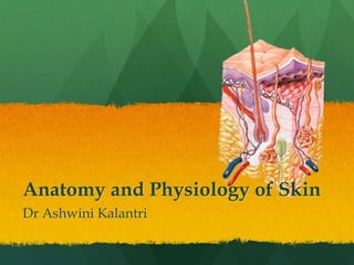
SlideShare a Scribd company logo
1 of 23
Anatomy and Physiology of Skin
Dr Ashwini Kalantri
Skin

 Largest organ of the body
 Completely covers the body
 Continuous with membranes lining body orifices
 Average thickness: 1-2 mm. (0.5mm eyelids, 6mm
  palms and soles)
 pH: 4-5.6
 Renewal of skin takes place in 28-50 days by
  shedding of the outer layer.
Structure of Skin

Layers
 1. Epidermis
 2. Dermis
Epidermis

 Most superficial layer of the skin composed of
  stratified epithelium

 No blood vessels

 Nutrition provided by the capillaries of dermis

 Approx. thickness 0.4-1.5mm, thickest on palms and
  soles.
Layers of Epidermis

   Stratum Corneum    {
      Stratum Lucidum   {


                   {
Stratum Granulosum


 Stratum Mucosum


Stratum Germinativm     {
Cells in the Epidermis

 Keratinocytes: Major building block of the
  epidermis

 Melanocytes: Large cells interspaced among
  keratinocytes, produce melanin.

 Langerhans Cells: Antigen presenting cells

 Merkel cells: Represent special nerve endings
  within epidermis

 Hair, Sebaceous glands and ducts of sweat glands.
Colour of the Skin

 Pigmentation of skin: Melanocytes contain the
  pigment melanin which darkens the skin on
  exposure to sunlight

 Hemoglobin content in blood: The level of
  oxygenation of blood and amount of blood
  circulating in the dermis play an important role in
  skin colouration.
Dermis

 Connective tissue layer made up of dense and stout
  collagen fibers, fibroblasts and histocytes. Collagen
  fibers have elastic property and are capable of
  storing water.

 Layers:
  1. Superficial Papillary Layer
  2. Reticular Layer
Superficial Papillary Layer

 Projects in to the epidermis

 Contains blood vessels, lymphatics and nerve fibers.

 Has some pigment containing cells called
  chromatophore.

 Dermal papillae are finger like projections arising
  from this layer
Reticular Layer

 Made up of reticular and elastic fibers

 These fibers are found around hair bulbs, sweat
  glands and sebaceous glands.

 Also contains mast cells, nerve
  endings, lymphatics, epidermal appendages and
  fibroblasts.

 The hair follicles with hairs, sweat
  glands, sebaceous glands and nails.
Glands of the Skin
 1. Sebaceous Glands
 2. Sweat Glands
Sebaceous Glands

 Structure: Ovoid or spherical in shape, developed
  from hair follicles and covered by connective tissue
  capsule

 Secretion: Secrete a oily substance called Sebum.

 Composition: Contains free fatty
  acids, sterols, paraffin, waxes, squalene and
  triglycerides.

 Functions: FFA has antibacterial and antifungal
  properties. Lipids keep skin smooth and
  oily, protecting from unnecessary desquamation
  and injury.
Sweat Glands - Eccrine

 Distributed throughout the body with exterior
  opening through sweat pore with watery and clear
  discharge.

 Temperature regulation

 Secretion increases with increase in temperature
  and emotions under nervous control

 Nerve supply by sympathetic cholinergic fibers.
Sweat Glands - Apocrine

 Distributed only in a limited area –
  Axilla, pubis, areola and umbilicus.

 Opens in to the hair follicle having thick and milky
  secretion.

 Starts functioning with puberty and has no role in
  temperature regulation

 Secretions increase under emotional conditions
  under hormonal control.

 Supply by sympathetic adrenergic fibers.
Functions of Skin
Protective

 Protection from bacteria and toxic substances by
  secreting lysozyme.

 Protection from mechanical blow

 Protection from UV rays with the help of melanin
  pigment.
Sensory

 Skin is considered the largest sensory organ

 Many nerve endings forming a specialized
  cutaneous receptors.
Storage

 Stores fat, water, chloride and sugar.

 Can also store blood with vasodilation of the
  cutaneous blood vessels
Synthetic

 Vitamin D3 is synthesized in the skin with the
  action of UV rays on cholesterol.
Temperature Regulation

 Excess heat is lost from the body by
  radiation, conduction, convection and evaporation.

 Active role in heat loss by secreting sweat.

 The lipid content of sebum prevents heat loss in
  cold temperature.
Water and Electrolyte
           balance
 Excretion: Waste materials like urea, salts and fatty
  substances are also excreted.

 Absorption: skin can absorb fat soluble substances
  and some ointments.

 Secretion: Sweat and sebum are secreted by the
  sweat and sebaceous glands. Helps in temperature
  and water balance. Sebum helps in keeping the skin
  smooth and provides protection.
Skin
Skin

More Related Content

What's hot

Integumentary system
Integumentary systemIntegumentary system
Integumentary system
MissReith
 
Skin functions
Skin functionsSkin functions
Skin functions
stewart_j
 
Integumentary system ppt
Integumentary system pptIntegumentary system ppt
Integumentary system ppt
nadhifah pratiwi
 
The skin
The skinThe skin
The skin
ssas1
 

What's hot (20)

The integumentary system (slide show)
The integumentary system (slide show)The integumentary system (slide show)
The integumentary system (slide show)
 
7. integumentary system
7. integumentary system7. integumentary system
7. integumentary system
 
Integumentary system
Integumentary systemIntegumentary system
Integumentary system
 
The integumentary system
The integumentary systemThe integumentary system
The integumentary system
 
Basic histopathology of skin
Basic histopathology of skinBasic histopathology of skin
Basic histopathology of skin
 
Skin functions
Skin functionsSkin functions
Skin functions
 
Integumentary system
Integumentary systemIntegumentary system
Integumentary system
 
Integumentary system ppt
Integumentary system pptIntegumentary system ppt
Integumentary system ppt
 
Structure of skin
Structure of skin Structure of skin
Structure of skin
 
The skin
The skinThe skin
The skin
 
The Integumentary System
The Integumentary SystemThe Integumentary System
The Integumentary System
 
Integumentary system ppt.
Integumentary system ppt.Integumentary system ppt.
Integumentary system ppt.
 
Skin presentation
Skin presentationSkin presentation
Skin presentation
 
Lymphatic system
Lymphatic systemLymphatic system
Lymphatic system
 
Unit II, Chapter-1-Integumentary System
Unit II, Chapter-1-Integumentary SystemUnit II, Chapter-1-Integumentary System
Unit II, Chapter-1-Integumentary System
 
Skin
SkinSkin
Skin
 
Anatomy and physiologyof skin
Anatomy and physiologyof skinAnatomy and physiologyof skin
Anatomy and physiologyof skin
 
skin ppt
skin pptskin ppt
skin ppt
 
Skin
SkinSkin
Skin
 
The skin & integumentary system. ppt
The skin & integumentary system. pptThe skin & integumentary system. ppt
The skin & integumentary system. ppt
 

Viewers also liked (14)

Anatomy of skin
Anatomy of skinAnatomy of skin
Anatomy of skin
 
Skin Structure
Skin StructureSkin Structure
Skin Structure
 
Anatomy Of The Skin Lecture
Anatomy Of The Skin LectureAnatomy Of The Skin Lecture
Anatomy Of The Skin Lecture
 
Skin.ppt
Skin.pptSkin.ppt
Skin.ppt
 
Benign skin lesions
Benign skin lesionsBenign skin lesions
Benign skin lesions
 
Integumentary System Notes
Integumentary System NotesIntegumentary System Notes
Integumentary System Notes
 
Physiology of Skin and Skeletal system
Physiology of Skin and Skeletal systemPhysiology of Skin and Skeletal system
Physiology of Skin and Skeletal system
 
BURNS
BURNSBURNS
BURNS
 
D anatomy and physiology of the hair and skin
D anatomy and physiology of the hair and skinD anatomy and physiology of the hair and skin
D anatomy and physiology of the hair and skin
 
Comparative Anatomy - Integumentary System
Comparative Anatomy - Integumentary SystemComparative Anatomy - Integumentary System
Comparative Anatomy - Integumentary System
 
Skin anatomy & physiology for ayush students
Skin anatomy & physiology for ayush studentsSkin anatomy & physiology for ayush students
Skin anatomy & physiology for ayush students
 
Ap skin-03
Ap skin-03Ap skin-03
Ap skin-03
 
Anatomy of skin by dr.sana
Anatomy of skin by dr.sanaAnatomy of skin by dr.sana
Anatomy of skin by dr.sana
 
The integumentary system
The integumentary systemThe integumentary system
The integumentary system
 

Similar to Skin

Integumentary system.pptx
Integumentary system.pptxIntegumentary system.pptx
Integumentary system.pptx
Dr Soumya Patil
 
2. Integumentary system .ppt
2. Integumentary system .ppt2. Integumentary system .ppt
2. Integumentary system .ppt
TigabuAgmas1
 
Integumentary System.pptx
Integumentary System.pptxIntegumentary System.pptx
Integumentary System.pptx
Sheetal Patil
 
Lect 5 intergumentary
Lect 5   intergumentaryLect 5   intergumentary
Lect 5 intergumentary
missazyaziz
 
5. The Integument and Related Structures
5. The Integument and Related Structures5. The Integument and Related Structures
5. The Integument and Related Structures
SUNY Ulster
 
Integumentary System Notes
Integumentary System NotesIntegumentary System Notes
Integumentary System Notes
rachelkucera
 

Similar to Skin (20)

Structure of skin.ppt
Structure of skin.pptStructure of skin.ppt
Structure of skin.ppt
 
Anatomy 1
 Anatomy 1 Anatomy 1
Anatomy 1
 
Integumentary system prep.pptx
Integumentary system prep.pptxIntegumentary system prep.pptx
Integumentary system prep.pptx
 
Integumentary system.pptx
Integumentary system.pptxIntegumentary system.pptx
Integumentary system.pptx
 
2. Integumentary system .ppt
2. Integumentary system .ppt2. Integumentary system .ppt
2. Integumentary system .ppt
 
Integumentary system
Integumentary systemIntegumentary system
Integumentary system
 
Dermatology 5th year, 1st lecture (Dr. Kazhan)
Dermatology 5th year, 1st lecture (Dr. Kazhan)Dermatology 5th year, 1st lecture (Dr. Kazhan)
Dermatology 5th year, 1st lecture (Dr. Kazhan)
 
Integumentary System.pptx
Integumentary System.pptxIntegumentary System.pptx
Integumentary System.pptx
 
Lect 5 intergumentary
Lect 5   intergumentaryLect 5   intergumentary
Lect 5 intergumentary
 
5. The Integument and Related Structures
5. The Integument and Related Structures5. The Integument and Related Structures
5. The Integument and Related Structures
 
Integumentary System Notes
Integumentary System NotesIntegumentary System Notes
Integumentary System Notes
 
Chapter 4 - Skin and Body Membranes
Chapter 4 - Skin and Body MembranesChapter 4 - Skin and Body Membranes
Chapter 4 - Skin and Body Membranes
 
Biosci
BiosciBiosci
Biosci
 
Skin and Hair
Skin and HairSkin and Hair
Skin and Hair
 
Skin and body membranes
Skin and body membranesSkin and body membranes
Skin and body membranes
 
Skin and body membranes
Skin and body membranesSkin and body membranes
Skin and body membranes
 
Integument
IntegumentIntegument
Integument
 
Anatomy of skin
Anatomy of skinAnatomy of skin
Anatomy of skin
 
EVA KIPTOO DENTAL PRESENTATION - Copy.pptx
EVA KIPTOO DENTAL PRESENTATION - Copy.pptxEVA KIPTOO DENTAL PRESENTATION - Copy.pptx
EVA KIPTOO DENTAL PRESENTATION - Copy.pptx
 
Integument ( Skin )
Integument ( Skin )Integument ( Skin )
Integument ( Skin )
 

More from Ashwini Kalantri (6)

shiny.pdf
shiny.pdfshiny.pdf
shiny.pdf
 
Urinary system
Urinary systemUrinary system
Urinary system
 
Typhoid
TyphoidTyphoid
Typhoid
 
Nervous system 2
Nervous system 2Nervous system 2
Nervous system 2
 
Nervous system
Nervous systemNervous system
Nervous system
 
Cholera
CholeraCholera
Cholera
 

Skin

  • 1. Anatomy and Physiology of Skin Dr Ashwini Kalantri
  • 2. Skin  Largest organ of the body  Completely covers the body  Continuous with membranes lining body orifices  Average thickness: 1-2 mm. (0.5mm eyelids, 6mm palms and soles)  pH: 4-5.6  Renewal of skin takes place in 28-50 days by shedding of the outer layer.
  • 3. Structure of Skin Layers 1. Epidermis 2. Dermis
  • 4. Epidermis  Most superficial layer of the skin composed of stratified epithelium  No blood vessels  Nutrition provided by the capillaries of dermis  Approx. thickness 0.4-1.5mm, thickest on palms and soles.
  • 5. Layers of Epidermis Stratum Corneum { Stratum Lucidum { { Stratum Granulosum Stratum Mucosum Stratum Germinativm {
  • 6. Cells in the Epidermis  Keratinocytes: Major building block of the epidermis  Melanocytes: Large cells interspaced among keratinocytes, produce melanin.  Langerhans Cells: Antigen presenting cells  Merkel cells: Represent special nerve endings within epidermis  Hair, Sebaceous glands and ducts of sweat glands.
  • 7. Colour of the Skin  Pigmentation of skin: Melanocytes contain the pigment melanin which darkens the skin on exposure to sunlight  Hemoglobin content in blood: The level of oxygenation of blood and amount of blood circulating in the dermis play an important role in skin colouration.
  • 8. Dermis  Connective tissue layer made up of dense and stout collagen fibers, fibroblasts and histocytes. Collagen fibers have elastic property and are capable of storing water.  Layers: 1. Superficial Papillary Layer 2. Reticular Layer
  • 9. Superficial Papillary Layer  Projects in to the epidermis  Contains blood vessels, lymphatics and nerve fibers.  Has some pigment containing cells called chromatophore.  Dermal papillae are finger like projections arising from this layer
  • 10. Reticular Layer  Made up of reticular and elastic fibers  These fibers are found around hair bulbs, sweat glands and sebaceous glands.  Also contains mast cells, nerve endings, lymphatics, epidermal appendages and fibroblasts.  The hair follicles with hairs, sweat glands, sebaceous glands and nails.
  • 11. Glands of the Skin 1. Sebaceous Glands 2. Sweat Glands
  • 12. Sebaceous Glands  Structure: Ovoid or spherical in shape, developed from hair follicles and covered by connective tissue capsule  Secretion: Secrete a oily substance called Sebum.  Composition: Contains free fatty acids, sterols, paraffin, waxes, squalene and triglycerides.  Functions: FFA has antibacterial and antifungal properties. Lipids keep skin smooth and oily, protecting from unnecessary desquamation and injury.
  • 13. Sweat Glands - Eccrine  Distributed throughout the body with exterior opening through sweat pore with watery and clear discharge.  Temperature regulation  Secretion increases with increase in temperature and emotions under nervous control  Nerve supply by sympathetic cholinergic fibers.
  • 14. Sweat Glands - Apocrine  Distributed only in a limited area – Axilla, pubis, areola and umbilicus.  Opens in to the hair follicle having thick and milky secretion.  Starts functioning with puberty and has no role in temperature regulation  Secretions increase under emotional conditions under hormonal control.  Supply by sympathetic adrenergic fibers.
  • 16. Protective  Protection from bacteria and toxic substances by secreting lysozyme.  Protection from mechanical blow  Protection from UV rays with the help of melanin pigment.
  • 17. Sensory  Skin is considered the largest sensory organ  Many nerve endings forming a specialized cutaneous receptors.
  • 18. Storage  Stores fat, water, chloride and sugar.  Can also store blood with vasodilation of the cutaneous blood vessels
  • 19. Synthetic  Vitamin D3 is synthesized in the skin with the action of UV rays on cholesterol.
  • 20. Temperature Regulation  Excess heat is lost from the body by radiation, conduction, convection and evaporation.  Active role in heat loss by secreting sweat.  The lipid content of sebum prevents heat loss in cold temperature.
  • 21. Water and Electrolyte balance  Excretion: Waste materials like urea, salts and fatty substances are also excreted.  Absorption: skin can absorb fat soluble substances and some ointments.  Secretion: Sweat and sebum are secreted by the sweat and sebaceous glands. Helps in temperature and water balance. Sebum helps in keeping the skin smooth and provides protection.

Editor's Notes

  1. Branched alveolar glands in the dermis