rprotein

This document summarizes recent applications of recombinant fusion proteins for tissue engineering. It discusses four main classes of recombinant fusion proteins: 1) Structure-based proteins like elastin-like polymers (ELPs) and silk-like polymers (SLPs) which can be designed to modulate mechanical properties and biodegradation. 2) Cell-bound growth factor fusion proteins which link growth factors like bFGF and EGF to promote cellular responses. 3) Hybrid systems combining recombinant proteins with synthetic polymers. 4) Cadherin-based fusion proteins which may be applicable for stem cell tissue engineering by facilitating cell-cell interactions. The document outlines examples of each type of fusion protein and their potential uses and advantages for tissue engineering applications.

Recommandé
Recommandé
Contenu connexe
Tendances
Tendances (20)
Similaire à rprotein
Similaire à rprotein (20)
Plus de Prasit Chanarat
Plus de Prasit Chanarat (20)
rprotein
- 1. Annals of Biomedical Engineering, Vol. 38, No. 3, March 2010 (Ó 2010) pp. 683–693 DOI: 10.1007/s10439-010-9935-3 Application of Recombinant Fusion Proteins for Tissue Engineering MASATO NAGAOKA,1 HU-LIN JIANG,2,3,5 TAKASHI HOSHIBA,4 TOSHIHIRO AKAIKE,1 and CHONG-SU CHO2,3 1 Graduate School of Bioscience and Biotechnology, Tokyo Institute of Technology, Yokohama 226-8501, Japan; 2Department of Agricultural Biotechnology, Seoul National University, Seoul 151-921, Korea; 3Research Institute for Agriculture and Life Sciences, Seoul National University, Seoul 151-921, Korea; 4Biomaterials Center, National Institute for Materials Science, Tsukuba 305-0044, Japan; and 5Brain Korea 21 Program for Veterinary Science, College of Veterinary Medicine, Seoul National University, Seoul 151-742, Korea (Received 9 May 2009; accepted 17 January 2010; published online 4 February 2010) Associate Editor Julia E. Babensee oversaw the review of this article. Abstract—Extracellular matrix (ECM) plays important roles INTRODUCTION in tissue engineering because cellular growth and differenti- ation, in the two-dimensional cell culture as well as in the Tissue engineering has attracted many scientists and three-dimensional space of the developing organism, require medical doctors with a hope to treat patients in a mini- ECM with which the cells can interact. Also, the develop- mally invasive and less painful way because it can be used ment of new synthetic ECMs is very important because ECMs facilitate the localization and delivery of cells to the to restore, maintain, or enhance tissues and organs66 and specific sites in the body. Therefore, the development of the engineered tissues could reduce or eliminate the need synthetic ECMs to replace the natural ECMs is increasingly for organ replacement.20 Tissue engineering approaches essential and promising in tissue engineering. Recombinant typically employ exogenous three-dimensional extracel- genetic engineering method has enabled the synthesis of lular matrix (ECM) to engineer new natural tissues from protein-based polymers with precisely controlled functional- ities for the development of new synthetic ECMs. In this natural cells.1 The exogenous ECMs are designed to review, the design and construction of structure-based bring the desired cell types into contact in an appropriate recombinant fusion proteins such as elastin-like polymers three-dimensional environment and also to provide (ELPs) and silk-like polymers (SLPs), cell-bound growth mechanical support until the newly formed tissues are factor-based recombinant fusion proteins such as basic structurally stabilized.58 Design of exogenous ECMs for fibroblast growth factor (bFGF) and epidermal growth factor (EGF), hybrid system composed of recombinant the tissue engineering is to mimic the functions of the protein and synthetic polymer, and E-cadherin-based fusion natural ECM molecules found in tissues because these protein by recombinant genetic engineering were explained natural ECMs act as a scaffold to bring cells together in a for application of the synthetic ECMs. Modulation of tissue, to control tissue structure, and to regulate cell mechanical properties, stimuli-sensitivity, biodegradation phenotype.62 Development of new artificial ECMs is and cell recognition can be achieved through precise control of sequence, length, hydrophobicity and cell binding domain very important because the ECMs facilitate localization by recombinant genetic engineering. and delivery of cells to specific sites in the body, they define and maintain a three-dimensional space for the Keywords—Extracellular matrix, Tissue engineering, Elastin- formation of new tissues with appropriate structure and like polymer, Silk-like polymer, E-cadherin. they guide development of new tissues with their appropriate functions.7 Typically, artificial ECM constructs, which form the backbone of most engineered tissues, are developed using either natural polymers such as collagen, algi- nate, chitosan, fibrin, hyaluronan, etc., or synthetic polymers such as PLA, PGA, PLGA, PEG, PCL, etc.17 Address correspondence to Toshihiro Akaike, Graduate School The collagen, fibrin and hyaluronic acid among the of Bioscience and Biotechnology, Tokyo Institute of Technology, natural polymers have been widely used as scaffold for Yokohama 226-8501, Japan. Electronic mail: takaike@bio.titech.ac.jp tissue engineering because their physiological proper- Address correspondence to Chong-Su Cho, Department of Agri- cultural Biotechnology, Seoul National University, Seoul 151-921, ties, biocompatibility and biodegradability are similar Korea. Electronic mail: chocs@plaza.snu.ac.kr to natural tissues. However, the low mechanical 683 0090-6964/10/0300-0683/0 Ó 2010 Biomedical Engineering Society
- 2. 684 NAGAOKA et al. properties, the risk of viral infection, the antigenicity, ECMs. However, these artificial ECMs are still far the instability of materials, deterioration and limited from showing the efficiency and rich complexity and versatility limit long-term implantation for the clinical functionality of the natural ECMs.18 application. Therefore, the development of artificial Recently, progress in recombinant DNA technology ECMs as alternatives to replace the natural polymers has allowed the synthesis of recombinant protein-based will be useful in tissue engineering. It will be expected polymers with precisely defined molecular weights, that development of new artificial ECMs for tissue compositions, sequences and stereochemistries.45 Such engineering plays an important role in providing an precise control over molecular structure of a polymer alternative to organ and tissue transplantation, both of allows similarly fine control over its physicochemical which suffer from a limitation of supply.17 characteristics and biological fate.16 Also, it enables This review summarizes the recent applications of construction of new tailor-made biomaterials with im- recombinant fusion proteins for tissue engineering. proved properties important for tissue engineering. This review mainly explains four recombinant ECMs A recombinant protein-based polymer is a polymer because it is too broad to summarize the structure– consisting of peptide sequence repeats, where each function relationships of other artificial ECMs such as repeating unit can be composed of as few as two or as collagen,31,41,44,74,84 fibrin,14,33 hyaluronic acid,9,13,85,86 many as hundreds of amino acid residues or without glycosaminoglycans82 and proteoglycans70 for tissue containing any repeating sequences, and may recur from engineering application although they can be prepared a few to hundreds of times.77 The key difference that by recombinant genetic engineering technique and separates genetically engineered polymers from poly some products are already commercial available. (amino acid)s and polypeptides is that they are synthe- However, some literatures are cited for the readers. sized by recombinant techniques.22 The entire amino One is structure-based recombinant fusion proteins acid sequence of recombinant polymers is controlled at such as elastin- and silk-like polymers well character- the DNA-level leading to polymers with precisely ized for the most part. Second is cell-bound growth defined, and potentially quite complex sequences and factor-based recombinant fusion proteins. Third is structures.8 Therefore, recombinant polymers can be hybrid system composed of recombinant protein and designed to incorporate a variety of functionalities, such synthetic polymer. Fourth is cadherin-based fusion as responsiveness to microenvironmental stimuli, con- protein because it can be applicable for stem cell tissue trolled biodegradation and presentation of informa- engineering. Also, it can be expected that the devel- tional motifs for cellular interaction.22 opment of recombinant fusion proteins will provide the promise of a safe, predictable and chemically defined source of ECMs for tissue engineering although CLASSES OF RECOMBINANT FUSION a prerequisite for the growth of applications based on PROTEINS recombinant fusion proteins is the improvement of the Mostly four recombinant fusion proteins have been production of larger amounts of recombinant proteins used in tissue engineering. One is structure-based and two-dimensional recombinant fusion proteins are recombinant fusion protein because several repeating almost applied in many cell culture studies by far. amino acid sequences or amino acid sequences without any repeating were constructed by recombinant tech- nique although elastin- and silk-like polymers are CHARACTERISTICS OF RECOMBINANT mainly covered in this review due to the limited appli- FUSION PROTEINS cations of other recombinant fusion proteins for tissue engineering. Second is cell-bound growth factor-based One of the parameters for the development of tissue recombinant fusion proteins because cell-bound engineering is to design and to produce materials for growth factor domain is mainly constructed by acting as an adequate scaffold in growing cells and recombinant technique. Third is hybrid system composed tissues.18 The development of artificial ECMs started of recombinant fusion protein and synthetic polymer. with the use of biocompatible and biodegradable Fourth is cadherin-based fusion protein because it can chemically synthetic polymers, which showed low cell be applicable for stem cell tissue engineering. attachment and spreading capabilities of a rather non- specific nature5 although these polymers were soon improved with more specific functionalities included in Structure-Based Recombinant Fusion Proteins their structure, specially peptide cell adhesive sequence Elastin-Like Polymers such as RGD (arg-gly-asp) tripeptide found in various natural ECM molecules.27 The use of these sequences Elastin is an ECM protein consisting of several significantly enhanced the performances of those repeating amino acid sequences, including VPGZG,
- 3. Application of Recombinant Fusion Proteins for Tissue Engineering 685 VPGVG, APGVGV, VPGFGCGAG and VPGG.64 specific cellular responses and direct new tissue for- The most renowned building block within this family is mation. The recombinant technique allows highly the VPGZG, where Z can be any natural or modified specialized artificial ECM proteins to be obtained in amino acid.76 Urry and co-workers performed a pio- good yield. This approach included RGD sequences,52 neer work for biomedical uses, and particularly in RGD and CS5 binding domains of fibronectin,26,42 tissue engineering78 because elastin-like polymers RGD binding domain of collagen11 and tenascin-C,32 (ELPs) showed an outstanding biocompatibility. heparin-binding domains,82 and RGD and heparin- Interestingly, Nicol et al. reported that ELPs induced binding domains.83 little or no immunogenic response in vivo.51,79 Also, their biodegradation proceeded by conventional met- Silk-Like Polymers abolic routes, yielding just natural amino acids.18 ELPs based on a repeating VPGVG are soluble in Silks are fibrous proteins composed of repeating aqueous medium below their inverse transition tem- sequences of both crystalline and amorphous perature (Tt). When this temperature is raised above domains.19 The silks are naturally produced by spiders the Tt, they aggregate by hydrophobic self-assembly and Lepdopterai. The primary amino acid components and undergo a phase transition.76 Reversibility of of silk proteins are glycine, alanine and other short physical changes in temperature-sensitive ELPs was chain amino acids. Synthesis of silk-like polymers applied for temporary functional scaffolds for tissue (SLPs) by recombinant technique is a powerful method restoration.78 Urry et al.80 reported that apparently for varying properties, through appropriate choice of natural tissue was generated with normal amounts of the different units, the number of units in each multi- collagen and elastic fibers, and red blood cells in mers, the spacing between them, and the number of arterioles were apparent as the darker bodies, indi- repeats of the multimer combination assembly.36 Also, cating vascularization of the new tissue bodies, when SLPs can comprise chemically active sites, enzymatic RGD-containing ELP was injected into guinea pig activity, receptor binding sites, and other functional- subcutaneously. Recently, temperature-sensitive ELPs ities. Especially, biomedical applications offer the were applied for cell sheet recovery because cell sheet highest potential for spider silk owing to the excellent engineering is a powerful technique in tissue engineer- mechanical properties, biocompatibility and biodeg- ing using poly (N-isopropylacrylamide) by Okano’s radation.2 Bini et al.6 included RGD cell-binding group.67 The cell sheets were obtained at 20 °C after domains into the recombinant spider silk. The results A549 cells were cultured on a culture dish coated with indicated that the recombinant spider silk with RGD ELP containing GVGVP repeating units, His tag and encoded into the protein supported enhanced differ- RGD sequence.46 entiation of human bone marrow-derived mesenchy- Injectable biomaterials are of interest for the rapid mal stem cells (hMSCs) to osteogenic outcomes when and in vivo formation of load bearing scaffolds for cultured to tissue culture plastic, suggestion of tissue engineering.35 This system is very attractive for potential application in stem cell tissue engineering. tissue engineering because cells and growth factors can Kaplan and coworkers focused carboxyl terminal be homogeneously mixed with a liquid and injected domain of dentin matrix protein 1 to exploit the self- into a defect site, followed by in situ formation of a assembly and physical properties of silk proteins with hydrogel.39 Also, the rapid formation of an elastomeric controlled hydroxyapatite (HA) formation for bioma- network can give the requisite mechanical provisional terial composites.28 The recombinant protein was scaffold with cells and growth factors in a three- mineralized using simulated body fluids and induced dimensional microenvironment.68 Several methods the formation of calcium-deficient carbonated HA, such as chemical cross-linking, enzymatic cross- suggestion of potential application in bone tissue linking, and physically cross-linked networks have engineering. While recombinant spider dragline silk been reported to prepare ELP hydrogels.12,37,43,49,55,75 displays superior mechanical properties for scaffolds, Recently, Chilkoti and coworkers reported that fibro- one of the technical hurdles is how to recapitulate these blasts embedded in the ELP hydrogels cross-linked by properties in the laboratory on a scalable basis.81 organophosphorous cross-linker survived during the Asakura et al.4 reported a silk-like hybrid protein cross-linking process and remained viable for at least consisting of polyalanine region of silk fibroin from 3 days in vitro, suggestion of controlling mechanical a wild silk worm, Samia cynthia ricini, and RDG properties and potentially functional outcomes in tis- sequence derived from fibronectin. The obtained silk- sue engineering by the ELPs.38 like hybrid protein film showed high cell adhesive and A common goal to biomaterial design involves growth activities of kidney VERO cells when com- creating biomimetic materials that resemble the ECM pared with those of collagen although strong acidic protein components or short peptide sequences to elicit solvents were used to make the film.
- 4. 686 NAGAOKA et al. While recombinant silk proteins have been success- two CBDs into the human bFGF to develop a collagen- fully synthesized, their low aqueous solubility limits based wound targeting repair system. The results biomedical applications. To enhance the aqueous sol- showed that the bFGF with the CBD derived from ubility of recombinant silks, differentially charged collagenase (C-bFGF) promoted vascularization at the analogues of block copolymers containing repeating implanted sites more effectively than bFGF with the sequences from silk (GAGAGS) and elastin (GVGVP) CBD derived from von Willeband’s factor (V-pFGF). were synthesized by recombinant technique by Sheng et al.65 constructed a fusion protein consisted of replacing a valine residue with glutamic acid.21 The bFGF fused to the C-terminus of glutathione S-trans- block copolymers showed potentials as injectable ure- ferase (GST). The GST-bFGF stimulated the growth of thral bulking agents for the treatment of female stress HUVECs to the same extent as recombinant bFGF, urinary incontinence due to pH- and temperature- whereas GST itself did not stimulate, suggesting that sensitive properties.50 The characteristics of structure- bFGF retains its biological activity when fused to GST based recombinant ECMs are summarized in Table 1. although GST-bFGF must be supplemented with bovine serum before storage to retain biological activity. Cell-Bound Growth Factor-Based Recombinant Fusion Epidermal Growth Factor Proteins Epidermal growth factor (EGF) is a 53-residue sin- Fibroblast Growth Factor gle chain polypeptide that plays important roles in tis- Basic fibroblast growth factor (bFGF) is a potent sue regeneration, stimulation of the initiation of DNA mitogen and a modulator for fibroblasts and vascular synthesis, cell replication, and activation of RNA, endothelial cells69 and plays an important role in tissue acceleration of wound healing, enhancement of prolif- regeneration and repair.72 The bFGF is found in eration and keratinization of epithelial tissues.10 The abundance in tissues such as brain, kidney and carti- EGFs have been constructed to use for EGF fusion lage. However, there are limitations in application to proteins consisted of EGF as the autocrine/paracrine synthetic ECMs because it would diffuse rapidly even peptide signaling molecules and synthetic ECMs. administered in topical side effects on the surrounding Kawase et al.34 constructed a fusion protein from tissues87 and free bFGF molecules possess a shorter EGF and cell-binding domain of fibronectin. The fusion half-life.59 Hashi et al.24 constructed a fusion protein of protein exhibited both cell-adhesive activity and growth the cell-binding domain of human fibronectin and factor activity, each of which was indistinguishable from human bFGF with both cell-adhesive activity and that of the corresponding unfused-protein. growth factor activity. The fusion protein adsorbed to Nishi et al.54 constructed fusion proteins consisting culture dishes stimulated the growth of human of EGF or bFGF and CBD derived from clostridium umbilical-vein endothelial cells (HUVECs) and the histolyticum collagenase. The fusion proteins tightly angiogenesis in chorioallantoic membranes of devel- bound to insoluble collagen and stimulated the growth oping chick embryos, suggestion of potential in neu- of BALB/c3T3 fibroblasts as much as the unfused roregenerative medicine. counterparts. Also, the CBDEGF remained at the sites Andrades et al.3 constructed a fusion protein, which of injection for up to 10 days when injected subcuta- contained bFGF and collagen-binding domain (CBD), neously into nude mice whereas EGF itself was not a decapeptide derived from von Willeband’s factor. The detectable at 24 h after injection, indicating that the recombinant protein increased affinity for collagen and fusion proteins are non-diffusible and long-standing enhanced wound healing. Also, Zhao et al.87 introduced in vivo. Also, Hayashi et al.25 constructed the fusion TABLE 1. Characteristics of structure-based recombinant fusion proteins. Protein Amino acid sequence Advantages Reference ELP VPGZG No immunogenic response in vivo Nicol et al.51 VPGZG + RGD Vascularization of new tissue bodies Urry et al.80 GVGVP + RGD Cell sheet formation Mie et al.46 VGA or KVF In situ hydrogel formation Lim et al.39 SLP (ASAAAAAA)m(GPGQQ)n Improved mechanical properties Lazaris et al.36 Spider silk + RGD Enhancement of differentiation of hMSCs Bini et al.6 Spider silk + dentin Formation of calcium-deficient carbonated HA Huang et al.28 Silk fibroin + RGD Higher cell adhesive and growth activities of kidney Asakura et al.4 VERO cells SELP Silk + elastin Chondrogenesis of mesenchymal stem cells Haider et al.21
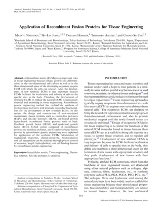
- 5. Application of Recombinant Fusion Proteins for Tissue Engineering 687 protein consisting of EGF and type III collagen. The fusion protein was shown to hold the triple helical conformation of collagen and the mitogenic activity of EGF and the fusion protein can be immobilized on tissue culture dishes as a fibrous form, suggestion of potential in application to tissue engineering. Further- more, Ishikawa et al.30 reported that the fusion protein consisting of EGF and fibronectin collagen-binding FIGURE 2. Sustained activation of MAPK. A431 cells were domain induced granulation tissue formation in the seeded onto EGF-Fc- or collagen-coated PS dishes and cul- tured in DMEM containing 0.5% FBS for various periods of wounds of kind limbs when applied with collagen gel. time. Cells cultured in collagen-coated PS dishes were stim- Ogiwara et al.56 constructed a fusion protein con- ulated with or without 100 ng mL21 EGF. Lysates were sub- sisting of EGF as a cell growth function and immu- jected to Western blotting with anti-phospho-MAPK antibody. From Ogiwara et al.56 noglobulin G (IgG) Fc region (EGF-Fc) to stably adsorb to a cell culture surface. Mouse fibroblast Swiss 3T3 cells adhered to EGF-Fc-coated surface, which is similar to collagen-coated one whereas the cells did not adhere to IgG-coated surface as shown in Fig. 1. Also, phosphorylation of EGF receptors was induced by the immobilized EGF-Fc as well as EGF itself, indicating that the immobilized EGF-Fc tranduced a signal to the cells through the phosphorylation of tyrosine residues on signal proteins. Furthermore, immobilized EGF-Fc continued to activate MARK after 4 h, whereas the activation of MARK in the cells cultured in the pres- ence of EGF itself rapidly decreased with time as shown in Fig. 2, suggesting that MARK activation induced by the immobilized EGF-Fc in the cells is continuous without internalization of growth factor and the constructed EGF-Fc can be used as a synthetic ECM. They also constructed recombinant photo- reactive EGF bearing p-azido phenylalanine at the C-terminal (HEGFP) to immobilize the HEGFP stably to biomaterial surface.57 The results indicated that almost same amounts of A 431 cells adhered to HEGFP-immobilized surface when compared with collagen-coated surface as shown in Fig. 3a. Also, as FIGURE 3. Cell adhesion to HEGFP: (a) A431 cells adhered to the HEGFP-immobilized and collagen-coated surfaces after 30 min of incubation. The data represent means 6 SD of experiments (n 5 3); (b) A431 cells pretreated with 100 ng/mL soluble EGF were seeded onto each surface. Adhesion ratio of the cells was measured after 30 min of incubation; (c) A431 FIGURE 1. Cellular adhesion to EGF-Fc. Swiss 3T3 cells cells suspended in PBS (2) were seeded onto each surface. adhered to the EGF-Fc- and collagen-coated dishes after 6 h As a positive control, the cells suspended in DMEM contain- of incubation. The data represent the mean 6 SD of experi- ing 0.5% FBS were seeded on collagen-coated surface. From ments (n 5 3). From Ogiwara et al.56 Ogiwara et al.57
- 6. 688 NAGAOKA et al. improved the bioactivity of fused NGF-b with enhancement of the nerve growth in vivo. Hybrid Systems Composed of Recombinant Fusion Protein and Synthetic Polymer Hybrid materials were prepared to mimic the nat- ural ECM. It will be expected that they represent a new and versatile class of biomimetic materials with clinical promise in serving as implants because they promote wound healing and tissue regeneration.63 Halstenberg et al.23 prepared hybrid system composed of recombinant protein containing an RGD integrin- FIGURE 4. Liver-specific functions and DNA uptake of binding motif, two plasmin degradation sites and a hepatocytes: hepatocytes were seeded onto HEGFP-immobi- heparin-binding site, and PEG chains with terminal lized, collagen-coated or PVLA-coated surface and cultured in WE for 3 days. Cells cultured on collagen were stimulated acrylate groups at the cysteine’s thiol groups, thereby with or without 100 ng/mL EGF. CYP1A2 and HNF-4a mRNA rendering the protein covalently photo-cross-linkable were detected by RT-PCR analysis. From Ogiwara et al.57 to form a hydrogel. The results indicated that it had specific integrin-binding capability with heparin bind- shown in Fig. 3b, the number of cells adhered onto ing and proteolytic penetration in 2D and 3D systems. HEGFP-immobilized surface remarkably decreased Hubbell and coworkers also synthesized hybrid whereas the number of cells adhered onto collagen- hydrogels of consisting of recombinant protein having coated one were not much changed, when A 431 cells the activity of vascular endothelial growth factor were pretreated with EGF, indication of receptor- (VEGF) to induce cell adhesion and to provide cell- mediated adhesion. Furthermore, as shown in Fig. 3c, mediated remodeling by cross-linking matrix metallo- Mg2+ does not play a prominent role in the receptor- proteinase (MMP), and reactive PEG.88 The results mediated interaction. Moreover, liver-specific func- indicated that the hybrid hydrogel matrices atop the tions, CYP1A2 and hepatocyte nuclear factor (HNF)- chick chorioallontoic membrane brought strong new 4a, were maintained as same level as hepatocytes blood vessel formation. When implanted subcutane- cultured on galactose-carrying polymer (PVLA) as ously in rats, these hybrid hydrogel matrices were shown in Fig. 4, suggesting that the HEGFP can be completely remodeled into native, vascularized tissue. used as the synthetic ECM for liver tissue engineering. Furthermore, they prepared hybrid hydrogels consist- Elloumi et al.15 constructed a fusion protein consisting ing of recombinant protein having cell adhesion motif of RGD sequence as a cell adhesive function, EGF as a RGD and degradation sites for plasmin and MMPs, cell growth function, and hydrophobic sequence as an and reactive PEG.60,61 The results showed that the efficient assembling function. The fusion protein coated hybrid hydrogels promoted specific cellular adhesion on hydrophobic surface retained both cell adhesive and exhibited degradability by the target enzymes. activity through the RGD sequence and cell growth Also, the hybrid hydrogels promoted healing of bone activity when A 549 cells were cultured, whereas EGF defects after treatment of rat calvarial defects, sug- itself had no growth activity even though it was gesting that the combination of recombinant genetic attached to a solid surface to some extent, suggesting technology and synthetic polymer emerges as a pow- that the hydrophobic sequence played roles of a cell erful for the development of artificial ECMs. adhesion function and stabilization of protein structure. Platelet-Derived Growth Factor E-CADHERIN Dai and coworkers constructed a fusion protein from platelet-derived growth factor (PDGF) and E-cadherin is a member of the intercellular adhesion CBD.40 The fusion protein promoted the binding of molecules and E-cadherin-mediated adhesion is regu- PDGF to collagen scaffold with enhanced vasculari- lated by Ca2+-dependent homophilic interaction.73 zation in vivo and caused more cells to proliferate on E-cadherin is essential for tissue morphogenesis and the collagen gel than native PDGF, suggesting that this maintenance of organized solid tissues.53 protein is effective for targeting tissue regeneration and Nagaoka et al.47 constructed a fusion protein wound repair. They also constructed a fusion protein consisting of E-cadherin extracellular domain and from nerve growth factor-b (NGF-b) and CBD.71 IgG Fc region (E-cad-Fc) to stably adsorb to a cell The fusion protein increased the expression level and culture surface as shown in Fig. 5. They reported that
- 7. Application of Recombinant Fusion Proteins for Tissue Engineering 689 FIGURE 5. Construction and expression of E-cadherin-IgG Fc fusion protein. (a) Generated segments of an extracellular domain of mouse E-cadherin and an IgG Fc region were subcloned into an eukaryotic expression vector pRC/CMV via HindIII-NotI site and NotI-XbaI site respectively. CMV pro- moter, human cytomegalovirus immediate-early promoter/ enhancer; BGH pA, bovine growth hormone polyadenylation signal; Neor, neomycin resistance gene for selection; Ampr, ampicillin resistance gene. (b) Conditioned media from transfected CHO-K1 cells were analyzed by Western blotting. Fusion protein was detected by either anti-E-cadherin or anti- mouse IgG antibody. Arrowhead, mature protein; dotted arrow, precursor. (c) Cell lysate and conditioned media of transfectant were analyzed to confirm the protein secretions by Western blotting using anti-mouse IgG antibody. From Nagaoka et al.47 FIGURE 7. Cell adhesion, morphology of ES cells on the E-cad-Fc fusion protein-immobilized surface. (a) ES cells (EB3) adhered to E-cad-Fc-coated dishes with equivalent efficiency as to 0.1% gelatin-coated dishes after 3 h of incubation. (b) ES cells (EB3) were cultured on E-cad-Fc-coated or fibronectin- coated dishes without serum. EGTA (5 mM) was added to the culture medium at 3 h after seeding (open bar). Detached cells were removed and remaining cells were counted using alamar Blue reagent. *p < 0.05, §p < 0.001 vs. no treated condition (closed bar). (c, d) Morphological observation of ES cells (EB3) on the two different matrices. ES cells were cultured on poly- styrene surfaces coated with 0.1% (wt/vol) gelatin (c), or 10 mg/ mL E-cad-Fc (d) in the presence of LIF for 2 days. High magni- fication images are shown in (c¢) and (d¢). From Nagaoka et al.48 hepatocytes adhered to the E-cad-Fc-coated surface were almost same as collagen-coated one and the adhesion was inhibited by pretreatment of hepatocytes with anti-E-cadherin antibody, whereas E-cadherin- deficient Hepa 1-6 cells did not adhere to the E-cad-Fc- coated surface as shown in Fig. 6, suggesting that the adhesion of hepatocytes to E-cad-Fc is mediated by E-cadherin. Also, they reported that hepatocytes adhered to E-cad-Fc-coated surface showed differenti- ated phenotypes such as low DNA synthesis activity and FIGURE 6. Adhesive activity to E-cad-Fc fusion protein. (a) maintenance of tryptophan oxygenase expression as a Primary hepatocytes could adhere to E-cad-Fc-coated dish as marker gene of differentiated hepatocytes,29 suggestion well as collagen-coated surface after 4 h. Inhibition assay was performed by incubating with neutralizing antibody (ECCD-1). of potential synthetic ECM in maintaining differentia- BSA was a negative control. (b) Adhesion of mouse hepatoma tion of hepatocytes for liver tissue engineering. cell line Hepa 1-6 to collagen or E-cad-Fc was analyzed after Also, Akaike and coworkers constructed E-cad-Fc 3 h culture. (c) The expression levels of E-cadherin in two types of liver-derived cells were assessed by western blotting. for culture of embryonic stem (ES) cells.48 They The data represent the mean 6 SEM of experiments (n 5 3). reported that EB 3 cells as the ES cell adhered to the From Nagaoka et al.47 E-cad-Fc-coated surface as similar with conventional
- 8. 690 NAGAOKA et al. FIGURE 8. ES cells show higher proliferation and higher transfection efficiency on the E-cad-Fc-coated surface. (a) The prolif- erative activity of ES cells on a gelatin- or E-cad-Fc-coated surface was evaluated. EB3 cells were seeded on gelatin-coated (open square) or E-cad-Fc-coated (filled square) dishes and the cell number was counted after staining with alamar Blue reagent. The data indicate means 6 SD of experiments (n 5 3). **p < 0.01 vs. gelatinized plates. (b) BrdU incorporation of EB3 cells under colony-forming (on gelatin) or scattering conditions (on E-cad-Fc). Relative BrdU incorporation value was evaluated. The data indicate means 6 SEM. §p < 0.001. (c) Transfection efficiency of ES3 cells cultured on gelatin- or E-cad-Fccoated surface. Relative expression of GFP was evaluated. The data indicate means 6 SEM. §p < 0.001 vs. gelatinized plates. From Nagaoka et al.48 TABLE 2. Characteristics of growth factor-based recombinant fusion proteins. Protein Natural Fused domain Advantages Reference FGF bFGF Fibronectin Stimulation of growth of HUVECs Hashi et al.24 bFGF Collagen Increased migration of fibroblasts Andrades et al.3 bFGF GST Stimulation of growth of HUVECs Sheng et al.65 EGF EGF Fibronectin Increased cell-adhesive activity of HB101 cells Kawase et al.34 EGF Collagen Stimulation of growth of BALB/c 3T3 fibroblasts Hayashi et al.,25 Nishi et al.54 and nondiffusible EGF Fibronectin Induction of granulation tissue formation in the wounds Ishikawa et al.30 EGF IgG Fc Longer activation of MARK of 3T3 fibroblasts Ogiwara et al.56 EGF p-Azido phenylalamine Stable immobilization of EGF Ogiwara et al.57 EGF RGD hydrophobic Retention of cell adhesive activity and stabilization Elloumi et al.15 sequences of protein structure PDGF PDGF Collagen Increased vascularization of collagen scaffold Lin et al.40 NGF NGF-b Collagen Enhancement of nerve growth Sun et al.71 gelatin-coated surface as shown in Fig. 7a. The cells dishes, suggesting that the E-cad-Fc will have a great adhered even in serum-free condition with Ca2+- potential as the artificial ECM for stem cell tissue dependency as shown in Fig. 7b. Interestingly, the engineering. The characteristics of growth factor-based adhered cells remained separated from each other even recombinant ECMs are summarized in Table 2. in the presence of leukemia inhibitory factor (LIF) with dendritic morphologies, whereas the cells adhered to gelatin-coated surface formed tightly aggregated SUMMARY AND FUTURE PERSPECTIVE colonies as shown in Figs. 7c and 7d, suggesting that blocking of close contact among cells in the aggregated Precise control over molecular weight, composition, colonies may not generate a heterogeneous environ- sequence and stereochemistry at the molecular level by ment within colonies, which potentially inhibit the recombinant genetic engineering method has created proliferation of ES cells and the distribution of soluble interest in the artificial ECMs of biomaterials for tissue factors. Also, the ES cells showed higher proliferation engineering. In this review, the design and construc- on the E-cad-Fc-coated surface than gelatin-coated tion of ELPs, SLPs, bFGF, EGF, E-cadherin, and one as shown in Figs. 8a and 8b and the cells adhered hybrid systems composed of recombinant protein and to the E-cad-Fc-coated surface showed higher trans- synthetic polymer by recombinant technique were fection efficiency than gelatin-coated one as shown in explained for the artificial ECMs. Modulation of the Fig. 8c. Furthermore, the expression of Oct-3/4 as the cell recognition is achieved through precise controls in undifferentiated marker of the ES cells was maintained sequence and length. While some examples of the for at least 3 days when cultured on E-cad-Fc-coated influence of sequence, length, hydrophobicity, and cell
- 9. Application of Recombinant Fusion Proteins for Tissue Engineering 691 8 binding domain on the cell recognition were reviewed in Cappello, J., and K. Park (Eds.). Controlled Drug Deliv- this article, medical applications are still on the horizon ery: Challenges and Strategies. Washington DC: American Chemical Society, 1997, p. 439. and the full potential of this recombinant in tissue 9 Chong, B. F., L. M. Blank, R. McLaughlin, and L. K. engineering has yet to materialize. The control of Nielsen. Microbial hyaluronic acid production. Appl. mechanical properties, biodegradation, biorecognition Microbiol. Biotechnol. 66:341–351, 2005. 10 and checking of no immunogenic response can signifi- Cohen, S. The epidermal growth factor (EGF). Cancer cantly expand the utility of these constructs in tissue 51:1787–1791, 1983. 11 Cutler, S. M., and A. J. Garcia. Engineering cell adhesive engineering although a prerequisite for the growth of surfaces that direct integrin alpha5beta1 binding using a applications based on recombinant fusion proteins is the recombinant fragment of fibronectin. Biomaterials 24: improvement of the production of larger amounts of 1759–1770, 2003. 12 recombinant proteins. Also, there will be increasing Di Zio, K., and D. A. Tirrell. Mechanical properties of demands that 3D recombinant fusion proteins provide artificial protein matrices engineered for control of cell and tissue behavior. Macromolecules 36:1553–1558, 2003. better model systems for physiologic situations because 13 Duan, X. J., H. X. Niu, W. S. Tan, and X. Zhang. 3D fusion proteins induce more effectively cellular Mechanism analysis of effect of oxygen on molecular functions than 2D ones although 2D in vitro assay on 2D weight of hyaluronic acid produced by Streptococcus zoo- recombinant fusion proteins are still applied in many cell epidemicus. J. Microbiol. Biotechnol. 19:299–306, 2009. 14 culture studies. Furthermore, future research will be Ehrbar, M., S. C. Rizzi, R. Hlushchuk, V. Djonov, A. H. Zisch, J. A. Hubbell, F. E. Weber, and M. P. Lutolf. aimed to the design of hybrid materials consisting of Enzymatic formation of modular cell-instructive fibrin intelligent recombinant proteins and other biomaterials analogs for tissue engineering. Biomaterials 28:3856–3866, with high mechanical characteristics for hard tissues. 2007. 15 Elloumi, I., R. Kobayashi, H. Funabashi, M. Mie, and E. Kobatake. Construction of epidermal growth factor fusion protein with cell adhesive activity. Biomaterials 27:3451–3458, 2006. ACKNOWLEDGMENT 16 Ghandehari, H., and J. Cappello. Genetic engineering of protein-based polymers: potential in controlled drug This work was supported by the funds provided by delivery. Pharm. Res. 15:813–815, 1998. Korea Research Foundation (KRF) (E00244). 17 Ghosh, K., and D. E. Ingber. Micromechanical control of cell and tissue development: implications for tissue engi- neering. Adv. Drug Deliv. Rev. 59:1306–1318, 2007. 18 Girotti, A., J. Reguera, J. C. Rodriguez-Cabello, F. J. Arias, M. Alonso, and A. M. Testera. Design and bio- production of a recombinant multi(bio)functional elastin- like protein polymer containing cell adhesion sequences for REFERENCES tissue engineering purposes. J. Mater. Sci. Mater. Med. 15:479–484, 2004. 1 19 Abatangelo, G., P. Brun, M. Radice, R. Cortivo, and Gosline, J. M., M. E. DeMont, and M. W. Denny. The M. K. H. Auth. Tissue engineering. In: Integrated Bio- structure and properties of spider silk. Endeavour 10:37–43, materials Science, edited by R. Barmucci. New York: 1986. 20 Kluwer Academic, 2001, p. 885. Griffith, L. G., and G. Naughton. Tissue engineering– 2 Altman, G. H., F. Diaz, C. Jakuba, T. Calabro, R. L. current challenges and expanding opportunities. Science Horan, J. Chen, H. Lu, J. Richmond, and D. L. Kaplan. 295:1009–1014, 2002. 21 Silk-based biomaterials. Biomaterials 24:401–416, 2003. Haider, M., J. Cappello, H. Ghandehari, and K. W. Leong. 3 Andrades, J. A., L. T. Wu, F. L. Hall, M. E. Nimmi, and In vitro chondrogenesis of mesenchymal stem cells in J. Becerra. Engineering, expression, and renaturation of a recombinant silk-elastinlike hydrogels. Pharm. Res. 25:692– collagen-targeted human bFGF fusion protein. Growth 699, 2008. 22 Factors 18:261–275, 2001. Haider, M., Z. Megeed, and H. Ghandehari. Genetically 4 Asakura, T., C. Tanaka, M. Yang, J. Yao, and engineered polymers: status and prospects for controlled M. Kurokawa. Production and characterization of a silk- release. J. Control Rel. 95:1–26, 2004. 23 like hybrid protein, based on the polyalanine region of Halstenberg, S., A. Panitch, S. Rizzi, H. Hall, and Samia cynthia ricini silk fibroin and a cell adhesive region J. A. Hubbell. Biologically engineered protein-graft- derived from fibronectin. Biomaterials 25:617–624, 2004. poly(ethylene glycol) hydrogels: a cell adhesive and plasmin- 5 Atala, A., and D. Mooney. Synthetic Biodegradable degradable biosynthetic material for tissue repair. Biomac- Polymer Scaffolds. Boston: Birkhauser, 1997. romolecules 3:710–723, 2002. 6 24 Bini, E., C. W. Foo, J. Huang, V. Karageorgiou, B. Kitchel, Hashi, H., M. Hatai, F. Kimizuka, I. Kato, and Y. Yaoi. and D. L. Kaplan. RGD-functionalized bioengineered Angiogenic activity of a fusion protein of the cell-binding spider dragline silk biomaterial. Biomacromolecules 7:3139– domain of fibronectin and basic fibroblast growth factor. 3145, 2006. Cell Struc. Func. 19:37–47, 1994. 7 25 Bouhadir, K. H., and D. J. Mooney. In vitro and in vivo Hayashi, M., M. Tomita, and K. Yoshizato. Production of models for the reconstruction of intercellular signaling. EGF-collagen chimeric protein which shows the mitogenic Ann. NY Acad. Sci. 842:188–194, 1998. activity. Biochem. Biophys. Acta 1528:187–195, 2001.
- 10. 692 NAGAOKA et al. 26 43 Heilshorn, S. C., K. A. Dizio, E. R. Welsh, and D. A. McHale, M. K., L. A. Setton, and A. Chilkoti. Synthesis Tirrell. Endothelial cell adhesion to the fibronectin CS5 and in vitro evaluation of enzymatically cross-linked elas- domain in artificial extracellular matrix proteins. Biomate- tin-like polypeptide gels for cartilaginous tissue repair. rials 24:4245–4252, 2003. Tissue Eng. 11:1768–1779, 2005. 27 44 Hersel, U., C. Dahmen, and H. Kessler. RGD modified Merrett, K., W. Liu, D. Mitra, K. D. Camm, C. R. polymers: biomaterials for stimulated cell adhesion and McLaughlin, Y. Liu, M. A. Watsky, F. Li, M. Griffith, and beyond. Biomaterials 24:4385–4415, 2003. D. E. Fogg. Synthetic neoglycopolymer-recombinant 28 Huang, J., C. Wong, A. George, and D. L. Kaplan. The human collagen hybrids as biomimetic crosslinking agents effect of genetically engineered spider silk-dentin matrix in corneal tissue engineering. Biomaterials 30:5403–5408, protein 1 chimeric protein on hydroxyapatite nucleation. 2009. 45 Biomaterials 28:2358–2367, 2007. Meyer, D. E., and A. Chilkoti. Genetically encoded syn- 29 Inoue, C., H. Yamamoto, T. Nakamura, A. Ichihara, and thesis of protein-based polymers with precisely specified H. Okamoto. Nicotinamide prolongs survival of primary molecular weight and sequence by recursive directional cultured hepatocytes without involving loss of hepatocyte- ligation: examples from the elastin-like polypeptide system. specific functions. J. Biol. Chem. 264:4747–4750, 1989. Biomacromolecules 3:357–367, 2002. 30 46 Ishikawa, T., H. Terai, T. Yamamoto, K. Harada, and Mie, M., Y. Mizushima, and E. Kobatake. Novel extra- T. Kitajima. Delivery of a growth factor fusion protein cellular matrix for cell sheet recovery using genetically having collagen-binding activity to wound tissues. Artif. engineered elastin-like protein. J. Biomed. Mater. Res. Part Organs 27:147–154, 2003. B: Appl. Biomater. 86B:283–290, 2008. 31 47 Ito, H., A. Steplewski, T. Alabyeva, and A. Gertala. Nagaoka, M., H. Ise, and T. Akaike. Immobilized E-cad- Testing the utility of rationally engineered recombinant herin model can enhance cell attachment and differentia- collagen-like proteins for applications in tissue engineering. tion of primary hepatocytes but not proliferation. J. Biomed. Mater. Res. A 76:551–560, 2006. Biotechnol. Lett. 24:1857–1862, 2002. 32 48 Jang, J. H., J. H. Hwang, and C. P. Chung. Production of Nagaoka, M., U. Koshimizu, S. Yuasa, F. Hattori, recombinant human tenascin-C module containing a cell H. Chen, T. Tanaka, M. Okabe, K. Fukuda, and adhesion recognition motif of RGD. Biotechnol. Lett. T. Akaike. E-cadherin-coated plates maintain pluripotent 26:1831–1835, 2004. ES cells without colony formation. PLoS One 1:e15, 2006. 33 49 Jing, P., J. S. Rudra, A. B. Herr, and J. H. Collier. Self- Nagapudi, K., W. T. Brinkman, B. S. Thomas, J. O. Park, assembling peptide-polymer hydrogels designed from the M. Srinivasarao, E. Wright, V. P. Conticello, and E. L. coiled coil region of fibrin. Biomacromolecules 9:2438–2446, Chaikof. Viscoelastic and mechanical behavior of 2008. recombinant protein elastomers. Biomaterials 26:4695– 34 Kawase, Y., Y. Ohdate, T. Shimojo, Y. Taguchi, 4706, 2005. 50 F. Kimizuka, and I. Kato. Construction and characteriza- Nagarsekar, A., J. Crissman, M. Crissman, F. Ferrari, tion of a fusion protein with epidermal growth factor and J. Cappello, and H. Ghandehari. Genetic engineering of the cell-binding domain of fibronectin. FEBS Lett. 298: stimuli-sensitive silkelastin-like protein block copolymers. 126–128, 1992. Biomacromolecules 4:602–607, 2003. 35 51 Langer, R., and D. A. Tirrell. Designing materials for Nicol, A., D. C. Gowda, T. M. Parker, and D. W. Urry. biology and medicine. Nature 428:487–492, 2004. Elastomeric polytetrapeptide matrices: hydrophobicity 36 Lazaris, A., S. Arcidiacono, Y. Huang, J. Zhou, F. Duguay, dependence of cell attachment from adhesive (GGIP)n to N. Chretien, E. A. Welsh, J. W. Soares, and C. N. Karatzas. nonadhesive (GGAP)n even in serum. J. Biomed. Mater. Spider silk fibers spun from soluble recombinant silk pro- Res. 27:801–810, 1993. 52 duced in mammalian cells. Science 295:472–476, 2002. Nicol, A., D. C. Gowda, and D. W. Urry. Cell adhesion 37 Lee, J., C. W. Macosko, and D. W. Urry. Elastomeric and growth on synthetic elastomeric matrices containing polypentapeptides cross-linked into matrixes and fibers. Arg-Gly-Asp-Ser-3. J. Biomed. Mater. Res. 26:393–413, Biomacromolecules. 2:170–179, 2001. 1992. 38 53 Lim, D. W., D. L. Nettles, L. A. Setton, and A. Chilkoti. Niewiadomska, P., D. Godt, and U. Tepass. DE-Cadherin Rapid cross-linking of elastin-like polypeptides with is required for intercellular motility during Drosophila (hydroxymethyl)phosphines in aqueous solution. Biomac- oogenesis. J. Cell Biol. 144:533–547, 1999. 54 romolecules 8:1463–1470, 2007. Nishi, N., O. Matsushita, K. Yuube, H. Miyanaka, 39 Lim, D. W., D. L. Nettles, L. A. Setton, and A. Chilkoti. In A. Okabe, and F. Wada. Collagen-binding growth factors: situ cross-linking of elastin-like polypeptide block copoly- production and characterization of functional fusion pro- mers for tissue repair. Biomacromolecules 9:222–230, 2008. teins having a collagen-binding domain. Proc. Natl. Acad. 40 Lin, H., B. Chen, W. Sun, W. Zhao, Y. Zhao, and J. Dai. Sci. USA 95:7018–7023, 1998. 55 The effect of collagen-targeting platelet-derived growth Nowatzki, P. J., and D. A. Tirrell. Physical properties of factor on cellularization and vascularization of collagen artificial extracellular matrix protein films prepared by scaffolds. Biomaterials 27:5708–5714, 2006. isocyanate crosslinking. Biomaterials 25:1261–1267, 2004. 41 56 Liu, W., K. Merrett, M. Griffith, P. Fagerholm, S. Dravida, Ogiwara, K., M. Nagaoka, C. S. Cho, and T. Akaike. B. Heyne, J. C. Scaiano, M. A. Watsky, N. Shinozatki, Construction of a novel extracellular matrix using a new N. Lagali, R. Munger, and F. Li. Recombinant human genetically engineered epidermal growth factor fused to collagen for tissue engineered corneal substitutes. Bioma- IgG-Fc. Biotechnol. Lett. 27:1633–1637, 2005. 57 terials 29:1147–1158, 2008. Ogiwara, K., M. Nagaoka, C. S. Cho, and T. Akaike. 42 Liu, J. C., and D. A. Tirrell. Cell response to RGD density Effect of photo-immobilization of epidermal growth factor in cross-linked artificial extracellular matrix protein films. on the cellular behaviors. Biochem. Biophys. Res. Commun. Biomacromolecules 9:2984–2988, 2008. 345:255–259, 2006.
- 11. Application of Recombinant Fusion Proteins for Tissue Engineering 693 58 Putnam, A. J., and D. J. Mooney. Tissue engineering using K. Yoshzato. Transgenic silkworms produce recombinant synthetic extracellular matrices. Nat. Med. 2:824–826, 1996. human type III procollagen in cocoons. Nat. Biotechnol. 59 Rapraeger, A. C., A. Krufka, and B. B. Olwin. Require- 21:52–56, 2003. 75 ment of heparan sulfate for bFGF-mediated fibroblast Trabbic-Carlson, K., L. A. Setton, and A. Chilkoti. growth and myoblast differentiation. Science 252:1705– Swelling and mechanical behaviors of chemically cross- 1708, 1991. linked hydrogels of elastin-like polypeptides. Biomacro- 60 Rizzi, S. C., M. Ehrbar, S. Halstenber, G. P. Raeber, H. G. molecules 4:572–580, 2003. 76 Schmoeke, H. Hagenmiiller, R. Muller, F. E. Weber, and Urry, D. W. Molecular machines: How motion and other J. A. Hubbell. Recombinant protein-co-PEG networks as functions of living organisms can result from reversible cell-adhesive and proteolytically degradable hydrogel chemical changes. Angew. Chem. Int. Ed. Eng. 32:819–841, matrixes. Part II: biofunctional characteristics. Biomacro- 1993. 77 molecules 7:3019–3029, 2006. Urry, D. W. Physical chemistry of biological free energy 61 Rizzi, S. C., and J. A. Hubbell. Recombinant protein- transduction as demonstrated by elastic protein-based co-PEG networks as cell-adhesive and proteolytically polymers. J. Phys. Chem. B 101:11007–11028, 1997. 78 degradable hydrogel matrixes. Part I: development and Urry, D. W. Elastic molecular machines in metabolism and physicochemical characteristics. Biomacromolecules 6: soft-tissue restoration. Trends Biotechnol. 17:249–257, 1226–1238, 2005. 1999. 62 79 Rosso, F., A. Giordano, M. Barbarisi, and A. Barbarisi. Urry, D. W., T. M. Parker, M. C. Reid, and D. C. Gowda. From cell-ECM interactions to tissue engineering. J. Cell Biocompatibility of the bioelastic materials, poly(GVGVP) Physiol. 199:174–180, 2004. and its irradiation cross-linked matrix: summary of generic 63 Rosso, F., G. Marino, A. Giordando, M. Barbarishi, biological test results. J. Bioact. Compat. Polym. 6:263–282, D. Parmeggiani, and A. Barbarishi. Smart materials as 1991. 80 scaffolds for tissue engineering. J. Cell Physiol. 203:465– Urry, D. W., A. Pattanaik, J. Xu, T. C. Woods, D. T. 470, 2005. McPherson, and T. M. Parker. Elastic protein-based 64 Sandberg, L. B., J. G. Leslie, C. T. Leach, V. L. Alvarez, polymers in soft tissue augmentation and generation. A. R. Torres, and D. W. Smith. Elastin covalent structure J. Biomater. Sci. Polym. Ed. 9:1015–1048, 1998. 81 as determined by solid phase amino acid sequencing. Wong, P., C. Foo, and D. L. Kaplan. Genetic engineering Pathol. Biol. 33:266–274, 1985. of fibrous proteins: spider dragline silk and collagen. Adv. 65 Sheng, Z., S. B. Chang, and W. J. Chirico. Expression and Drug Deliv. Rev. 54:1131–1143, 2002. 82 purification of a biologically active basic fibroblast growth Wu, S. C., J. R. Chiang, and C. W. Lin. Novel cell adhesive factor fusion protein. Protein Expr. Purif. 27:267–271, glycosaminoglycan-binding proteins of Japanese encepha- 2003. litis virus. Biomacromolecules 5:2160–2164, 2004. 66 83 Shin, H., S. Jo, and A. G. Mikos. Biomimetic materials for Wu, S. C., J. C. Yu, S. H. Hsu, and D. C. Chen. Artificial tissue engineering. Biomaterials 24:4353–4364, 2003. extracellular matrix proteins contain heparin-binding and 67 Shiroyanagi, Y., M. Yamato, Y. Yamazaki, H. Toma, and RGD-containing domains to improve osteoblast-like cell T. Okano. Transplantable urothelial cell sheets harvested attachment and growth. J. Biomed. Mater. Res. A 79:557– noninvasively from temperature-responsive culture surfaces 565, 2006. 84 by reducing temperature. Tissue Eng. 9:1005–1012, 2003. Yang, C., P. J. Hillas, J. A. Baez, M. Nokelainen, J. Balan, 68 Silva, G. A., C. Czeisler, K. L. Niece, E. Beniash, D. A. J. Tang, R. Spiro, and J. W. Polarek. The application of Harrington, J. A. Kessler, and S. I. Stupp. Selective dif- recombinant human collagen in tissue engineering. Bio- ferentiation of neural progenitor cells by high-epitope Drugs 18:103–119, 2004. 85 density nanofibers. Science 303:1352–1355, 2004. Yu, H., and G. Stephanopoulos. Metabolic engineering of 69 Squires, C. H., J. Childs, S. P. Eisenberg, P. J. Polverini, Escherichia coli for biosynthesis of hyaluronic acid. Metab. and A. Sommer. Production and characterization of Eng. 10:24–32, 2008. 86 human basic fibroblast growth factor from Escherichia coli. Yu, H., K. Tyo, H. Alper, D. Klein-Marcuschamer, and J. Biol. Chem. 263:16297–16302, 1988. G. Stephanopoulas. A high-throughput screen for hyalu- 70 Sugahara, K., and H. Kitagawa. Heparin and heparan ronic acid accumulation in recombinant Escherichia coli sulfate biosynthesis. IUBMB Life 54:163–175, 2002. transformed by libraries of engineered sigma factors. 71 Sun, W., H. Lin, B. Chen, W. Zhao, Y. Zhao, and J. Dai. Biotechnol. Bioeng. 101:788–796, 2008. 87 Promotion of peripheral nerve growth by collagen scaffolds Zhao, W., B. Chen, X. Li, H. Lin, W. Sun, Y. Zhao, loaded with collagen-targeting human nerve growth factor- B. Wang, Y. Zhao, Z. Han, and J. Dai. Vascularization and beta. J. Biomed. Mater. Res. 83:1054–1061, 2007. cellularization of collagen scaffolds incorporated with two 72 Tabata, Y., and Y. Ikada. Vascularization effect of basic different collagen-targeting human basic fibroblast growth fibroblast growth factor released from gelatin hydrogels factors. J. Biomed. Mater. Res. A 82:630–636, 2007. 88 with different biodegradabilities. Biomaterials 20:2169– Zisch, A. H., M. P. Lutolf, M. Ehrbar, G. P. Raeber, 2175, 1999. S. C. Rizzi, N. Davies, H. Schmokel, D. Bezuidenhout, 73 Takeichi, M. Morphogenetic roles of classic cadherins. V. Djonov, P. Zilla, and J. A. Hubbell. Cell-demanded Curr. Opin. Cell Biol. 7:619–627, 1995. release of VEGF from synthetic, biointeractive cell 74 Tomita, M., H. Munetsuna, T. Sato, T. Adachi, R. Hino, ingrowth matrices for vascularized tissue growth. FASEB M. Hayashi, K. Shimizu, N. Nakamura, T. Tamura, and J. 17:2260–2262, 2003.