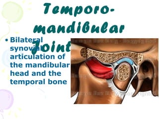
2. Temporomandibular Joint : TMJ
Synovial articulation between the mandible and the cranium
Allows considerable movement and has a joint cavity filled with synovial
fluid and is surrounded by a joint capsule
Bones of the joint-1
articular disc-2
divides
the joint cavity
into
upper
lower
compartment
posterior
bilaminar
zone
wraps around the joint
compartment
anterior
bilaminar
zone
:Articular capsule-3
3. :Joint description
• Type of joint
• Bones involved , Ligaments
• Disc
• Capsule
• Synovial membrane
• Innervation
• Blood supply
• Muscles bringing about
movement
9. • The disc is divided into three zones:
• Anterior band – split into 2 lamellae:
• -Upper lamella attached to articular
eminence
• -Lower lamella attached to condylar head
• fibers of superior head of the lateral pterygoid
inserted in between
10. –: Intermediate band
•
Thinnest central region which is avascular and
. has no innervation
This zone is composed of fibrous connective •
. tissue and is devoid of cells
11.
12. : Posterior band •
•
: This region attaches posteriorly by a
( superior lamella(retrodiscal pad •
attaches superiorly to the capsule and temporal - •
; bone
highly vascular, innervated . (loose - •
( CT ,elastic fs., BV., nerves
inferior lamella •
attaches to the posterior aspect of the neck of - •
.the condyle
consists mainly collagenous fibers with no - •
. elastic fibers
16. Histological Structure of TMJ
.fibrous C.T
.fibrous C.T
inner zone
intermediate z
outer zone
synovial
membrane
descending
limb
Trabeculae radiate from the center of condyle and reach the surface at right angles
19. Articular Capsule
outer fibrous layer
inner Synovial membrane
•thin C.T.
•lined by synovial cells
•intermingled with C.T. fibers
• fibroblasts,histiocytes,lymphocytes
•EM.:TypeA secrete hyaluronic acid
Type B pr.rich secretion
Synovial
folds and villi
20. : The capsule consists of •
Outer fibrous layer •
.dense fibrous collagenous connective tissue-
•
(Inner synovial layer (synovial membrane
lines the inner aspect of the capsule facing the. .two synovial spaces,has synovial villi
•
•
21. The Synovial Fluid
• Clear, straw colored, viscous fluid
• Infiltrate of the blood
• Contains hyaluronic acid sectreted by synovial
cells.
• Contains free macrophages.
Functions
• Nutriant for the avascular tissue of the articulation
• Lubricant for the articulating surfaces
• Clears the tissue debris caused by the normal wear
and tear of the articulating surfaces- from the
articular cavity
22. Thick fibrous
covering of the
condyle with
condroid
changes
Synovial folds
become
fibrotic with
thick
underlying
b.m
Age Changes of
TMJ
Flattening of the condyle
Thin articular
disc with
hyalinization
and chondroid
changes
Osteoporosis of
the underlying
bone
Thining of the
cartilagenous zone
of the condyle
decreased synovial
fluid
loss of lubrication
Dysfunction of TMJ in old age
Decreased
extensibility of
the disc and
capsule
Decreased
nerves in disc
and capsule
23. Innervation
: of TMJ
Blood Supply
of TMJ
- Trigeminal
n.- mandibular
Auriculotemporal
Masseteric n
Internal maxillary artery
Deep auricular
Superfecial temporal
Pterygoid plexus- venous drainage
Vascular plexus in the wall of the capsule- production of synovial fluid