Femoroacetabular impingement
•
28 j'aime•8,922 vues

The document discusses femoroacetabular impingement (FAI), a cause of hip pain and damage in athletes. FAI occurs when the femoral head and acetabulum abnormally contact each other, either from bone growth (CAM impingement) or acetabular overcoverage (pincer impingement). Surgery aims to correct the impingement through osteoplasty of the femoral head or acetabulum. While conservative care is sometimes attempted, surgery best addresses the underlying biomechanical issue causing FAI and progression of damage.

Recommandé
Contenu connexe
Tendances
Tendances (20)
En vedette
En vedette (15)
Similaire à Femoroacetabular impingement
Similaire à Femoroacetabular impingement (20)
Plus de Advanced Physiotherapy
Plus de Advanced Physiotherapy (10)
Dernier
Dernier (20)
Femoroacetabular impingement
- 2. Hip and groin injuries are said to account for 5-6% of all adult athletic injuries and are a significant cause of morbidity in athletes. FAI was first reported in 1957 by Paterson and has more recently been recognized as a site of pathology in symptomatic hips. Awareness of this condition has significantly increased in the last decade.
- 3. Currently research is showing greater reporting of acetabular labral tears and intra-articular cartilage damage in athletes, and a cause of both these problems is femoroacetabular impingement( FAI). There are two different aetiologies, termed CAM and Pincer impingement. They can occur in isolation or together.
- 4. Hip is a ball and socket joint consisting of the femoral head and the acetabulum of the pelvis, it enables a wide range of movement and three degrees of freedom. Articular cartilage is prodominently composed of type II collagen. The central acetabular floor is non-articular, being a fatty layer. The ligamentum teres joins the femoral head to the acetabulum, and may play a role in stability.
- 5. Three major ligaments Ischiofemoral Ligament surround the hip: – Runs horizontally and posteriorlly. Fibres Iliofemoral Ligament – tighten with extension Lies anterior and tightens and limit internal with hip extension. rotation. Pubofemoral ligament – Lies inferomedially which tightens in hip extension and abduction.
- 6. The acetabular labrum is a fibrocartilagenous rim, which is attached to the bony edge of the acetabulum. The labrum is completed by the transverse ligament of the acetabulum which acts as a heavy fibrous band in continuity with the labrum. Labrum’s main functions include: Deepening the socket. Enhancing stability by providing a negative intra- articular pressure. It is also suggested that the labrum is involved nocioception and proprioception.
- 7. FAI usually presents in young and middle aged adults, typically men with insidious onset of groin pain that may have been preceded by minor trauma. Although many patients will report no specific incident or injury. FAI is a process by which morphological abnormalities of the hip cause’s damage to the surrounding acetabular labrum and cartilage of the hip.
- 8. Patients will often report the following: Pain with sitting, especially long periods Catching pain in the groin and some time they will report a click or catching discomfort. Difficulty with end range movements.
- 9. The aetiology of the condition is still unclear, they are unsure whether certain sports can induce femoral neck abnormality through osteophyte-type formation or simply through exaggerating a problem with specific range of motion movements. Other conditions will increase this risk of FAI and I will discuss this later. The pain produced with internal rotation in flexion is due to the abutment and impingement of the femoral neck against the acetabular labrum.
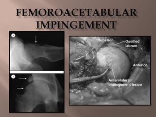
- 10. CAM Is impingement that occurs outside of the spherical head of the femur, caused by bone metaplastic overgrowth at the head neck junction, that damages the articular cartilage from overload and shear as it rotates in the socket. The result is a spectrum of degenerative changes in the labrum and in acetabular cartilage. Pathology ranges from softening and blistering of anterior acetabular articular cartilage in the earliest stages, to labrocartilaginous junctional degeneration and tearing or full- carpet delamination from the bone in late stages.
- 11. Acetabular Overgrowth Cartilage Delamination
- 12. Risk factors for developing CAM impingement include: Excessive movement in end range of motion Other predisposing conditions that result in anatomical deformity of the femoral neck can result in CAM impingement. Such as: Rotational malunion due to previous femoral neck fracture.’ Flattening of the femoral head due to femoral head necrosis Reported to be involved in some people with slipped capital femoral epiphysis Perthe’s Elliptical Femoral head (decreased epiphyseal height) Decreased femoral anteversion
- 13. Occurs with acetabular overcoverage, which limits range of motion and leads to a conflict between the acetabulum and the femur. The result is more labral damage then acetabular articular changes. In cases of Pincer impingement, the femoral head may be morphologically normal; however abutment results from an acetabular abnormality.
- 14. A common cause is acetabular overcoverage of the femoral head known as coxa profunda. In this instance, the centre-edge or Wiberg’s angle, which measures the lateral covering of the femoral head by the acetabular roof is greater than 40 degrees with a normal range for adults stated as being 20-46 degrees. Wiberg’s Angle
- 15. Repetitive micro-trauma between the femoral neck and the acetabular rim may cause multiple cleavage planes within the labrum leading to labral hypertrophy, intrasubstance cyst formation, calcification and labral degeneration, thus leading to worsening of acetabular overcoverage. Other causes of Pincer impingement are protrusion acetabuli, in which the medial wall of the acetabulum invades the pelvic cavity with associated medial displacement of the femoral head. This is associated with a variety of conditions, most noticeably Marfan’s Syndrome.
- 16. It has been shown that separation of the acetabular cartilage from the labrum occurs in the anterior superior region, this is the most common site of pathology. Maximal damage occurs at the 1 0’clock position to a mean depth on average around 11mm, this is roughly one-third of the depth of the cartilage at this point.
- 17. The pathological findings can be explained by looking at the structure of a normal acetabulum. In a normal hip, the acetabulum labrum merges with the acetabular cartilage through a transition zone of roughly 1-2mm, but most patients with CAM impingement, separation of the acetabular cartilage from the labrum is usually seen (the delaminating effect). As the labrum has a stable fixation to the acetabular rim, it appears that the abutment of the CAM causes a pathological under surface separation of the cartilage from the labrum across the ‘transition zone’.
- 18. In cases of Pincer pathology a wider pattern of disease is usually noted in this type of impingement it has been noted that some patients have additional posterior lesions associated with the disease. Labral lesions were shown to be maximal between the 11 and 1 o’clock positions, although maximal damage is usually seen at 12 o’clock, with the mean depth of the cartilage lesion usually around 4mm.
- 19. It has been shown that 31% of Pincer impinged patients showed posterior-inferior roughening of the femoral head, and 62% had cartilage damage posteriorlly. This occurs due to the posterior subluxation of the femoral head once further flexion occurs on an already impinging anterior superior rim. A study by Seldes in 2001, showed endochondral ossification within the labrum and they proposed that repeated micro-trauma induces bone growth at the base of the labrum, further deepening the acetabulum and compounding the problem.
- 20. Acetabular Labrum Normal acetabular labrum is composed of circumferential collagen fibres running parallel to the acetabular rim, together with small flat, inactive fibroblasts. Studies have shown that aging produces labral tears with internal cleavage being demonstrated on histological examination.
- 21. Conversely, labral histological examination with patients with FAI showed no mechanical lesions, in the matrix, the pathological process is consistent with a chronic degenerative process, showing only thickened and disorganised occasionally cystic matrix with no signs of inflammation. This shows that FAI is a chronic process that gradually produces a degenerative reaction at the site of the impingement.
- 22. Cartilage Cartilage in patients with FAI has shown to be coarse with relatively low levels of proteoglycans. The heterotrophic bone shows large amounts of unmineralized osteoid formation with pseudocysts, this is attributed to increased osteoblastic activity at the site.
- 23. Bone Routine radiographic investigations should include the following: AP view of the pelvis with the patient lying supine with the leg in 15 degrees of internal rotation. Cross-table lateral view, with the leg internally rotated. Alternative view is the Dunn/Rippstein, the hip is placed in 45 degrees of flexion to show anterior femoral head-neck junction abnormality.
- 25. Findings on radiographs with patients with FAI include the following: Reduced head neck offset Herniation Pits Pistol grip deformity (femoral head OA, head of the femur loses it non-spherical appearance). Subclinical slipped capital femoral epiphysis Coxa profunda Protrusion acetabuli Coxa vara or extreme coxa valga
- 26. Additionally imaging modalities may be obtained to further define femoracetabular impingement abnormalities and to characterise associated intra- articular disease. MRI provides detailed information regarding the anatomy of the femoral head-neck junction, presence of labral disease and integrity of articular cartilage. CT scan offers more detail of bony anatomy and may assist in analysing acetabular version, head-neck junction offset, femoral head shape, joint congruency and subchondral degenerative cystic changes.
- 27. Conservative (Non-surgical Modalities) Non-operative measures encompass relative rest, activity modification, NSAID’s, local and global strengthening and corticosteroid injections. This success of conservative management is highly dependent on the severity of the structural impingement, stage of secondary OA, patient age and patient activity level. There efficacy of using this type of treatment in research has shown to be questionable as it does not address the underlying pathomechanics of FAI, and it has been shown in research that disease progression is very common.
- 28. Surgical Options Surgical treatment for FAI is dictated by the anatomic location of the offending lesion, associated intra- articular problems and the extent of the joint degeneration. The goal of surgery in FAI is to improve the clearance of the head-neck junction within the acetabulum and to eliminate the abnormal contact between the proximal femur and the acetabular rim. The technical goals are to treat the diseased elements of the acetabular labrum and articular cartilage. Both open and arthroscopic techniques have been developed to improve clearance.
- 29. CAM Open procedures have long been the recommended type of surgery used to treat FAI as it allows an unobstructed view of the femoral head and acetabulum.
- 30. For CAM impingement: Surgical dislocation is performed. Followed by excision osteoplasty of small ‘sleeves’ of bone from the impinging section of the femoral head. Varying amounts of the anterolateral portion of the femoral neck junction is removed to alleviate the symptoms, however removing more than 30% results in an increased rate of fracture in response to axial loading, and is therefore to be avoided.
- 31. For Pincer Impingement: If the abnormality lies with the acetabulum, then a resection osteoplasty of the excessive acetabular rim may be undertaken. Or if the acetabulum is retroverted a peri-acetabular osteotomy is usually considered.
- 32. Arthroscope is starting to become more popular in the surgical treatment of FAI. With the arthroscope technique the intra-articular pathology is usually addressed first, including chondral defects and labral repair. Following this an osteplasty in usually performed.